While some people know what Ehlers-Danlos Syndrome (EDS) is, not many know that it is actually a group of 13 different disorders or types. One of the most prevalent types is Hypermobile Ehlers-Danlos Syndrome (hEDS), accounting for 80-90% of EDS cases [1]. The Ehlers-Danlos Syndromes are defined as inherited connective tissue disorders, affecting structural proteins (namely collagen) that leads to joint hypermobility, skin hyperextensibility, and tissue fragility [2]. hEDS is the only EDS subtype that does not have a currently known genetic basis, though it is a hereditary disorder with autosomal dominant inheritance pattern [2]. Therefore, hEDS is in part a diagnosis of exclusion, as genetic testing can be done for the other subtypes.
The Ehlers-Danlos Syndromes as whole are related to the Joint Hypermobility Spectrum, a spectrum of disorders intended to classify different severities of Hypermobility Spectrum Disorders (HSD). At the most extreme end of the hypermobile spectrum lies hEDS [3]. hEDS differs from the other EDS subtypes in its presentation as well. hEDS includes general joint hypermobility (GJH) but has less severe skin involvement compared to classical or vascular EDS types [1]. Easy bruising and impaired wound healing are also common. The definition of hEDS has evolved to include chronic pain and chronic fatigue as common presentations in those affected by the disease. The current diagnostic criteria include confirmation of GJH using the Beighton scoring system, positive musculoskeletal and pain symptoms or family history, and exclusion of other EDS subtypes or HSDs [4].
(Visit this link for the full diagnostic criteria of hEDS)
Apart from acute complications such as dislocation and subluxation, hEDs treatment revolves around chronic pain management and prevention of complications [1]. Acute exacerbations should be treated accordingly using joint reduction techniques and acute pain management. Physical therapy can be used to increase joint stability using low resistance exercises and stretching to increase muscle tone, thereby reducing the chance of acute joint injury. Patients should generally avoid hyperextension and high impact exercise. Oral acetaminophen, NSAIDs, and COX-2 inhibitors can be used as analgesics for chronic pain, as well as after acute injury. Cannabinoids can be considered for chronic pain, but opiates should rarely be used. If an hEDS patient must undergo surgery, careful technique should be used to minimize the wound site and promote healing. In summary, it is important to understand hEDS and how it differs from EDS and other HSDs to properly diagnose and treat patients.
References
1.Tinkle B, Castori M, Berglund B, Cohen H, Grahame R, Kazkaz H, et al. Hypermobile Ehlers-Danlos syndrome (a.k.a. Ehlers-Danlos syndrome Type III and Ehlers-Danlos syndrome hypermobility type): Clinical description and natural history. American Journal of Medical Genetics Part C: Seminars in Medical Genetics. 2017 Feb 1;175(1):48–69.
2.The Types of EDS [Internet]. The Ehlers Danlos Society. 2017. Available from: https://www.ehlers-danlos.com/eds-types/
3.What are the hypermobility spectrum disorders? [Internet]. The Ehlers Danlos Society. 2017. Available from: https://www.ehlers-danlos.com/what-is-hsd/
4.Forghani I. Updates in Clinical and Genetics Aspects of Hypermobile Ehlers Danlos Syndrome. Balkan Medical Journal. 2019 Jan 10;36(1):12–6.
Written by Jonathan Hunyadi with Dr. Nabil Ebraheim
Chronic exertional compartment syndrome (CECS) a pathology in runners usually involving the anterior compartment of the leg. It is believed to result from swelling and hypoperfusion of muscle and nerve during physical activity. Patients typically present with anterior burning leg pain that is exacerbated by exercise and is greatly reduced or completely subsides 15 to 30 minutes after exercise.
The condition can be diagnosed by measuring the pressure of the affected compartment one and five minutes after exercise. A pressure of 30 mmHg one minute and a pressure of 20 mmHg five minutes after exercise is considered diagnostic. Patients with CECS usually have a resting intra-compartment pressure greater than 15 mmHg which greatly increases during running. This typically produces a burning, cramping or aching pain after about 10 minutes of running resulting in cessation of exercise. Additionally, patients sometimes report tingling over the dorsal aspect of the foot while running.
Patients often present following stretching and strengthening therapy without relief. On physical exam, the patient will present with diffuse, nonspecific tenderness over the anterolateral leg without focal tenderness over bone. Pulses and x-ray will be normal and bone scan or MRI will be negative for stress fractures. Classic findings of acute compartment syndrome such as pain with passive toe dorsiflexion and sensory loss in the first web space, are typically absent.
Following diagnosis, treatment consists of the surgical release of affected compartments. During lateral compartment release, the superficial peroneal nerve, which pierces the fascia 10cm to 12cm proximal to the tip of the lateral malleolus, must be avoided. Surgical fasciotomy is usually successful but with a relatively high recurrence rate of approximately 20%. Recurrence typically occurs around two years following the initial procedure and is due to fibrosis within the compartment, causing return of symptoms and potential nerve entrapment. Additional causes of recurrences are inadequate release, failure to recognize and release all compartments, and misdiagnosis.
The differential diagnosis for CECS is large with overlap of symptoms. A common example is medial tibial stress syndrome. With this condition, bony tenderness along the posteromedial tibia will be present. Popliteal artery entrapment, a dynamic exercise related vascular phenomenon, is another condition in the differential. CECS can be distinguished by its predictable exercise related onset, relief of symptoms at rest and by being present for a long time.
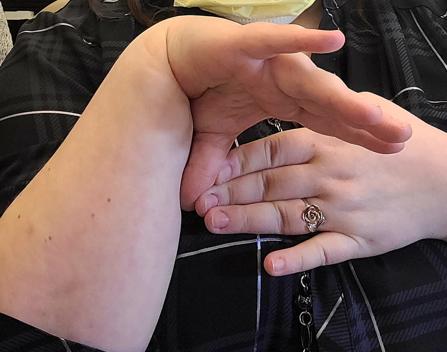
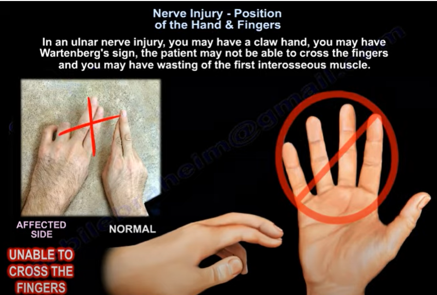
The presentation of a patient’s hand may provide insight to which nerve is damaged and the approximate location of the damage. When presenting with ulnar n. damage, a patient may show a claw hand, Wartenberg’s sign, or Froment’s sign. Claw hand will present with clawing of the fourth and fifth digits due to the inability to extend the fingers specifically at the interphalangeal joints. This presentation is due to the lack of innervation to the intrinsic muscles of the hands, and the unopposed action of the flexor digitorum profundus m. (Moore et al., 2018). This indicates damage near the distal end of the ulnar n., below the elbow, and potentially near the wrist. Wartenberg’s sign will be seen as the inability to adduct the fifth digit when extended as well as an inability to cross the second and third digit. This is due to ulnar n. injury leading to wasting of the fifth interosseous m. This causes the fifth digit to rest in a more abducted position due to the unbalanced action of the extensor digiti minimi m. (Ebraheim, 2021). There will also be loss of function of the lumbrical m. in the fourth and fifth digits. A Froment’s test will detect palsy of the ulnar n. resulting from compression in the cubital tunnel. When asking the patient to pinch a piece of paper between their thumb and second digit, a positive Froment’s Sign will show as the patient flexing their thumb’s interphalangeal joint to grip the paper as the paper is pulled away (Attum, 2021). This will result from a weak adductor pollicis m. due to ulnar n. palsy.
Depending on the location of a lesion to the median n., the patient will present with one of three signs. A positive Benedictine sign will be caused by proximal median n. damage. It will be seen as paralysis of the first and second digit, with weakness to the third digit. A proximal lesion would lead to paralysis of several muscles (FDS, FPL, FPB, and the radial half of FDP), leaving the ulnar half of the flexor digitorum profundus m. as the only remaining flexor (Ebraheim, 2021). When the patient is asked to make a fist, the hand will resemble the similar position taken during a blessing. A positive Benedict sign will also resemble the ulnar claw hand. However, the Benedict sign will present when the patient is flexing, not extending the fingers (Ebraheim, 2021). A median n. injury affecting the anterior interosseous n. branch will present as an inability to do the OK sign. This occurs due to paralysis of the flexor pollicus longus m. and the lateral part of the flexor digitorum m. (Moore et al., 2018). A positive Ape hand (Simian hand) is caused by paralysis of only the thenar m. from damage to the recurrent branch of the median n. to the thenar m. The thumb will be seen in the same plane as the other digits due the thumb being pulled more dorsal by the action of the adductor pollicis m., which is innervated by the ulnar n. (Moore et al., 2018).
Finally, proximal radial n. damage will be seen as wrist drop. This is usually caused by fractures of the distal third of the humeral shaft (Holstein-Lewis Fracture) and caused paralysis of the wrist and fingers extensors (Ebraheim, 2010). Lower radial n. injury will present in the patient as the ability to extend the wrist, but the loss of finger extension. There will be no wrist drop, but the patient would not be able to make a hitchhiking sign.
Reference List
1. Attum B. Physical exam of the hand [Internet].
Orthobullets. Lineage Medical, Inc.; 2021 [cited 2021Oct21]. Available from:
https://www.orthobullets.com/hand/6008/physical-exam-of-the-hand
2. Ebraheim N. Anterior Interosseous Nerve Injury -
Everything You Need To Know - Dr. Nabil Ebraheim [Internet]. YouTube. 2021
[cited 2021Oct21]. Available from: https://www.youtube.com/watch?v=M9y-iDKLDPE
3. Ebraheim N. Claw Hand, Ulnar Claw Hand - Everything You
Need To Know - Dr. Nabil Ebraheim [Internet]. YouTube. 2017 [cited 2021Oct21].
Available from: https://www.youtube.com/watch?v=GyqaKGg3HmM
4. Ebraheim N. Nerve Injury Position of the Hand &
Fingers - Everything You Need To Know - Dr. Nabil Ebraheim [Internet]. YouTube.
2021 [cited 2021Oct21]. Available from:
https://www.youtube.com/watch?v=nwd1h0Dfo5o
5. Ebraheim N. Radial Nerve Palsy, injury - WRIST DROP .
Everything You Need To Know - Dr. Nabil Ebraheim [Internet]. YouTube. 2010 [cited
2021Oct21]. Available from: https://www.youtube.com/watch?v=_Cu6ttAhe8Y
6. Moore KL, Dalley AF, Agur A. Clinically Oriented Anatomy.
8th ed. Philadelphia, PA: Lippincott Williams and Wilkins; 2018.
Lateral
epicondylitis, also known as tennis elbow, is an overuse injury that results in
inflammation, tendinosis, and lateral elbow pain. It is the most common cause
of pain in the lateral elbow, affecting between 1 and 3% of the general
population (1). The primary structure impacted in tennis elbow is the extensor carpi
radialis brevis, which originates at the lateral epicondyle (2). This condition is primarily seen in middle-aged individuals,
especially those between the ages of 40 and 50 (3). Tennis players (up to 50% of regular players) and workers who engage
in heavy lifting or repetitive gripping are more likely to develop tennis elbow
(2). Other conditions, such as rotator cuff pathology or De Quervain’s
diseases, and lifestyle factors, such as smoking, are associated risk factors
for lateral epicondylitis (4). Rotator cuff pathology could be a risk factor because lateral
epicondylitis can also be caused by biomechanical stress, but it is unclear
exactly why it and other conditions are associated with each other (4). In terms of histology, disorganized collagen, dense fibroblasts, and
vascular hyperplasia are primarily seen (2). Immature fibroblastic and vascular infiltration of the origin of the
extensor carpi radialis brevis has consistently been identified during surgery (5). A physical exam and history are typically used to diagnose this
condition. Clinical tests to assist in diagnosis include grip strength,
Cozen’s, Maudsley’s, and Mill’s tests (3). Lateral epicondylitis is indicated if the previous tests are
positive along with reduced grip strength or reproduced pain (3). If necessary, diagnostic scans can be obtained. Majority of patients
show altered signal around the lateral epicondyle on MRI scans and hot focus on
infrared thermography (6, 7). Radial
tunnel syndrome is a differential diagnosis of tennis elbow. This syndrome is
seen in 5% of patients who have compression of the posterior interosseous
nerve. The pain associated with radial tunnel syndrome is approximately 3-4 cm
distal and anterior to the lateral epicondyle, which differentiates it from
tennis elbow.
Non-surgical
treatments are the primary mode of treatment and there is a 95% success rate
with treatments to relieve pain. The most frequently used treatment is
corticosteroid injection (2). Oral or topical non-steroidal anti-inflammatory drugs (NSAIDs) can
also be prescribed, but their effectiveness is variable (8). Patients can also undergo physical therapy to relieve their
symptoms. Eccentric exercises have been shown to be especially effective in
pain management (9). Using an inelastic, nonarticular proximal forearm brace could also
be recommended (10). Two relatively newer treatments for tennis elbow are ultrasonic (US)
and extracorporeal shock wave therapy (ESWT). There are minimal side effects to
US and ESWT, thus making them preferable for patients and clinicians (11). Even though there is no difference between US and ESWT in elbow
function evaluation scores, ESWT has been shown to have greater efficacy in
pain relief (12). Surgical procedures are a last resort for treatment of tennis elbow
and only indicated if patients are unresponsive to conservative treatments
after an extended period of time. Debridement is the most common surgical
intervention, but it can result in injury of the lateral collateral ligament
and subsequent posterolateral rotary instability of the elbow.
References
1. Shiri R, Viikari-Juntura
E, Varonen H, Heliovaara M. Prevalence and Determinants of Lateral and Medial
Epicondylitis: A Population Study. American Journal of Epidemiology.
2006;164(11):1065-74. doi: 10.1093/aje/kwj325.
2. Cutts S, Gangoo S, Modi N, Pasapula C.
Tennis elbow: A clinical review article. Journal of Orthopaedics.
2020;17:203-7. doi: 10.1016/j.jor.2019.08.005.
3. Speers CJ, Bhogal GS, Collins R.
Lateral elbow tendinosis: a review of diagnosis and management in general
practice. British Journal of General Practice. 2018;68(676):548-9. doi:
10.3399/bjgp18x699725.
4. Titchener AG, Fakis A, Tambe AA, Smith
C, Hubbard RB, Clark DI. Risk factors in lateral epicondylitis (tennis elbow):
a case-control study. Journal of Hand Surgery (European Volume).
2013;38(2):159-64. doi: 10.1177/1753193412442464.
5. Nirschl RP, Pettrone FA. Tennis elbow.
The surgical treatment of lateral epicondylitis. The Journal of bone and joint
surgery American volume. 1979;61(6A):832-9. PubMed PMID: 479229.
6. Steinborn M, Heuck A, Jessel C, Bonel
H, Reiser M. Magnetic resonance imaging of lateral epicondylitis of the elbow
with a 0.2-T dedicated system. European Radiology. 1999;9(7):1376-80. doi:
10.1007/s003300050851.
7. Thomas D, Siahamis G, Marion M, Boyle
C. Computerised infrared thermography and isotopic bone scanning in tennis
elbow. Annals of the Rheumatic Diseases. 1992;51(1):103. doi:
10.1136/ard.51.1.103.
8. Pattanittum P, Turner T, Green S,
Buchbinder R. Non‐steroidal anti‐inflammatory drugs (NSAIDs) for treating
lateral elbow pain in adults. Cochrane Database of Systematic Reviews. 2013(5).
doi: 10.1002/14651858.CD003686.pub2. PubMed PMID: CD003686.
9. Croisier J-L, Foidart-Dessalle M,
Tinant F, Crielaard J-M, Forthomme B. An isokinetic eccentric programme for the
management of chronic lateral epicondylar tendinopathy. British Journal of
Sports Medicine. 2007;41(4):269. doi: 10.1136/bjsm.2006.033324.
10. Johnson GW, Cadwallader K, Scheffel SB,
Epperly TD. Treatment of lateral epicondylitis. Am Fam Physician.
2007;76(6):843-8. Epub 2007/10/04. PubMed PMID: 17910298.
11. Coombes BK, Connelly L, Bisset L,
Vicenzino B. Economic evaluation favours physiotherapy but not corticosteroid
injection as a first-line intervention for chronic lateral epicondylalgia:
evidence from a randomised clinical trial. British Journal of Sports Medicine.
2016;50(22):1400-5. doi: 10.1136/bjsports-2015-094729.
12. Yan C, Xiong Y, Chen L, Endo Y, Hu L, Liu
M, et al. A comparative study of the efficacy of ultrasonics and extracorporeal
shock wave in the treatment of tennis elbow: a meta-analysis of randomized controlled
trials. Journal of Orthopaedic Surgery and Research. 2019;14(1). doi:
10.1186/s13018-019-1290-y.
Bursitis of the Knee, Hip, Elbow and Shoulder - Everything You Need to Know Written by Andrew Kelley with Dr. Nabil Ebraheim
Prepatellar Bursitis of the Knee Prepatellar bursitis, also known as housemaid’s, carpet layer’s,
and carpenter’s knee, is a superficial bursitis caused by inflammation of the
bursa separating the patellar bone and the skin (1). Patients with prepatellar
bursitis will normally present with knee pain and swelling (2). Prepatellar bursitis
is mostly caused by long-term repetitive mini trauma from kneeling and crawling
on hard surfaces. Other causes include acute injury, infection, gout, and
rheumatoid arthritis (2). Its annual incidence is 10/100,000 per year with 80%
of those affected being males age 40-60 (1). In cases of non-traumatic prepatellar
bursitis, treatment is dependent on resolution of the underlying condition.
Early differentiation between septic and non-septic bursitis is important in
the early presentation in order to improve patient outcomes. Acute bursitis
normally responds well to conservative treatment such as rest, ice, activity
modification, NSAIDs, and fluid aspiration. Chronic bursitis due to mini
traumas is treated similarly but may require additional corticosteroid therapy
(1).
Olecranon Bursitis of the Elbow Olecranon Bursitis, also known as student’s elbow and plumber’s
elbow, is caused by inflammation of the bursa overlaying the olecranon process
of the ulnar bone at the tip of the elbow. This bursa allows for smooth motion
of the olecranon process against the superficial tissue at the tip of the
elbow. Affected patients normally present with swelling at the bend of the
elbow. A characteristic “golf ball” shape of swelling can be seen, and a fully
intact range of motion of the elbow can differentiate it from elbow joint
injuries (3). Olecranon Bursitis most commonly affects men age 30-60. Most
cases are due to repeated minor trauma and sports (4). Treatment is focused on resolving
the underlying cause of inflammation. Conservative treatment includes ice and
rest along with NSAIDs for symptomatic relief are indicated. While aspiration
and corticosteroid injection are proven relief interventions, they carry an
increased risk for infection (4).
Greater Trochanteric Bursitis of the Hip Greater trochanteric bursitis, or greater trochanteric pain
syndrome (GTPS), is caused by inflammation of the bursa laying deep to the
iliotibial band and superficial to the greater trochanter of the femur. It acts
as a lubricant for the gluteal tendons. Patients with hip bursitis normally
present with chronic intermittent pain of the lateral hip, thigh, and buttock
(6). This bursitis normally affects women age 40-60. The increased pelvic width
of women relative to their body may predispose them to increased iliotibial
band tension on the bursa (6). The cause of hip bursitis can be repetitive
microtrauma, blunt trauma, or idiopathic. Movements requiring repetitive hip
abduction like stair climbing and bicycling, direct traumatic falls, and
sedentary lifestyles are common causes of this condition (5). Common treatments for this bursitis include
NSAIDs, physical therapy, and corticosteroid injection. Surgery is a rare
treatment option for bursitis resistant to conservative treatment options (5).
Subacromial Bursitis of the Shoulder Subacromial bursitis is caused by inflammation to the bursa just
below the acromion process. The subacromial bursa acts as a lubricating medium between
the acromion process superiorly and the muscles of the rotator cup inferiorly. Subacromial bursitis normally presents as
anterolateral shoulder pain, especially during overhead activities. This chronic
inflammation of the shoulder bursa can eventually lead to weakness and rupture
of the surrounding ligaments and tendons (7). Older individuals are more likely
to experience shoulder bursitis due to years of overuse. Most patients present due
to direct trauma to the shoulder or repetitive overhead activities (7).
Treatment includes rest, NSAIDs, physical therapy, and corticosteroid
injections. Surgical therapy is reserved for cases unresponsive to conservative
therapy (7).
References: 1.Rishor-Olney CR, Pozun A. Prepatellar Bursitis. [Updated 2021
Sep 2]. In: StatPearls [Internet]. Treasure Island (FL): StatPearls Publishing;
2021 Jan-. 2.J.
Dean Cole MD. Causes of knee bursitis (prepatellar bursitis) [Internet].
Arthritis. Arthritis-health; [cited 2021Oct28]. Available from:
https://www.arthritis-health.com/types/bursitis/causes-knee-bursitis-prepatellar-bursitis 3.Pangia
J. Olecranon bursitis [Internet]. StatPearls [Internet]. U.S. National Library
of Medicine; 2021 [cited 2021Oct28]. Available from:
https://www.ncbi.nlm.nih.gov/books/NBK470291/ 4.Blackwell JR, Hay BA, Bolt AM, Hay SM. Olecranon bursitis: a
systematic overview. Shoulder Elbow. 2014 Jul;6(3):182-90. doi:
10.1177/1758573214532787. Epub 2014 May 6. PMID: 27582935; PMCID: PMC4935058. 5.Seidman
AJ. Trochanteric bursitis [Internet]. StatPearls [Internet]. U.S. National
Library of Medicine; 2021 [cited 2021Oct28]. Available from:
https://www.ncbi.nlm.nih.gov/books/NBK538503/ 6.Reid
D. The management of Greater Trochanteric pain syndrome: A systematic
literature review [Internet]. Journal of orthopaedics. Elsevier; 2016 [cited
2021Oct28]. Available from:
https://www.ncbi.nlm.nih.gov/pmc/articles/PMC4761624/ 7.Faruqi
T. Subacromial bursitis [Internet]. StatPearls [Internet]. U.S. National
Library of Medicine; 2021 [cited 2021Oct29]. Available from:
https://www.ncbi.nlm.nih.gov/books/NBK541096/
Fractures of the Calcaneus: Everything You Need to Know
Written by Dominic Ruwe and Dr. Nabil Ebraheim
Fractures of the calcaneus can be open or closed.1
Open fractures are more serious than closed fractures.1 The primary
fracture line is caused by an axial load injury.1 The primary
fracture line goes from anterolateral to posteromedial.1 The primary
fracture line divides the calcaneus into two main fragments: the superomedial
fragment which is also called the constant or sustentacular (SAS) fragment and
the superolateral or tuberosity fragment.1 The superomedial fragment
includes the sustentaculum tali and is stabilized to the talus by ligaments.
So, the talus is attached to the constant fragment.1 The sustentacular
fragment is a useful reference point for fracture reduction.2 The
flexor hallucis longus tendon lies underneath the sustentaculum. If screw
placement to the sustentacular fragment is too long, the flexor hallucis longus
tendon could be affected, causing fixed flexion of the big toe.3
The
Essex-Lopresti classification system is a useful way to differentiate between
different joint fractures. There are two types of Essex-Lopresti fractures: a
tongue-type fracture and a joint depression type fracture.1 In the
tongue-type, the posterior facet is attached to the tuberosity. In the joint
depression type, the posterior facet is not attached to the tuberosity.4
In the tongue-type, the primary fracture line exits anterolaterally and
posteromedially.5 The secondary fracture line appears beneath the
posterior facet and exits posteriorly through the tuberosity.5 The
superolateral fragment and posterior facet are attached to the tuberosity. The
tongue-type fracture can be treated with open reduction and internal fixation.6
In
the joint depression type, the primary fracture line splits the calcaneus
obliquely through the posterior facet and exits anterolaterally and
posteromedially.1 The secondary fracture line exits superiorly just
behind the posterior facet.1 The posterior facet is a free fragment.
The lateral portion of the posterior facet is usually involved and depressed.4
The Sander’s classification of calcaneal fractures is
used to guide the treatment and predict the outcome of the treatment. This
classification system is based on the number of posterior facet fracture
fragments seen on a coronal CT scan.7 Type I is a nondisplaced
fracture which requires nonoperative treatment.7 Type II is a
two-part fracture of the posterior facet.7 Type III is a three-part
fracture of the posterior facet.7 Type II and III calcaneal
fractures benefit from surgery of reduction and fixation.1 Type III
fractures normally result in more arthritis because it has more fracture
fragments and may end by fusion.8 Type IV fractures are highly
comminuted.9 They may require primary subtalar arthrodesis.1
Calcaneal avulsion fractures are typically serious. These
types of fractures require urgent reduction and internal fixation to prevent
skin complications.10 In joint depression fractures of the
calcaneus, the swelling must go down before surgery. Avulsion fractures of the
calcaneus are emergencies, so emergency surgery is performed before the
swelling goes down. Open reduction and internal fixation of the calcaneus is
generally delayed for 1-2 weeks to allow for improvement of the soft tissue
swelling, except with avulsion fractures.1 Avulsion fractures can
cause skin tenting and urgent reduction is recommended.10
There are many associated conditions with calcaneal
fractures. Ten percent are associated with spinal fractures.11 Ten
percent are associated with compartment syndrome of the foot.12 If
this is neglected, it will lead to claw toes due to contracture of the
intrinsic flexor muscles.12 Approximately ten percent are associated
with bilateral fractures.13 Sixty percent are associated with
calcaneocuboid joint fractures.14 Calcaneal fractures may also be
associated with peroneal tendon subluxation. Peroneal tendon subluxation may be
detected on axial CT scans or it may be seen as an avulsion fracture of the
fibula on x-rays.15
Complication
rates for calcaneal fractures are high. Factors associated with poor outcomes are
age greater than 50, smoking, early surgery, history of a fall, heavy manual
labor, males, bilateral injury, workman’s compensation, and peripheral vascular
disease.1,16,17 Men do worse with calcaneal fractures than women.
Calcaneal fractures in men are normally associated with workman’s compensation,
heavy labor, and a 0˚ Bohler angle.1 These fractures typically need
subtalar fusion.18 Calcaneal fractures in females have a simple
fracture pattern. Since calcaneal fractures in males are usually more severe,
it follows that better outcomes are seen in females with calcaneal fractures.19
The
Bohler angle is measured on lateral x-rays.1 This angle is normally
between 20˚-40˚.1 The Bohler angle is formed by a line drawn from
the highest point of the anterior process of the calcaneus to the highest point
of the posterior facet and a line drawn tangential to the superior edge of the
tuberosity.1 A decrease in this angle indicates a collapse of the
posterior facet.1 When viewing calcaneal fractures with the Harris
view, the calcaneus appears to be shortened and widened with varus.1
When viewing calcaneal fractures through CT scans, the axial cut shows the
calcaneocuboid joint and peroneal tendon subluxation.1,20 The
sagittal view shows the subtalar joint and its depression.21 The
coronal view shows the displacement of the posterior facet.22
Coronal CT scans can also show the number of the joint fracture fragments.1
The surgical outcome of calcaneal fractures correlate with the number of the
joint fracture fragments and the quality of reduction.1 MR imaging shows
stress fractures of the calcaneus and the integrity of the peroneal tendons.23,24
Stress fractures of the calcaneus may be misdiagnosed as
plantar fasciitis.25 Stress fractures usually occur in female
runners.26 It is characterized by swelling and tenderness with
medial and lateral compression of the hindfoot during the squeeze test.27 If
the X-ray is negative, an MRI should be obtained. The fracture will be seen in
T1 MR imaging as a linear streak or a band of low signal intensity in the
posterior calcaneal tuberosity.28 In T2 imaging, the signal will be
increased.28
There
are several complications with calcaneal fractures. Wound-related complications
are the most common complication.29 Wound-related complications
occur more in smokers, diabetics, and patients with open fractures.1
Open fractures of the calcaneus is another common complication. Open fractures
of the calcaneus can lead to amputation.30 There is also a high risk
of infection with open fractures.30 Grade I and Grade II open
fractures have wounds that open medially. Open reduction and internal fixation
(ORIF) can be done to treat this complication.30 Open reduction and
internal fixation should not be done in Grade III medial wounds and in most
lateral wounds.30 Another complication is malunion of the calcaneus.31
This is characterized by widening of the heel, varus deformity, and loss
of height.31 The talus is dorsiflexed, limiting dorsiflexion of the
ankle.31 Peroneal tendon irritation and impingement from the lateral
wall is another complication.32
Surgery
on the calcaneus decreases the risk of post-traumatic arthritis.33 Tongue-type
and joint depression type fractures may benefit from open reduction and
internal fixation.6 Subtalar distraction arthrodesis is a good
operation to treat calcaneal fractures associated with loss of height and
limited dorsiflexion of the ankle.31 This operation improves talar
inclination and decreases anterior ankle impingement.31
Additionally, it takes care of arthritis in the subtalar joint.31
Another surgical approach is extensile lateral approach. The lateral calcaneal
artery provides blood supply to the lateral flap associated with the calcaneal
extensile approach.34 It is important to be aware that the Sural
nerve is in the vicinity of the surgical area.35 Delayed wound
healing is a common complication in the extensile lateral approach.35
References:
1. Trompeter A, Razik A, Harris M. Calcaneal fractures:
Where are we now? Strategies in Trauma and Limb Reconstruction.
2017;13(1):1–11.
2. Berberian W, Sood A, Karanfilian B, Najarian R, Lin S,
Liporace F. Displacement of the SUSTENTACULAR fragment in INTRA-ARTICULAR
CALCANEAL FRACTURES. Journal of Bone and Joint Surgery. 2013;95(11):995–1000.
3. Carr JB. Complications of CALCANEUS fractures
entrapment of the Flexor hallucis longus. Journal of Orthopaedic Trauma.
1990;4(2):166–8.
5. White EA, Skalski MR, Matcuk GR, Heckmann N, Tomasian
A, Gross JS, et al. Intra-articular tongue-type fractures of the calcaneus:
Anatomy, injury patterns, and an approach to management. Emergency Radiology.
2018;26(1):67–74.
6. Chhabra N, Sherman SC, Szatkowski JP. Tongue-type
calcaneus fractures: a threat to skin. The American Journal of Emergency
Medicine. 2013;31(7).
7. Jiménez-Almonte JH, King JD, Luo TD, Aneja A,
Moghadamian E. Classifications in Brief: Sanders classification OF
INTRAARTICULAR fractures of the calcaneus. Clinical Orthopaedics & Related
Research. 2018;477(2):467–71.
8. Rammelt S, Marx C. Managing severely malunited
calcaneal fractures and fracture-dislocations. Foot and Ankle Clinics.
2020;25(2):239–56.
9. Piovesana LG, Lopes HC, Pacca DM, Ninomiya AF, Dinato
MC, Pagnano RG. Assessment of reproducibility of sanders classification for
calcaneal fractures. Acta Ortopédica Brasileira. 2016;24(2):90–3.
10. Berringer R. Avulsion fracture of the calcaneus.
Canadian Medical Association Journal. 2018;190(45).
11. Rowe CR. Fractures of the os calcis. JAMA.
1963;184(12):920.
12. Myerson Mark, Manoli Arthur. Compartment syndromes of
the foot after calcaneal fractures. Clinical Orthopaedics and Related Research.
1993;&NA;(290).
13. Popelka V.
Súčasné trendy v liečbe intraartikulárnych zlomenín pätovej kosti [Current
Concepts in the Treatment of Intra-Articular Calcaneal Fractures]. Acta Chir
Orthop Traumatol Cech. 2019;86(1):58-64. Slovak. PMID: 30843515.14.
14. Kinner B, Schieder S, Müller F, Pannek A, Roll C.
Calcaneocuboid joint involvement IN CALCANEAL FRACTURES. Journal of Trauma:
Injury, Infection & Critical Care. 2010;68(5):1192–9.
15. Park C-H, Gwak H-C, Kim J-H, Lee C-R, Kim D-H, Park
C-S. Peroneal tendon Subluxation and dislocation In CALCANEUS FRACTURES. The
Journal of Foot and Ankle Surgery. 2021;60(2):233–6.
16. Su J, Cao X. Can operations achieve good outcomes in
elderly patients with SANDERS II–III calcaneal fractures? Medicine.
2017;96(29).
17. Clare MP, Crawford WS. Managing complications of
CALCANEUS FRACTURES. Foot and Ankle Clinics. 2017;22(1):105–16.
18. Csizy M, Buckley R, Tough S, Leighton R, Smith J,
McCormack R, et al. Displaced Intra-articular CALCANEAL FRACTURES. Journal of
Orthopaedic Trauma. 2003;17(2):106–12.
20. Toussaint RJ, Lin D, Ehrlichman LK, Ellington JK,
Strasser N, Kwon JY. Peroneal tendon DISPLACEMENT Accompanying INTRA-ARTICULAR
CALCANEAL FRACTURES. Journal of Bone and Joint Surgery. 2014;96(4):310–5.
21. Badillo K, Pacheco JA, Padua SO, Gomez AA, Colon E,
Vidal JA. Multidetector CT evaluation Of CALCANEAL FRACTURES. RadioGraphics.
2011;31(1):81–92.
22. Buckley R. Displaced fracture of the calcaneus body
[Internet]. AO Foundation Surgery Reference. [cited 2021Sep29]. Available from:
https://surgeryreference.aofoundation.org/orthopedic-trauma/adult-trauma/calcaneous/displaced-body-fractures/definition
23. Kato M, Warashina H, Kataoka A, Ando T, Mitamura S.
Calcaneal insufficiency fractures following ipsilateral total knee
arthroplasty. Injury. 2021;52(7):1978–84.
24. Park HJ, Cha SD, Kim HS, Chung ST, Park NH, Yoo JH,
et al. Reliability of MRI findings OF PERONEAL Tendinopathy in patients with
LATERAL CHRONIC Ankle Instability. Clinics in Orthopedic Surgery.
2010Nov5;2(4):237.
25. Weber JM, Vidt LG, Gehl RS, Montgomery T. Calcaneal
stress fractures. Clinics in Podiatric Medicine and Surgery. 2005;22(1):45–54.
26. Labronici P, Pires RE, Amorim L. Calcaneal stress
fractures in civilian patients. Journal of the Foot & Ankle.
2021;15(1):54–9.
27. Kiel J,
Kaiser K. Stress Reaction and Fractures. 2021 Aug 4. In: StatPearls [Internet].
Treasure Island (FL): StatPearls Publishing; 2021 Jan–. PMID: 29939612.
28. Lawrence DA, Rolen MF, Morshed KA, Moukaddam H. MRI of
heel pain. American Journal of Roentgenology. 2013Apr18;200(4):845–55.
29. Ding L, He Z, Xiao H, Chai L, Xue F. Risk factors for
postoperative wound complications of calcaneal fractures following plate
fixation. Foot & Ankle International. 2013;34(9):1238–44.
30. Heier KA, Infante AF, Walling AK, Sanders RW. Open
fractures of THE Calcaneus: Soft-tissue Injury DETERMINES OUTCOME. The Journal
of Bone and Joint Surgery-American Volume. 2004;86(11):2569.
31. Guang-Rong Y, Xiao Y. Surgical management Of
Calcaneal Malunion. Journal of Orthopaedics, Trauma and Rehabilitation.
2013;17(1):2–8.
32. Davis D,
Seaman TJ, Newton EJ. Calcaneus Fractures. 2021 Aug 9. In: StatPearls
[Internet]. Treasure Island (FL): StatPearls Publishing; 2021 Jan–. PMID:
28613611.
33. Vilá-Rico J, Ojeda-Thies C, Mellado-Romero MÁ,
Sánchez-Morata EJ, Ramos-Pascua LR. Arthroscopic posterior subtalar arthrodesis
for salvage of posttraumatic arthritis following calcaneal fractures. Injury.
2018;49.
34. Mehta CR, An VV, Phan K, Sivakumar B, Kanawati AJ,
Suthersan M. Extensile lateral versus sinus Tarsi approach For displaced,
intra-articular Calcaneal Fractures: A meta-analysis. Journal of Orthopaedic
Surgery and Research. 2018;13(1).
35. Buckley R. Extended lateral approach to the calcaneus
[Internet]. AO Foundation Surgery Reference. [cited 2021Sep29]. Available from:
https://surgeryreference.aofoundation.org/orthopedic-trauma/adult-trauma/calcaneous/approach/extended-lateral-approach-to-the-calcaneus