Showing posts with label causes and treatment. Show all posts

Tuesday, September 18, 2018

Ganglion Cyst of the Shoulder


Ganglion cysts can be important when they are located around the shoulder, especially when they are located in the suprascapular notch and the spinoglenoid notch. The suprascapular nerve passes under the transverse scapular ligament at the suprascapular notch. The transverse scapular artery runs above the transverse scapular ligament. The artery and nerve joint and then pass through the spinoglenoid notch under the inferior scapular ligament. The suprascapular nerve gives branches to the supraspinatus muscle and branches to the infraspinatus muscle.

Nerve compression from a ganglion cyst at the suprascapular notch affects both the supraspinatus and infraspinatus muscles, causing a decrease in abduction and loss of external rotation of the shoulder. Nerve compression at the spinoglenoid notch affects only infraspinatus muscle, causing loss of external rotation of the shoulder with the arm to the side. Spinoglenoid notch compression is usually associated with cysts and ganglia. In addition to compression of the suprascapular nerve, these patients may also have associated posterior labral tears.

Tuesday, July 3, 2018

Hip Dislocation Following Total Hip Surgery


There are two types of hip joint dislocations: posterior and anterior. The position of the leg is important in determining the type of hip dislocation. When the hip is dislocated, the leg is usually shortened and it assumes a different position than the normal leg (the other leg). If the dislocation is posterior, the leg will be in adduction and internal rotation. If the dislocation is anterior, the leg will be in abduction and external rotation. Notice that the affected extremity is shortened and externally rotated. Leg shortening can also be seen in hip fractures and the leg will be shortened and externally rotated.


Dislocation of the hip following total hip surgery may require revision surgery, but it is rare. The majority of hip dislocations after total hip dislocations are posterior, and they are usually treated without surgery. Most occur within the first month of THA; 1-4% in primary, 16% in revision. There is more incidence of dislocation in revision hip replacement.


Causes & Risk Factors:

  • Posterior Approach (try to repair the capsule adequately)
  • Malposition of the component
    • Ideally, the normal cup component will be in 20° of anteversion and 40° of abduction
    • When the hip dislocates posterior, always check for retroversion of the cup.

  • Prior hip fracture surgery, especially in the elderly
  • Weakness of the abductor muscle—must achieve soft tissue tension and function
  • Alcohol abuse
  • Improper neck length—looseness of the hip

The patient should be careful to avoid all activities that cause dislocation after total hip surgery. The patient should use a pillow between the legs while sleeping on their back and they should be careful to not cross their legs in their sleep. Patients cannot sleep on their sides as well. The patient should not bend the body at the waist farther than 90°. When sitting, the patient must avoid chairs that make it difficult to stand up, and sit at more than a 90° angle. The patient must not sit with their legs crossed in the chair. The patient must be made aware that if the leg is changed from its usual position, or becomes shortened, then the hip is probably dislocated and their doctor should be consulted.


X-rays of the dislocated total hip should include AP and lateral views. Look for eccentric wear and look for the position of the prosthesis. CT scans may be needed before or after reduction of the dislocation to check the version of the components. Treatment is variable and depends on the situation. The treatment should be tailored for each case. The majority of these cases with early dislocations can be treated successfully with closed reduction and immobilization.

The treatment should start with closed reduction of the total hip and immobilization. Hip stability is checked after reduction of the dislocation. Immobilization can be done by a brace or a hip spica. Trochanteric osteotomy and advancement of the trochanter and tensioning the abductor muscle. Screws or wires can be used. The prosthesis must be in good alignment for this procedure to work. Constrained acetabular components are used when the abductor muscle is deficient and the component position is good. Revision total hip is done in recurrent dislocation with malposition of the component or polyethylene wear.

Tuesday, June 19, 2018

Isolated Fibular Fractures


Fibular fractures are usually associated with a complex injury, however they can be an isolated fracture. Complex injuries where a fibula fracture can occur include: fracture of the fibula and tibia, ankle fracture, pilon fracture, and Maisonneuve fractures.

Maisonneuve fractures involve a fracture of the proximal fibula associated with an occult injury of the ankle. Isolated fibular fractures are rare and usually the result of direct trauma. The fibula carries about 15% of the axial load and is the site of muscle attachment for the peroneus muscles and the flexor hallucis longus muscle. Check the patient who has a fibular fracture and no other fracture involving the tibia to rule out a possible Maisonneuve fracture, especially if there is no history of direct trauma to the leg. A high index of suspicion is necessary to diagnose and treat this injury. For high fibular fractures, the physician should look for signs of syndesmotic injury. Syndesmotic injury may include an unexplained increase in the medial clear space or the tibiofibular clear space is widened (should be less than 5mm). The x-ray will show the fracture to be rotational or oblique. Maisonneuve fractures require surgery to fix the syndesmosis.


Treatment will consist of reduction and fixation. It is important to determine if the injury is a Maisonneuve fracture or an isolated fibular fracture. An isolated fibular fracture will not need surgery.

Friday, March 2, 2018

Clay Shoveler’s Fracture


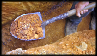
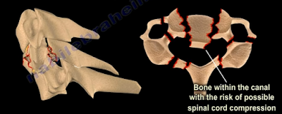
A clay shoveler’s fracture is a stable fracture through the spinous process of the vertebra C6-T1 and typically occurs at the level of C6 or C7. Clay shoveler’s fracture gets its name from the common injury suffered by men diffing deep ditches in Australia during the 1930s.
Powerful hyperflexion of the neck combined with contraction of the paraspinous muscles during shoveling is the typical mechanism of injury. The tremendous force pulls on the spinous process, producing an avulsion fracture.
The fracture is best seen on a lateral view x-ray. There will be a ghost sign on AP view x-rays—double spinal process of C6 or C7, resulting from a displaced fractured spinous process.
Stable fractures can be treated nonoperatively, utilizing a collar and physical therapy.

Tuesday, February 6, 2018

Stem Cells and Orthopaedics


Stem cells may help tissues that are injured or damaged to renew and regenerate themselves. Depending on the treatment and medium, stem cells have the ability to become different types of cells such as bone, cartilage, and blood vessels. There are several conditions in which stem cells are used as treatment, including: avascular necrosis, arthritis, and nonunion.
When Avascular Necrosis of the femoral head occurs due to the diminished blood supply, there is a death of a segment of bone, which is considered necrotic. The surgeon can inject stem cells into this area to revive this area by drilling into the bone. When using stem cells to treat AVN, the surgeon will need to create a channel for new blood vessels to form into the area that lacks blood supply. After the channel is created, the stem cells are injected into the necrotic femoral head.
Stem cell treatments for joint pain and arthritis is not proven to be effective. However, there is some use in knee arthritis for cartilage regeneration.
 

The best use of stem cells in Orthopaedics is its treatment for nonunion fractures. A nonunion fracture is classified as a fracture that does not heal after a reasonable period of time or a fixation failure. Nonunion may also be due to motion of the bony ends and incomplete healing of the fracture; fractures of this nature will need a lot of assistance. Two elements are needed for treatment of nonunions: vascularity—which improve the local conditions to facilitate healing; and stability—in the form of fixation such as a rod or plate.

The most common causes of nonunion are smoking (5 times more common), diabetes, obesity, osteoporosis, unstable fixation, infection (most common), open fractures, and the severe displacement of the fracture.

 Options available for treatment:

  1. Bone Morphogenetic Protein—very expensive
  2. Bone Graft—donor site morbidity
  3. Stem Cells

Stem cells must be extracted from the bone marrow and are aspirated and harvested from the anterior iliac crest. This procedure is performed with an outlet view under fluoroscopy. Once extracted, the bone marrow is prepared to be centrifuged. After centrifuging the bone marrow, a good sample is extracted for injection.

The surgeon will mark and localize the area for injection and the trocar is placed. The sample will then be injected into the fracture area—occasionally, two areas of nonunion are treated. Adult mesenchymal stem cells are special cells that can copy themselves, divide, and multiply. They can differentiate into bone cells that heal the nonunion and lay down new bone. This process can be monitored by alkaline phosphatase activity or by the genes of the stem cells. The whole cellular mechanism can help increase the vascularity of the nonunion.


It is important to note that adult mesenchymal stem cells are not embryonic stem cells. There is a large amount of information in regards to stem cells that is lacking or misleading. Cells should probably be combined with some type of matrix. Additionally, surgeons need a better delivery system and localization during the injection of the stem cells due to the fact that the dye kills the cells. It is beneficial to allow the cells to expand and grow in the culture prior to injection. Moreover, the effect of certain medications such as aspirin, Plavix, and Coumadin, should be studied further.


Tuesday, October 10, 2017

Tibial Plateau Fracture Balloon Osteoplasty


A tibial plateau depression fracture occurs when axial forces from the femoral condyles against the articular surface of the tibia cause the injury. The compression fracture may also be associated with other fractures. Several techniques are used for the treatment of this fracture. Balloon osteoplasty is proposed as a useful tool for reduction and elevation of the tibial plateau depression fracture.

The technique of balloon osteoplasty can be done with or without the supplementation of screws and plate fixation. Trajectory lines are drawn on the medial side of the knee for access to the fracture area. A small incision is made at the point where the lines cross. A cannula and trocar tip stylet is used to access the fractured area under radiographic guidance. The cannula and trocar is advanced under the area of depression using a mallet or drill when needed. Once inserted, the trocar is then removed from the cannula.
 The precision drill is then inserted into the cannula and advanced until it passes under the depressed area of bone. Then, the precision drill is removed from the cannula and the inflatable bone tamp is inserted through the cannula to the area of the depressed fragments. The inflatable bone tamp is centered under the area of depression and then inflated to reduce the depression fracture. Once the depression has been adequately reduced, the inflatable bone tamp can be deflated and removed from the cannula.
The void that has been left by the inflatable bone tamp is now ready to be replaced with bone filler. The device loaded with injectable material is inserted and used until the void is completely filled. Finally, you will remove the injectable material device and the cannula. Plate fixation may be added before or after the injectable material is inserted.

Friday, September 8, 2017

Orthopaedic Emergencies Part IV


In our final blog post regarding Orthopaedic Emergencies, we will review:

  1. Transverse Atlantal Ligament Rupture
  2. Bilateral Cervical Facet Dislocation
  3. Spinal Cord Compression
  4. Cauda Equina Syndrome


Transverse Atlantal Ligament Rupture


The normal Atlanto-Dental Interval is less than 3mm. An A.D.I measuring greater than 3mm will be translationally unstable in the sagittal plane due to transverse atlantal ligament rupture. This is usually apparent on x-rays or CT scan. If the condition is not diagnosed, it can result in spinal cord compression, respiratory arrest, and a catastrophic outcome. Treatment typically requires a posterior atlanto-axial arthrodesis.


Bilateral Cervical Facet Dislocation


Facet dislocations of the cervical spine:

  1. Unilateral Facet Dislocation
    1. Displacement is less than 50% of the vertebral body width
    2. May need surgery
  2. Bilateral Facet Dislocation
    1. Displacement greater than 50% of the vertebral body width
    2. Usually needs surgery
    3. Exclude disc herniation

Obtain a preoperative MRI to rule our disc herniation associated with facet dislocations.


Spinal Cord Compression


Spinal cord compression is more common with cervical spine injuries and thoracic spine injuries. Neurogenic shock resulting from spinal cord injury may complicate resuscitation of the patient and should be differentiated from hypovolemic shock. It is important to look for hypotension and bradycardia as well as thoracolumbar fractures which could be missed. Treatment consists of emergency management involving resuscitation and hemodynamic stabilization with concurrent neurologic examination. Protocol requires steroids given early. Definitive treatment consists of stabilization of unstable spinal injuries.


Cauda Equina Syndrome


Central disc herniation compressing the cauda equine. It results from injury to the lumbosacral nerve roots within the spinal canal. This syndrome presents with involvement of the bladder, bowel, and lower limbs and usually results from central disc herniation or fractures. Central disc herniation or bony fragments results in the compression of the nerve roots. Early diagnosis is imperative to find the cause of the compression on the nerve roots. Urgent decompression by the removal of the central disc herniation or stabilization of the fracture is necessary for treatment.

Thursday, June 22, 2017

Congenital Muscular Torticollis


Congenital Muscular Torticollis is usually caused by contracture of the sternocleidomastoid muscle and usually occurs in infants. The cause of congenital muscular torticollis is unknown. It may be caused form pressure on the muscle or compartment syndrome of the muscle. The child holds the head towards the affected side with the chin rotated towards the opposite side. There is difficulty in turning the head due to a tight and shortened sternocleidomastoid muscle. It is a common neck problem in childhood and the condition usually resolves itself spontaneously over a period of several months.


Conditions associated with congenital muscle torticollis:

Molding disorder or packaging deformity such as hip dysplasia (DDH) and metatarsus adductus (up to 20%). Usually delivery is traumatic and probably breach.

Child will have a frim palpable mass within the first four weeks of life and a head tilt.

X-rays of the cervical spine are needed to exclude other conditions such as rotatory C1-C2 instability and Klippel Feil syndrome.


Ultrasound is important as it can differentiate between mild cases and severe fibrosis.

Differential diagnosis of Congenital Muscular Torticollis:

Rotatory Atlanto-axial instability/ Grisel’s disease and Klippel Feil syndrome. These conditions are serious.

Congenital Muscular Torticollis usually resolves spontaneously in about 90% of infants by passive stretching in the first year. Passive stretching technique should include lateral head tilt away from the affected side and chin rotation towards the affected side.
Treatment may be guided by the findings in ultrasound examination of the muscle. Surgical treatment by Z plasty is done if the child is older than 1 year with severe limitation or rotation.