Monday, November 29, 2021

Hypermobile Ehlers-Danlos Syndrome (hEDS)

Hypermobile Ehlers-Danlos Syndrome

Written by Sarit Dhar with Dr. Nabil Ebraheim

While some people know what Ehlers-Danlos Syndrome (EDS) is, not many know that it is actually a group of 13 different disorders or types. One of the most prevalent types is Hypermobile Ehlers-Danlos Syndrome (hEDS), accounting for 80-90% of EDS cases [1]. The Ehlers-Danlos Syndromes are defined as inherited connective tissue disorders, affecting structural proteins (namely collagen) that leads to joint hypermobility, skin hyperextensibility, and tissue fragility [2]. hEDS is the only EDS subtype that does not have a currently known genetic basis, though it is a hereditary disorder with autosomal dominant inheritance pattern [2]. Therefore, hEDS is in part a diagnosis of exclusion, as genetic testing can be done for the other subtypes. 

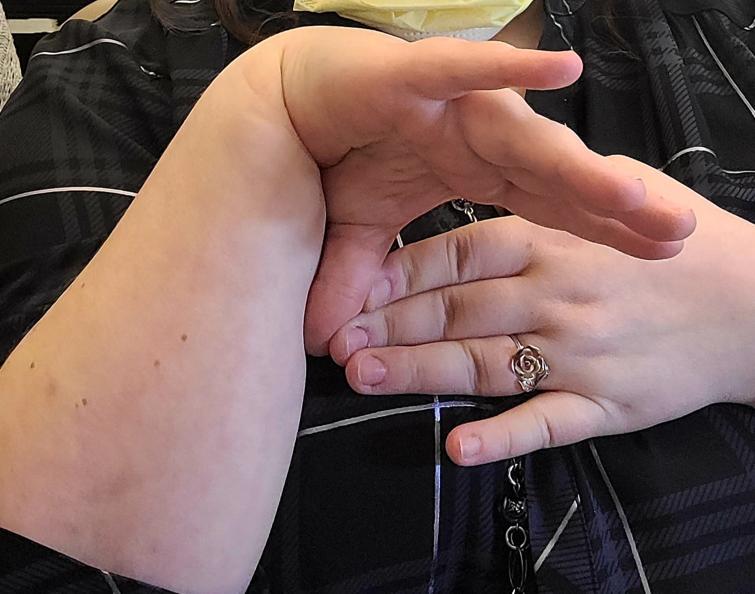
The Ehlers-Danlos Syndromes as whole are related to the Joint Hypermobility Spectrum, a spectrum of disorders intended to classify different severities of Hypermobility Spectrum Disorders (HSD). At the most extreme end of the hypermobile spectrum lies hEDS [3]. hEDS differs from the other EDS subtypes in its presentation as well. hEDS includes general joint hypermobility (GJH) but has less severe skin involvement compared to classical or vascular EDS types [1]. Easy bruising and impaired wound healing are also common. The definition of hEDS has evolved to include chronic pain and chronic fatigue as common presentations in those affected by the disease. The current diagnostic criteria include confirmation of GJH using the Beighton scoring system, positive musculoskeletal and pain symptoms or family history, and exclusion of other EDS subtypes or HSDs [4]. 

(Visit this link for the full diagnostic criteria of hEDS)

Apart from acute complications such as dislocation and subluxation, hEDs treatment revolves around chronic pain management and prevention of complications [1]. Acute exacerbations should be treated accordingly using joint reduction techniques and acute pain management. Physical therapy can be used to increase joint stability using low resistance exercises and stretching to increase muscle tone, thereby reducing the chance of acute joint injury. Patients should generally avoid hyperextension and high impact exercise. Oral acetaminophen, NSAIDs, and COX-2 inhibitors can be used as analgesics for chronic pain, as well as after acute injury. Cannabinoids can be considered for chronic pain, but opiates should rarely be used. If an hEDS patient must undergo surgery, careful technique should be used to minimize the wound site and promote healing. In summary, it is important to understand hEDS and how it differs from EDS and other HSDs to properly diagnose and treat patients.


References

1. Tinkle B, Castori M, Berglund B, Cohen H, Grahame R, Kazkaz H, et al. Hypermobile Ehlers-Danlos syndrome (a.k.a. Ehlers-Danlos syndrome Type III and Ehlers-Danlos syndrome hypermobility type): Clinical description and natural history. American Journal of Medical Genetics Part C: Seminars in Medical Genetics. 2017 Feb 1;175(1):48–69.

2. The Types of EDS [Internet]. The Ehlers Danlos Society. 2017. Available from: https://www.ehlers-danlos.com/eds-types/

3. What are the hypermobility spectrum disorders? [Internet]. The Ehlers Danlos Society. 2017. Available from: https://www.ehlers-danlos.com/what-is-hsd/

4. Forghani I. Updates in Clinical and Genetics Aspects of Hypermobile Ehlers Danlos Syndrome. Balkan Medical Journal. 2019 Jan 10;36(1):12–6.


Monday, November 22, 2021

Chronic Exertional Compartment Syndrome

Chronic Exertional Compartment Syndrome
Written by Jonathan Hunyadi with Dr. Nabil Ebraheim

Chronic exertional compartment syndrome (CECS) a pathology in runners usually involving the anterior compartment of the leg. It is believed to result from swelling and hypoperfusion of muscle and nerve during physical activity. Patients typically present with anterior burning leg pain that is exacerbated by exercise and is greatly reduced or completely subsides 15 to 30 minutes after exercise.

The condition can be diagnosed by measuring the pressure of the affected compartment one and five minutes after exercise. A pressure of 30 mmHg one minute and a pressure of 20 mmHg five minutes after exercise is considered diagnostic. Patients with CECS usually have a resting intra-compartment pressure greater than 15 mmHg which greatly increases during running. This typically produces a burning, cramping or aching pain after about 10 minutes of running resulting in cessation of exercise. Additionally, patients sometimes report tingling over the dorsal aspect of the foot while running.

Patients often present following stretching and strengthening therapy without relief. On physical exam, the patient will present with diffuse, nonspecific tenderness over the anterolateral leg without focal tenderness over bone. Pulses and x-ray will be normal and bone scan or MRI will be negative for stress fractures. Classic findings of acute compartment syndrome such as pain with passive toe dorsiflexion and sensory loss in the first web space, are typically absent.

Following diagnosis, treatment consists of the surgical release of affected compartments. During lateral compartment release, the superficial peroneal nerve, which pierces the fascia 10cm to 12cm proximal to the tip of the lateral malleolus, must be avoided. Surgical fasciotomy is usually successful but with a relatively high recurrence rate of approximately 20%. Recurrence typically occurs around two years following the initial procedure and is due to fibrosis within the compartment, causing return of symptoms and potential nerve entrapment. Additional causes of recurrences are inadequate release, failure to recognize and release all compartments, and misdiagnosis.


The differential diagnosis for CECS is large with overlap of symptoms. A common example is medial tibial stress syndrome. With this condition, bony tenderness along the posteromedial tibia will be present. Popliteal artery entrapment, a dynamic exercise related vascular phenomenon, is another condition in the differential. CECS can be distinguished by its predictable exercise related onset, relief of symptoms at rest and by being present for a long time.


Monday, November 15, 2021

Nerve Injury Positions of the Hands and Fingers

Nerve Injury Positions of the Hands and Fingers

Written by: Alec Bryson with Dr. Nabil Ebraheim

 The presentation of a patient’s hand may provide insight to which nerve is damaged and the approximate location of the damage. When presenting with ulnar n. damage, a patient may show a claw hand, Wartenberg’s sign, or Froment’s sign. Claw hand will present with clawing of the fourth and fifth digits due to the inability to extend the fingers specifically at the interphalangeal joints. This presentation is due to the lack of innervation to the intrinsic muscles of the hands, and the unopposed action of the flexor digitorum profundus m. (Moore et al., 2018). This indicates damage near the distal end of the ulnar n., below the elbow, and potentially near the wrist. Wartenberg’s sign will be seen as the inability to adduct the fifth digit when extended as well as an inability to cross the second and third digit. This is due to ulnar n. injury leading to wasting of the fifth interosseous m. This causes the fifth digit to rest in a more abducted position due to the unbalanced action of the extensor digiti minimi m. (Ebraheim, 2021). There will also be loss of function of the lumbrical m. in the fourth and fifth digits. A Froment’s test will detect palsy of the ulnar n. resulting from compression in the cubital tunnel. When asking the patient to pinch a piece of paper between their thumb and second digit, a positive Froment’s Sign will show as the patient flexing their thumb’s interphalangeal joint to grip the paper as the paper is pulled away (Attum, 2021). This will result from a weak adductor pollicis m. due to ulnar n. palsy. 

Depending on the location of a lesion to the median n., the patient will present with one of three signs. A positive Benedictine sign will be caused by proximal median n. damage. It will be seen as paralysis of the first and second digit, with weakness to the third digit. A proximal lesion would lead to paralysis of several muscles (FDS, FPL, FPB, and the radial half of FDP), leaving the ulnar half of the flexor digitorum profundus m. as the only remaining flexor (Ebraheim, 2021). When the patient is asked to make a fist, the hand will resemble the similar position taken during a blessing. A positive Benedict sign will also resemble the ulnar claw hand. However, the Benedict sign will present when the patient is flexing, not extending the fingers (Ebraheim, 2021). A median n. injury affecting the anterior interosseous n. branch will present as an inability to do the OK sign. This occurs due to paralysis of the flexor pollicus longus m. and the lateral part of the flexor digitorum m. (Moore et al., 2018). A positive Ape hand (Simian hand) is caused by paralysis of only the thenar m. from damage to the recurrent branch of the median n. to the thenar m. The thumb will be seen in the same plane as the other digits due the thumb being pulled more dorsal by the action of the adductor pollicis m., which is innervated by the ulnar n. (Moore et al., 2018). 

Finally, proximal radial n. damage will be seen as wrist drop. This is usually caused by fractures of the distal third of the humeral shaft (Holstein-Lewis Fracture) and caused paralysis of the wrist and fingers extensors (Ebraheim, 2010). Lower radial n. injury will present in the patient as the ability to extend the wrist, but the loss of finger extension. There will be no wrist drop, but the patient would not be able to make a hitchhiking sign.

Reference List

1. Attum B. Physical exam of the hand [Internet]. Orthobullets. Lineage Medical, Inc.; 2021 [cited 2021Oct21]. Available from: https://www.orthobullets.com/hand/6008/physical-exam-of-the-hand

2. Ebraheim N. Anterior Interosseous Nerve Injury - Everything You Need To Know - Dr. Nabil Ebraheim [Internet]. YouTube. 2021 [cited 2021Oct21]. Available from: https://www.youtube.com/watch?v=M9y-iDKLDPE

3. Ebraheim N. Claw Hand, Ulnar Claw Hand - Everything You Need To Know - Dr. Nabil Ebraheim [Internet]. YouTube. 2017 [cited 2021Oct21]. Available from: https://www.youtube.com/watch?v=GyqaKGg3HmM

4. Ebraheim N. Nerve Injury Position of the Hand & Fingers - Everything You Need To Know - Dr. Nabil Ebraheim [Internet]. YouTube. 2021 [cited 2021Oct21]. Available from: https://www.youtube.com/watch?v=nwd1h0Dfo5o

5. Ebraheim N. Radial Nerve Palsy, injury - WRIST DROP . Everything You Need To Know - Dr. Nabil Ebraheim [Internet]. YouTube. 2010 [cited 2021Oct21]. Available from: https://www.youtube.com/watch?v=_Cu6ttAhe8Y

6. Moore KL, Dalley AF, Agur A. Clinically Oriented Anatomy. 8th ed. Philadelphia, PA: Lippincott Williams and Wilkins; 2018.


Monday, November 8, 2021

Tennis Elbow - Everything You Need to Know

Tennis Elbow

Written by Devon Patel with Dr. Ebraheim

Lateral epicondylitis, also known as tennis elbow, is an overuse injury that results in inflammation, tendinosis, and lateral elbow pain. It is the most common cause of pain in the lateral elbow, affecting between 1 and 3% of the general population (1). The primary structure impacted in tennis elbow is the extensor carpi radialis brevis, which originates at the lateral epicondyle (2). This condition is primarily seen in middle-aged individuals, especially those between the ages of 40 and 50 (3). Tennis players (up to 50% of regular players) and workers who engage in heavy lifting or repetitive gripping are more likely to develop tennis elbow (2). Other conditions, such as rotator cuff pathology or De Quervain’s diseases, and lifestyle factors, such as smoking, are associated risk factors for lateral epicondylitis (4). Rotator cuff pathology could be a risk factor because lateral epicondylitis can also be caused by biomechanical stress, but it is unclear exactly why it and other conditions are associated with each other (4). In terms of histology, disorganized collagen, dense fibroblasts, and vascular hyperplasia are primarily seen (2). Immature fibroblastic and vascular infiltration of the origin of the extensor carpi radialis brevis has consistently been identified during surgery (5). A physical exam and history are typically used to diagnose this condition. Clinical tests to assist in diagnosis include grip strength, Cozen’s, Maudsley’s, and Mill’s tests (3). Lateral epicondylitis is indicated if the previous tests are positive along with reduced grip strength or reproduced pain (3). If necessary, diagnostic scans can be obtained. Majority of patients show altered signal around the lateral epicondyle on MRI scans and hot focus on infrared thermography (6, 7). Radial tunnel syndrome is a differential diagnosis of tennis elbow. This syndrome is seen in 5% of patients who have compression of the posterior interosseous nerve. The pain associated with radial tunnel syndrome is approximately 3-4 cm distal and anterior to the lateral epicondyle, which differentiates it from tennis elbow.

Non-surgical treatments are the primary mode of treatment and there is a 95% success rate with treatments to relieve pain. The most frequently used treatment is corticosteroid injection (2). Oral or topical non-steroidal anti-inflammatory drugs (NSAIDs) can also be prescribed, but their effectiveness is variable (8). Patients can also undergo physical therapy to relieve their symptoms. Eccentric exercises have been shown to be especially effective in pain management (9). Using an inelastic, nonarticular proximal forearm brace could also be recommended (10). Two relatively newer treatments for tennis elbow are ultrasonic (US) and extracorporeal shock wave therapy (ESWT). There are minimal side effects to US and ESWT, thus making them preferable for patients and clinicians (11). Even though there is no difference between US and ESWT in elbow function evaluation scores, ESWT has been shown to have greater efficacy in pain relief (12). Surgical procedures are a last resort for treatment of tennis elbow and only indicated if patients are unresponsive to conservative treatments after an extended period of time. Debridement is the most common surgical intervention, but it can result in injury of the lateral collateral ligament and subsequent posterolateral rotary instability of the elbow.


References

1.         Shiri R, Viikari-Juntura E, Varonen H, Heliovaara M. Prevalence and Determinants of Lateral and Medial Epicondylitis: A Population Study. American Journal of Epidemiology. 2006;164(11):1065-74. doi: 10.1093/aje/kwj325.

2.         Cutts S, Gangoo S, Modi N, Pasapula C. Tennis elbow: A clinical review article. Journal of Orthopaedics. 2020;17:203-7. doi: 10.1016/j.jor.2019.08.005.

3.         Speers CJ, Bhogal GS, Collins R. Lateral elbow tendinosis: a review of diagnosis and management in general practice. British Journal of General Practice. 2018;68(676):548-9. doi: 10.3399/bjgp18x699725.

4.         Titchener AG, Fakis A, Tambe AA, Smith C, Hubbard RB, Clark DI. Risk factors in lateral epicondylitis (tennis elbow): a case-control study. Journal of Hand Surgery (European Volume). 2013;38(2):159-64. doi: 10.1177/1753193412442464.

5.         Nirschl RP, Pettrone FA. Tennis elbow. The surgical treatment of lateral epicondylitis. The Journal of bone and joint surgery American volume. 1979;61(6A):832-9. PubMed PMID: 479229.

6.         Steinborn M, Heuck A, Jessel C, Bonel H, Reiser M. Magnetic resonance imaging of lateral epicondylitis of the elbow with a 0.2-T dedicated system. European Radiology. 1999;9(7):1376-80. doi: 10.1007/s003300050851.

7.         Thomas D, Siahamis G, Marion M, Boyle C. Computerised infrared thermography and isotopic bone scanning in tennis elbow. Annals of the Rheumatic Diseases. 1992;51(1):103. doi: 10.1136/ard.51.1.103.

8.         Pattanittum P, Turner T, Green S, Buchbinder R. Non‐steroidal anti‐inflammatory drugs (NSAIDs) for treating lateral elbow pain in adults. Cochrane Database of Systematic Reviews. 2013(5). doi: 10.1002/14651858.CD003686.pub2. PubMed PMID: CD003686.

9.         Croisier J-L, Foidart-Dessalle M, Tinant F, Crielaard J-M, Forthomme B. An isokinetic eccentric programme for the management of chronic lateral epicondylar tendinopathy. British Journal of Sports Medicine. 2007;41(4):269. doi: 10.1136/bjsm.2006.033324.

10.       Johnson GW, Cadwallader K, Scheffel SB, Epperly TD. Treatment of lateral epicondylitis. Am Fam Physician. 2007;76(6):843-8. Epub 2007/10/04. PubMed PMID: 17910298.

11.       Coombes BK, Connelly L, Bisset L, Vicenzino B. Economic evaluation favours physiotherapy but not corticosteroid injection as a first-line intervention for chronic lateral epicondylalgia: evidence from a randomised clinical trial. British Journal of Sports Medicine. 2016;50(22):1400-5. doi: 10.1136/bjsports-2015-094729.

12.       Yan C, Xiong Y, Chen L, Endo Y, Hu L, Liu M, et al. A comparative study of the efficacy of ultrasonics and extracorporeal shock wave in the treatment of tennis elbow: a meta-analysis of randomized controlled trials. Journal of Orthopaedic Surgery and Research. 2019;14(1). doi: 10.1186/s13018-019-1290-y.

Monday, November 1, 2021

Bursitis of the Knee, Hip, Elbow and Shoulder - Everything You Need to Know

 Bursitis of the Knee, Hip, Elbow and Shoulder - Everything You Need to Know
Written by Andrew Kelley with Dr. Nabil Ebraheim

Prepatellar Bursitis of the Knee
Prepatellar bursitis, also known as housemaid’s, carpet layer’s, and carpenter’s knee, is a superficial bursitis caused by inflammation of the bursa separating the patellar bone and the skin (1). Patients with prepatellar bursitis will normally present with knee pain and swelling (2). Prepatellar bursitis is mostly caused by long-term repetitive mini trauma from kneeling and crawling on hard surfaces. Other causes include acute injury, infection, gout, and rheumatoid arthritis (2). Its annual incidence is 10/100,000 per year with 80% of those affected being males age 40-60 (1). In cases of non-traumatic prepatellar bursitis, treatment is dependent on resolution of the underlying condition. Early differentiation between septic and non-septic bursitis is important in the early presentation in order to improve patient outcomes. Acute bursitis normally responds well to conservative treatment such as rest, ice, activity modification, NSAIDs, and fluid aspiration. Chronic bursitis due to mini traumas is treated similarly but may require additional corticosteroid therapy (1).


Olecranon Bursitis of the Elbow
Olecranon Bursitis, also known as student’s elbow and plumber’s elbow, is caused by inflammation of the bursa overlaying the olecranon process of the ulnar bone at the tip of the elbow. This bursa allows for smooth motion of the olecranon process against the superficial tissue at the tip of the elbow. Affected patients normally present with swelling at the bend of the elbow. A characteristic “golf ball” shape of swelling can be seen, and a fully intact range of motion of the elbow can differentiate it from elbow joint injuries (3). Olecranon Bursitis most commonly affects men age 30-60. Most cases are due to repeated minor trauma and sports (4). Treatment is focused on resolving the underlying cause of inflammation. Conservative treatment includes ice and rest along with NSAIDs for symptomatic relief are indicated. While aspiration and corticosteroid injection are proven relief interventions, they carry an increased risk for infection (4).


Greater Trochanteric Bursitis of the Hip
Greater trochanteric bursitis, or greater trochanteric pain syndrome (GTPS), is caused by inflammation of the bursa laying deep to the iliotibial band and superficial to the greater trochanter of the femur. It acts as a lubricant for the gluteal tendons. Patients with hip bursitis normally present with chronic intermittent pain of the lateral hip, thigh, and buttock (6). This bursitis normally affects women age 40-60. The increased pelvic width of women relative to their body may predispose them to increased iliotibial band tension on the bursa (6). The cause of hip bursitis can be repetitive microtrauma, blunt trauma, or idiopathic. Movements requiring repetitive hip abduction like stair climbing and bicycling, direct traumatic falls, and sedentary lifestyles are common causes of this condition (5).  Common treatments for this bursitis include NSAIDs, physical therapy, and corticosteroid injection. Surgery is a rare treatment option for bursitis resistant to conservative treatment options (5).


Subacromial Bursitis of the Shoulder
Subacromial bursitis is caused by inflammation to the bursa just below the acromion process. The subacromial bursa acts as a lubricating medium between the acromion process superiorly and the muscles of the rotator cup inferiorly.  Subacromial bursitis normally presents as anterolateral shoulder pain, especially during overhead activities. This chronic inflammation of the shoulder bursa can eventually lead to weakness and rupture of the surrounding ligaments and tendons (7). Older individuals are more likely to experience shoulder bursitis due to years of overuse. Most patients present due to direct trauma to the shoulder or repetitive overhead activities (7). Treatment includes rest, NSAIDs, physical therapy, and corticosteroid injections. Surgical therapy is reserved for cases unresponsive to conservative therapy (7).


References:  
1.  Rishor-Olney CR, Pozun A. Prepatellar Bursitis. [Updated 2021 Sep 2]. In: StatPearls [Internet]. Treasure Island (FL): StatPearls Publishing; 2021 Jan-.
2.  J. Dean Cole MD. Causes of knee bursitis (prepatellar bursitis) [Internet]. Arthritis. Arthritis-health; [cited 2021Oct28]. Available from: https://www.arthritis-health.com/types/bursitis/causes-knee-bursitis-prepatellar-bursitis
3.  Pangia J. Olecranon bursitis [Internet]. StatPearls [Internet]. U.S. National Library of Medicine; 2021 [cited 2021Oct28]. Available from: https://www.ncbi.nlm.nih.gov/books/NBK470291/
4.  Blackwell JR, Hay BA, Bolt AM, Hay SM. Olecranon bursitis: a systematic overview. Shoulder Elbow. 2014 Jul;6(3):182-90. doi: 10.1177/1758573214532787. Epub 2014 May 6. PMID: 27582935; PMCID: PMC4935058.
5.  Seidman AJ. Trochanteric bursitis [Internet]. StatPearls [Internet]. U.S. National Library of Medicine; 2021 [cited 2021Oct28]. Available from: https://www.ncbi.nlm.nih.gov/books/NBK538503/
6.  Reid D. The management of Greater Trochanteric pain syndrome: A systematic literature review [Internet]. Journal of orthopaedics. Elsevier; 2016 [cited 2021Oct28]. Available from: https://www.ncbi.nlm.nih.gov/pmc/articles/PMC4761624/
7.  Faruqi T. Subacromial bursitis [Internet]. StatPearls [Internet]. U.S. National Library of Medicine; 2021 [cited 2021Oct29]. Available from: https://www.ncbi.nlm.nih.gov/books/NBK541096/