Wednesday, September 26, 2018

Adhesive Capsulitis, Frozen Shoulder


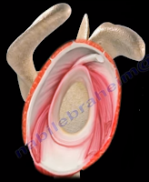
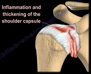
Adhesive Capsulitis, or frozen shoulder, is a painful progressive loss of shoulder motion. It affects both active and passive movement of the shoulder joint. The shoulder will be stiff and painful and occurs due to inflammation, fibrosis, scarring, and contraction of the capsule. A normal shoulder joint capsule is elastic and allows great range of motion. Inflammation and thickening of the shoulder capsule and may lead to adhesive capsulitis. Frozen shoulder may occur without any specific cause, however it may be triggered by a mild trauma to the shoulder.


This condition develops slowly and goes through three phases:

  1. Pain and freezing
  2. Stiffness or frozen
  3. Resolution

During the pain and freezing phase, the pain is worse at night and increases with any movement. This phase will last several months. During the second phase, range of motion is limited as pain is diminishing. This may last up to one year. The resolution phase may begin overtime and may last up to three years.


Conditions associated with frozen shoulder include:

  • Diabetes
  • Thyroid problems
  • Auto immune disease
  • Stroke
  • Rheumatoid arthritis
  • Trauma or post-surgery

A patient with frozen shoulder will have loss of both active (movement without assistance) and passive (movement with assistance) motion. External rotation of the shoulder is very limited and the condition is self-limiting and may resolve on its own. X-rays are needed to rule out degenerative arthritis.  An MRI or arthrogram will show small fluid in joint cavity. Rotator cuff may be normal and synovitis and narrowing of the rotator cuff interval is usually seen.

Treatment consists of anti-inflammatory medications, physical therapy, injections, and manipulation under anesthesia. Surgery will be done in the form of a release of the capsule when nonoperative methods fail. The physician should always check the patient for diabetes.  
Tuesday, September 25, 2018

Profunda Femoris Artery


The profunda femoris artery is the main blood supply of the thigh. The profunda femoris artery arises from the posterolateral aspect of the femoral artery about 4 cm below the inguinal ligament. The profunda femoris artery crosses the pectineus muscle and the adductor brevis muscles, and runs under the adductor longus muscle. Then it runs between the adductor magnus and the adductor longus muscles. When it reaches the adductor magnus, it gives three perforating branches and it ends by perforating the adductor magnus as the fourth perforating branch. The perforating arteries are called “perforating” because they perforate the insertion of the adductor margnus in order to reach the back of the thigh. The first three perforating arteries are branches of the profunda femoris itself, while the fourth perforating artery is a continuation of the profunda femoris artery itself. During posterior exposure of the hip, partial section of more than 2cm of the gluteus maximus tendon attachment on the femur can be associated with a risk of injury to the first perforating branch. The profunda femoris artery gives the medial circumflex femoral artery and the lateral circumflex femoral artery. The MCFA will give the ascending, the acetabular, and transverse branches. The LCFA will give the ascending, the descending, and transverse branches.  

Tuesday, September 18, 2018

Ganglion Cyst of the Shoulder


Ganglion cysts can be important when they are located around the shoulder, especially when they are located in the suprascapular notch and the spinoglenoid notch. The suprascapular nerve passes under the transverse scapular ligament at the suprascapular notch. The transverse scapular artery runs above the transverse scapular ligament. The artery and nerve joint and then pass through the spinoglenoid notch under the inferior scapular ligament. The suprascapular nerve gives branches to the supraspinatus muscle and branches to the infraspinatus muscle.

Nerve compression from a ganglion cyst at the suprascapular notch affects both the supraspinatus and infraspinatus muscles, causing a decrease in abduction and loss of external rotation of the shoulder. Nerve compression at the spinoglenoid notch affects only infraspinatus muscle, causing loss of external rotation of the shoulder with the arm to the side. Spinoglenoid notch compression is usually associated with cysts and ganglia. In addition to compression of the suprascapular nerve, these patients may also have associated posterior labral tears.

Tuesday, September 11, 2018

Anatomy of the Posterior Cutaneous Nerve of the Thigh


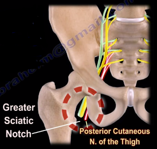
The posterior cutaneous nerve of the thigh (small sciatic nerve) arises from the sacral plexus from S1-S3. The posterior cutaneous nerve of the thigh exits from the pelvis through the greater sciatic notch below the piriformis muscle. The nerve descends below the gluteus maximus muscle along with the inferior gluteal artery. It runs into the back of the thigh beneath the fascia lata and over the long head of the biceps femoris muscle to the back of the knee. The nerve then pierces the deep fascia and accompanies the short saphenous vein to the middle of the back of the leg. The posterior cutaneous nerve of the thigh innervates the distal part of the gluteal region, the skin of the perineum and the posterior part of the thigh.
The nerve can become compressed when passing through the tunnel below the piriformis muscle and under the gluteus maximus muscle. This may result in sensitivity disturbances of the innervation area of the nerve. Causes of the syndrome may be hypertrophy or abnormality of the piriformis muscle such as entrapment below the piriformis which compresses the nerve. Compression of the nerve can also occur due to prolonged sitting. During the clinical examination, pain and sensitivity will be evident. Pain and sensitivity disturbances are characteristic of the nerve distribution site in the posterior part of the thigh down the knee. This disturbance can be from hyperesthesia to hypoesthesia or burning sensation similar to meralgia paresthetica of the lateral cutaneous nerve of the thigh.

Differential diagnosis include piriformis syndrome. The patient should avoid sitting for long periods of time, especially on a hard base. Treatment consists of physical therapy, massage, and injection. Surgery is rarely needed.

Tuesday, September 4, 2018

SLAP Tear- Symptoms, Diagnosis, and Treatment


A SLAP tear is a tear that occurs where the biceps tendon inserts into the superior labrum. A SLAP tear is different from a Bankart lesion. SLAP tears are not common and can be hard to diagnose. Symptoms of a SLAP tear include: pain deep within the shoulder or in the back of the shoulder, as well as catching, popping, or clicking sensations. The patient may also experience pain when throwing a ball with a decrease in velocity and the feeling of having a dead arm after pitching. Patients will also experience pain with overhead activity which mimics impingement syndrome. This typically affects throwing athletes. When the biceps tendon is involved, pain may also be located at the front of the shoulder. A SLAP tear can be an isolated lesion or it can be associated with internal impingement, articular sided cuff tear, or instability.
A SLAP tear is diagnosed with a clinical examination and testing. The O’Brien’s test is the most commonly used test. Multiple tests are usually used including the anterior slide test and the clunk test. An MRI with contrast is the best imaging technique. When performing the O’Brien’s test, the patient is standing or sitting with the arm at 90° of flexion, 10° of adduction, and full internal rotation with the forearm pronated.  The examiner applies pressure to the forearm and instructs the patient to resist the applied downward force. Pain at the shoulder joint suggests a SLAP lesion. Decrease in pain of the shoulder joint on supination of the arm is suggestive of a SLAP tear.
Treatment consists of physical therapy, anti-inflammatory medications, injections, and surgery (when conservative treatment fails). If surgery is necessary, a labral debridement will be performed for minor tearing and fraying. Biceps Tenodesis is becoming popular, as it is a procedure that cuts the biceps tendon where it attaches to the labrum and reinserts it in another area, usually in front of the shoulder. A biceps tenotomy is a procedure that cuts the biceps tendon from the glenoid, releasing the long head of the biceps tendon from its attachment allowing it to fall into the upper arm out of the shoulder joint. A biceps tenotomy is probably best suitable for some elderly patients. A SLAP repair is a procedure which uses sutures to anchor the torn labrum to the glenoid. This repair is usually done for athletes and patients under the age of 40 years.