Platelet Rich Plasma, or PRP, is a volume of the plasma of
autologous blood having a platelet concentration above the baseline. Platelets
facilitate healing by stimulating the release of different growth factors. The
growth factors recruit stem cells that assist with healing, repair, or
regeneration of the injured tissue. PRP is injected directly into the injured
tissue, stimulating a healing response in a more powerful form.
 |
| Red Blood Cell |
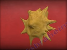 |
| Active Platelet |
Platelets aid in hemostasis (stop the bleeding) and in
building new tissues—they act as a scaffold for tissue regeneration. Platelets
aid in the attraction and binding of stem cells. Platelets act as the directors
while stem cells work. Platelets divide, multiply and differentiate to become
the healing cells for injured tissue. The platelets become activated by
thrombin and other factors which cause a change in their morphology and the
release of multiple growth factors. These growth factors bind themselves to the
receptors of the cell causing intracellular changes down to the nucleus and affecting
its DNA. The result is a change in the
performance and function of the cell.

In order to produce PRP, 30-60mLs of the patient’s own
venous blood is drawn from the antecubital vein. The blood is then placed into
a device to be centrifuged which separates the blood into platelet poor plasma
(PPP), red blood cells (RBC), and platelet rich plasma (PRP). The blood is then
placed in a centrifuge for 15 minutes at 3,200 rpm. The centrifuge spins and
separates the platelets from the rest of the blood components and increases the
concentration of platelets and growth factors. The more platelet concentration,
the greater the healing power. After the centrifuge process is complete, the
plasma has been separated from the blood producing the PRP and the platelet
poor plasma is withdrawn to be discarded. Platelet rich plasma is withdrawn for
injection. Sodium Bicarbonate is used to neutralize the acidity of the sample.
The more platelet concentration, the more growth factors and healing power the
sample has.

Ultrasounds help deliver a concentrated sample of platelets
to the injured tissue. Ultrasound increases the accuracy of delivering the
sample to the injured tissue. Preferably, injections should be performed with
the aid of ultrasound imaging. Needling induced injury releases thrombin which
activates the platelets. Platelets help in hemostasis and produce growth
factors as well as chemotactic factors. Platelets act as a scaffold for
mesenchymal stem cells which start the process of tissue regeneration. Patients
will typically experience minimal to moderate discomfort which may last up to
one week following the injection. Avoid the use of anti-inflammatory medications
for 6 weeks after the injection.


