Tuesday, January 18, 2022

Frozen Shoulder Adhesive Capsulitis - Everything You Need To Know

Frozen Shoulder Adhesive Capsulitis - Everything You Need To Know

https://www.youtube.com/watch?v=VhfaPe8f7g0 

Frozen shoulder (adhesive capsulitis)

The frozen shoulder can be associated with diabetes or thyroid disease.  It may be the initial presenting symptom for these conditions.  The exact details of this relationship remain poorly understood.  Ever patient with a frozen shoulder should have the HbA1c and TSH levels tested.  Also, check for arthritis, rheumatoid factors and antinuclear antibodies.  Most patients with frozen shoulder are female between the ages of 40 and 60 years old.  And frozen shoulder, the patient will lose both the active and the passive range of motion of the shoulder.  The patient develops pain, which means that there is inflammation with early fibrosis of the joint capsule, leading to joint stiffness.  The active and passive global motion, especially external rotation, will be reduced compared to the other side.  The shoulder pain and motion loss is usually not related to trauma.  It is an idiopathic process that results in shoulder pain and loss of motion due to contracture of the capsule.  The essential lesion involves the coracohumeral ligament and the rotator interval.  The synovial inflammation and capsular fibrosis results in pain and joint volume loss.  Check for previous trauma or fractures.  Rule out shoulder joint arthritis and rule out posterior dislocation of the shoulder.  It can also occur post-surgery from a rotator cuff tear.  It may also be associated with dupuytren disease and cervical disc disease.  Check for medical comorbidities such as stroke or cardiac diseases.  The x-ray will exclude trauma, malignancy, arthritis, calcific tendinitis, impingement, and AC joint arthritis.  And frozen shoulder, the humeral head will remain in its normal location.


MRI

There is a space reduction in the axillary recess.  Rotator cuff strength is normal by exam and on the MRI.  The pain and stiffness lasts beyond 6 months, then you can do manipulation of the shoulder under anesthesia.  There is a 50% failure rate in diabetics.  The diabetes is associated with a much worse prognosis and poor outcome for surgical and nonsurgical treatment.


Treatment

·         Nonoperative

o   This should be done for at least 3 to 6 months.

o   Supervised or home-based capsular stretching program +/-intra-articular steroid joint injection.

o   Nonsteroidal anti-inflammatory medication

·         Surgery

o   Capsular release (arthroscopic or open) and release the intra-articular and subacromial adhesions.

o   The axillary nerve may be injured during release of the capsule.

o   Utilize surgery in patients that have failure of initial conservative treatment for 3 months, and the patient remains functionally limited.


Monday, January 10, 2022

Ankle Arthritis - Everything You Need to Know

Ankle Arthritis - Everything You Need To Know - Dr. Nabil Ebraheim

https://www.youtube.com/watch?v=ysx-FT6fllA

The patient will have longstanding global pain that is inside the ankle.  The patient will have antalgic gait with swelling of the ankle and decreased ankle motion.  You want to check the sensation.  Lack of sensation with Simmons–Weinstein 5.07 monofilament testing is important because insensate patients are poor candidates for ankle joint replacement.  The patient will usually have a trial of nonsurgical treatment first.
Treatment

  •          Anti-inflammatory medication
  •         Activity modification
  •         Ankle orthosis/bracing
  •          Injections


Treatment Orthosis

·       Single rocker sole shoe modification and custom Arizona brace.  You may give the patient a cane.

With failed conservative treatment, the patient may need arthrodesis of the ankle, which is fusion of the ankle, or an ankle replacement.  If you are going to do arthrodesis of the ankle, it will be arthrodesis of the tibiotalar joint.  In ankle arthrodesis, the fixation can be done by multiple techniques.  The whole idea is to obtain rigid fixation.  You can use plates, screws, or combination of both.

How to you position arthrodesis?

·         Neutral flexion

·         The gastroc recession or TAL may be needed if we cannot achieve neutral dorsiflexion.  From 0–5 hindfoot valgus when 5–10 external rotation of the foot.


Arthrodesis

·         80% will have difficulty on even ground.

·         75% will have difficulty with stairs.

If you find a person with an ankle fusion that when they walk, they have knee hyperextension when the heel comes off the ground during the stance phase, then that person's ankle is fused and some plantar flexion.  The plantar flexion position will create forced recurvatum of the knee.  The ankle should be fused in neutral flexion.  If the person has arthritis of the ankle joint and the subtalar joint, then you will do tibiocalcaneal arthrodesis.  Some people elect to go for total ankle replacement.


What are the contraindications for total ankle replacement?

  •  Severe deformity
  • Charcot joint
  • Avascular necrosis of the talus
  • Soft tissue compromise
  • Active ankle infection


What is the relative contraindication for ankle joint replacement?

  • Ligament instability
  • Diabetes
  •  Smoking
  •  Osteoporosis
  • Morbid obesity


When do you do distraction arthroplasty?

            You do this in younger patients with some motion and less severe joint destruction.


Arthritis of the ankle can be osteoarthritis, posttraumatic arthritis, or inflammatory arthritis as rheumatoid arthritis or gout.  Posttraumatic arthritis accounts for 80% of all ankle arthritis.  The primary osteoarthritis is about 10% only.  Pain with weightbearing, swelling, decreased range of motion compared to the other side, and you will be able to see the arthritis on the x-ray.  Ankle arthrodesis will be done in younger patients with a high demand, or if the patient has comorbidities such as diabetes and obesity.  10 years after ankle arthrodesis, 50% of the patients will have subtalar arthritis.  Some of the patients may get nonunion. Revision of the nonunion can lead to fusion in about 85%.  A young, active worker is a contraindication to total ankle arthroplasty.  You will do the arthrodesis and end-stage ankle arthritis and the union rate is about 90%.  The arthrodesis gives us a reliable pain control but will also give us adjacent joint arthritis.  The entire foot and the patient's comorbidities must be evaluated to choose the proper surgical technique.  Total ankle arthroplasty has superior gait mechanics compared to ankle arthrodesis.  Ankle arthrodesis is done 6 times more than a total ankle replacement.  If you have an elderly patient with no comorbidities and you want to do a motion preserving procedure, then this would be total ankle arthroplasty.  Revision of total ankle occurs due to loosening of the implant or subsidence, especially in patients less than 55 years old.  In a patient with rheumatoid arthritis with end-stage arthritis, total ankle may be a reasonable option.  In older patients with low demand, you will give total ankle arthroplasty.  In younger, active patients he will do a fusion.  The total ankle arthroplasty is gaining a lot of popularity.  For patients with nonunion, you need to evaluate for infection and metabolic bone disease.  You may also need a CT scan.  Preservation of the fibula can help the potential conversion to a total ankle at a later date.  When you compare patient with an ankle prosthesis to a patient with thin ankle fusion, there is more complication rate and revision rate for a patient with total ankle and more increased wound complications for people with rheumatoid arthritis.  If you are in doubt, use ankle arthrodesis.


Monday, January 3, 2022

Differential Diagnosis Of Spinal Stenosis

Written by: Vihan DeSilva with Dr. Nabil Ebraheim

Spinal stenosis is a diagnosis mainly made through history and CT/MRI imaging as physical exam findings can be normal namely in lumbar spinal stenoses. A common finding along with painful extension of the spine and decreased lumbar lordosis is narrowing of the spinal foramina, but diagnosis is made when patients present with neurogenic claudication and/or cervical myelopathy. The cause may be congenital or acquired through, for example, endocrinopathies, calcium metabolism disorders, inflammatory diseases, and infectious diseases.1,2 There 
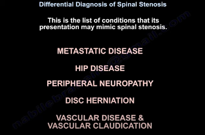
are other diseases that produce similar symptoms that should be considered in the differential diagnosis of spinal stenosis including metastatic disease, hip disease, peripheral neuropathy, disc herniation, and vascular disease/ vascular claudication.2 The rest of this article will be a discussion on how to differentiate spinal stenosis from these other conditions.
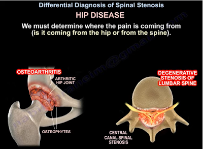

Patients’ pain may be metastatic if it is constant and worse at night or is unresolved even after previous attempts at treatment. Metastatic origin of pain should also be considered in patients who have a cancer/ cancer treatment history or experience fatigue, malaise, unintentional weight loss, or nonspecific symptoms.2,3

Distinguishing hip disease (ie. osteoarthritis) pain from lumbar stenosis pain can be challenging because the two ailments can coexist in a condition known as hip-spine syndrome. The location of the pain can be helpful in identifying the primary pain generator: hip pain can be felt in the groin, lateral hip, posterior hip, or near the spine and SI joint. Internal rotation of the hip can also be compromised in hip disease.2,4 Another way to isolate the pain generator is to inject the hip with steroid and observe. Worsening symptoms could indicate pain coming from lumbar stenosis. However, increased activity of the patient may also cause pain in related structures after the initial injection if it was successful in treating pain from underlying hip osteoarthritis.2

Peripheral neuropathy can also coexist with lumbar stenosis and may further complicate the differential diagnosis of spinal stenosis. EMG studies could aid in discerning stenosis from peripheral neuropathy and motor neuron disease.5 Certain clinical findings may also be useful. Bilateral burning foot pain at night is a distinguishing feature of peripheral neuropathy whereas unilateral leg pain with activity that is relieved by sitting is characteristic of lumbar stenosis/ radiculopathy. Additionally, sensory testing that demonstrates a dermatomal pattern indicates a problem in the spinal root whereas a glove and stock pattern would hint towards peripheral neuropathy.1,2
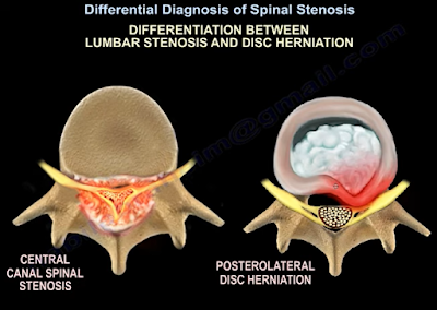
Disc herniation should also be considered against spinal stenosis as a possible source of pain. Location of pain can once again help in identifying the correct source. According to one comparative study, herniations tend to produce leg pain in the anterior thigh, anterior knee, and shin whereas posterior knee pain was common with stenosis.6 Furthermore, characteristics of stenosis include bilateral, nonspecific leg pain that is generally above the knee and rarely produces a positive straight-leg test. In contrast, herniation causes unilateral pain along the dermatome of the affected nerve root along with a positive straight-leg test.2

Vascular disease should be ruled out as well when considering a spinal stenosis diagnosis. One main distinguishing principle is that vascular disease produces vascular claudication whereas spinal stenosis produces neurogenic claudication. These two different types of claudication have different clinical sequelae. For instance, the distance a patient can walk before feeling symptoms is more variable with neurogenic claudication than with vascular claudication and uphill walking is better tolerated only with neurogenic claudication. Sitting attenuates neurogenic claudication symptoms whereas both sitting and standing still may ease vascular claudication symptoms.7 Vascular pain travels from distal sites to proximal ones whereas neurogenic pain goes from proximal to distal. Unlike vascular claudication, bilateral pedal pulses are normal with neurogenic claudication. Unlike spinal stenosis, vascular disease may produce lower extremity ulcers, hair loss, edema, and skin changes.2 Lastly, postural adjustments, such as flexion of the spine, ease stenosis claudication symptoms due to relief of pressure on the nerve roots. This is not true in vascular disease. This is also why a bicycle test relieves stenosis pain while making vascular pain worse.8

                Other rarer considerations include spinal arteriovenous malformations, tumors of the cauda equina, and differential diagnosis of myelopathy (ALS, multiple sclerosis, or subacute combined degeneration).1 Once a spinal stenosis diagnosis is made, the condition can be managed non-surgically with drugs, physiotherapy, and injections or surgically through decompression, spinal fusion, or interspinous spacer devices.9 Evidence is still being gathered on effectiveness and outcomes for all these non-surgical and surgical treatment options though.


References

1. Melancia JL, Francisco AF, Antunes JL. Spinal stenosis. Handb Clin Neurol. 2014;119:541-9.

2. Ebraheim N. Differential Diagnosis of Spinal Stenosis [Internet]. Toledo (OH): University of Toledo Medical Center, Department of Orthopedic Surgery; 2021 Jun 25. Available from: https://www.youtube.com/watch?v=eYxPmrnfjfA&ab_channel=nabilebraheim.

3. Dodwad SM, Savage J, Scharschmidt TJ, Patel A. Evaluation and treatment of spinal metastatic disease. Cancer Treat Res. 2014 Jul 29;162:131-50.

4. Brown MD, Gomez-Marin O, Brookfield KF, Li PS. Differential diagnosis of hip disease versus spine disease. Clin Orthop Relat Res. 2004 Feb;419:280-4.

5. Plastaras CT. Electrodiagnostic challenges in the evaluation of lumbar spinal stenosis. Phys Med Rehabil Clin N Am. 2003 Feb;14(1):57-69.

6. Rainville J, Lopez E. Comparison of radicular symptoms caused by lumbar disc herniation and lumbar spinal stenosis in the elderly. Spine (Phila Pa 1976). 2013 Jul 1;38(15):1282-7.

7. Genevay S, Atlas SJ. Lumbar spinal stenosis. Best Pract Res Clin Rheumatol. 2010 Apr;24(2):253-65.

8. Binder DK, Schmidt MH, Weinstein PR. Lumbar spinal stenosis. Semin Neurol. 2002 Jun;22(2):157-66.

9. Lurie J, Tomkins-Lane C. Management of lumbar spinal stenosis. BMJ. 2016 Jan 4;352:h6234.