Monday, March 25, 2019

Lisfranc Injury


Lisfranc Injury


Lisfranc injury is an important topic. If Lisfranc injury is not diagnosed and treated properly, it can lead to an altered gait, midfoot arthritis, and long term disability. Lisfranc injury indicated disruption between the base of the 2nd metatarsal and the medial cuneiform. Lisfranc injuries are a spectrum of injuries of the tarsometatarsal joints. Diagnosing Lisfranc injury is important. Diagnosis is missed in about 20%-30% of cases especially in multiple trauma patients. A high index suspicion is needed to prevent progression of the foot deformity, chronic pain, and dysfunction. You may need weight-bearing films for diagnosis of Lisfranc injury. Lisfranc injury may also be associated with compartment syndrome. Lisfranc injury could be purely ligamentous or can be associated with fractures. ORIF is better in cases of fractures. Arthrodesis is better in cases of purely ligamentous injury. In general, ligamentous injury does worse than fractures. The Lisfranc ligament is a large oblique ligament that extends from the plantar aspect of the medial cuneiform to the base of the second metatarsal. The Lisfranc ligament stabilizes the 2nd metatarsal and maintains the midfoot arch. Osseous stability is provided by the roman arch of the metatarsals and the recessed keystone of the 2nd metatarsal base. Tarsometatarsal joint complex is divided into three units: medial, middle, and lateral. The medial is the 1st metatarsal joint at 6o mobility. The middle is the 2nd and 3rd tarsometatarsal joints, and it is rigid. The lateral is the 4th and 5th tarsometatarsal joints; it is mobile which is why you do not fuse the 4th and 5th tarsometatarsal joints. The dorsalis pedis artery and the deep peroneal nerve both run between the first and second metatarsal bases. A direct injury with a plantar displacement is more common. Indirect injuries are more common than direct injuries. They result from axial loading or twisting on a plantar flexed midfoot. Dorsal displacement of the 2nd metatarsal is more common. Check the alignment of the dorsum of the 2nd metatarsal with the middle cuneiform. Associated fractures are typically tarsal fractures, especially a cuboid fracture. A “Nutcracker” fracture results from twisting injury causing forceful abduction of the forefoot. It is a fracture of the base of the 2nd metatarsal and compression fracture of the cuboid. nd metatarsal, at the navicular, and cuboid. Check for widening between the first and second ray (more than 2 mm is an indication for surgery). In the lateral view, check the dorsal displacement or subluxation of a metatarsal. It should be at the level of the corresponding cuneiform. Check for the FLECK sign (bony fragment). Avulsion fragment of the Lisfranc ligament from the base of the 2nd metatarsal. The medial side of the fourth metatarsal should line up with the medial side of the cuboid on the oblique view (30o). CT scan can be useful and MRI can confirm purely ligamentous injury. These injuries should be treated with a cast. For a dorsal sprain and no instability, the patient can be treated with non-weight bearing cast for 6 weeks and return to activity gradually. Surgery can be done for instability. Open reduction internal fixation with cortical screws if there is bony fractures. When you do ORIF- you need anatomic reduction. Hardware removal between 5-6 months (some surgeons leave the hardware in place indefinitely). Arthrodesis if the injury is purely ligamentous. Healing of the ligaments is less reliable than bony healing. Purely ligamentous injury needs primary arthrodesis. Arthrodesis is also done in old injuries if there is delay in treatment for if there is failure of open reduction and internal fixation of Lisfranc injury. Midfoot arthrodesis is also used for chronic Lisfranc injury that leads to severe midfoot arthritis with progressive arch collapse and midfoot abduction. Fusion of the medial and middle column; first, second, and third tarsometatarsal joints. Do not fuse the lateral column (lateral column is mobile). For the lateral column, do reduction and stabilization by k-wire fixation. Post-traumatic arthritis occurs in up to 50% of patients. Patient may have altered gait and long term disability. Purely ligamentous injury has a worse prognosis than injuries with fractures. Malalignment of the fractures usually lead to arthritis.
Lisfranc classifications are not useful in deciding the treatment or the prognosis of the injury. Severe injuries are obvious, easily diagnosed, and may develop compartment syndrome of the foot. Injuries with minimal displacement could be missed, and they will need surgery regardless of the classification. Arthritis may develop even with minimal displacement. In general, there are three patterns of injury: total incongruity, partial incongruity, and divergent. Total incongruity occurs when all five metatarsals are displaced in the same direction. Total incongruity occurs lateral or medial, with lateral being more common. Partial incongruity occurs when one or two metatarsals are displaced from the others. Divergent occurs when the lateral displacement of the lesser metatarsals with medial displacement of the first metatarsal. The one thing all these injuries have in common is disruption of the tarsometatarsal joint complex. The patient has severe pain in the midfoot and is unable to bear weight. There may be some swelling in the midfoot dorsally. Plantar bruising may be present, especially medially. Tenderness over the tarsometatarsal joint. Check the skin condition and rule out compartment syndrome. Check the neurovascular status of the foot. Plantar ecchymosis is a classic clinical sign of potential Lisfranc injury. Wight bearing standing x-rays with comparison views if x-rays are normal and if the physician clinically suspects a Lisfranc injury. Another alternative is to get physician assisted midfoot stress radiograph. Obtain three views: AP, oblique, and lateral. Medial border of the second metatarsal should line up with the medial border of the middle cuneiform on both the AP and the oblique view. Check for fractures, especially at the base of the 2

Monday, March 18, 2019

Odontoid Fractures


Odontoid Fractures

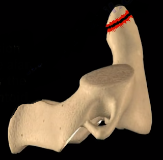
There are three types of odontoid fractures.
Type I fractures are a stable avulsion fracture of the alar ligament near the tip of the odontoid. A soft collar can be used to treat Type I fractures. Be aware of significant ligamentous injuries. Type II fractures are at the base of the odontoid process. Type II are the most common and are troublesome. The nonunion rate is about 20-80% due to interruption of the blood supply. The risk factors of nonunion include if the patient is over the age of 60 years old, if the patient has more than 6mm of displacement, smoking and diabetes, and you are unable to achieve reduction. In posterior displacement, extension injury (rare type) the anterior displacement is more common (flexion injury). Delay in treatment also increases the rate of nonunion. For treatment of young patients with no nonunion risks use a halo. The patient is younger than 60 years. The fracture is minimally displaced. Initial dens displacement is less than 6 mm, and the reduction is within one week of the injury. Healing will occur in the majority of cases.
If the patient has a nonunion risk, or when reduction of the fracture cannot be achieved or maintained, then we need to think about surgery and the fracture pattern. When the fracture pattern allows, you can put an anterior screw into the odontoid (to preserve the motion of C1/C2). Odontoid screw is used in younger patients instead of fusion to avoid loss of 50% of the neck rotation). Do not use the anterior screw fixation in patients with osteoporosis, in older patients, or in patients with a short neck. Another scenario is, if the patient has nonunion risks but the fracture pattern does not allow you to place an anterior odontoid screw, then you are going to fuse C1 to C2 (this will lose 50% of neck rotation). In general, C1/C2 fusion is used in cases of nonunion or it is used in cases of displaced fracture in the older patient and it can also be used if there is a failure of treatment with a halo. C1/C2 fusion can also be used if the fracture is comminuted and unstable. Posterior C1/C2 fusion can be done with different screw or wire constructs. A vascular watershed area exists between the apex of the odontoid, which is supplied by branches of the internal carotid artery and the base of the odontoid, which is supplied by branches of the vertebral artery. Type II fracture of the odontoid may get nonunion due to cortical bone and poor blood supply.
Type III fractures extend through the body of C2. This area is rich in blood supply and the fracture heals in the majority of cases. Treatment for Type III odontoid fractures includes external cervical orthosis (especially in the elderly patient) and a halo (if the fracture is displaced) (do not use in elderly patients). Odontoid fractures in the elderly can occur due to a simple fall and usually the diagnosis is missed. It is associated with increased complications and mortality. Do not use a halo in elderly patients. Use an external cervical orthosis of some sort. Fibrous union might be adequate if the fracture is not badly displaced, otherwise you will do fusion of C1/C2. For example, an 80 year old patient with osteoporosis, who is a smoker and has a displaced odontoid fracture that cannot be reduced, then this fracture will lead to nonunion and more complications. You need to do posterior C1/C2 arthrodesis. In general, if the elderly patient with an odontoid fracture is not a good surgical candidate, then you will give the patient a cervical orthosis. You can do the C1/C2 fusion by using transarticular screws, which you are not going to do if you have an aberrant vertebral artery. Another technique can be done for the fusion where fusion between C1/C2 is done with the screw placed into the C1 lateral mass and the C2 pedicle, plus a bone graft. There is increased survival for the elderly patient that undergoes surgery for Type II odontoid fracture. This may be a selection bias, because they have healthier patients who are physiologically active and young who are fit for surgery. The synchondrosis between the odontoid and the C2 body fuses by the age of 6 years.
Odontoid fracture in young children usually occurs by the age of 4 years. Physicians may confuse the synchondrosis with a fracture. The treatment of odontoid fracture in children is done with a Minerva brace or halo vest, if the fracture is displaced. You will use more pins and less torque. Finger tighten the pins. The Os Odontoideum looks like a fracture. It is oval shaped, it has sclerotic edges, and Os is smaller than the normal dens. The Os Odontoideum is a congenital process. The mechanism that causes the Os Odontoideum is unknown, but it is probably developmental or it can result from an old trauma.

Monday, March 11, 2019

Shoulder Dislocation, Posterior


Shoulder Dislocation, Posterior

The usual story is that the patient visits the emergency room and comes back to see the doctor because the patient is having constant shoulder pain and is unable to move the shoulder. When examining the patient, the patient will have limitation of external rotation of the shoulder. You may be shown an x-ray, an AP view of the shoulder, and the interpretation of the x-ray is that the shoulder appears normal. You need to get two x-ray views (orthogonal views): AP view and axillary view. An AP view x-ray alone will not diagnose posterior shoulder dislocation. When you have posterior dislocation of the shoulder, the AP x-ray view will show the classic “lightbulb” humeral head due to internal rotation of the shoulder.
The humeral head takes on a rounded appearance. The axillary view x-ray will show dislocation of the shoulder posteriorly. It is the best view to show the posterior shoulder dislocation. After reduction, always get an axillary view and check concentric reduction. Locate the coracoid (anteriorly) and outline it. Locate the acromion (posteriorly). Then locate the glenoid and determine whether the dislocation is posterior or anterior. In posterior dislocation of the shoulder, the axillary view will show the humeral head going posteriorly away from the coracoid and in the direction of the acromion. With posterior shoulder dislocation, the shoulder is locked in the internal rotation position with prominence of the posterior shoulder, prominence of the coracoid process, and flattening of the anterior shoulder. Posterior shoulder dislocation may be associated with fracture of the lesser tuberosity. 50% of posterior shoulder dislocations will have a Reverse Hill-sachs lesion or impaction fracture next to the lesser tuberosity. When you examine the patient and you see limitation of the range of motion, especially external rotation of the shoulder, you may think it is adhesive capsulitis (frozen shoulder). Frozen shoulder can start by limiting the external rotation, however it is usually a global restriction of the range of motion.
Posterior dislocation of the shoulder is rare (about 5%) and it is usually stable after reduction if no fracture is present. Posterior dislocation of the shoulder usually occurs after seizures or electric shock. Why is it that dislocation of the shoulder most commonly occurs as a posterior shoulder dislocation with seizures and electric shock? This is a controversial subject. Some physicians believe that this is due to the fact that the shoulder internal rotator muscles (pectoralis major, latissimus dorsi, and subscapularis) are stronger than the external rotator muscles. Up to 50% of posterior dislocations of the shoulder can go undiagnosed when the patient is examined in the emergency room, especially if dislocation results from seizures. If posterior dislocation of the shoulder occurs due to seizures, the patient should be examined carefully and neurology consult should be done to control the patient’s seizures. Any future treatment of posterior dislocation of the shoulder may fail due to lack of controlling seizures. Closed reduction is not difficult in the acute setting and can be done up to 3 months. Instability is rare with absence of fracture. Immobilize the arm in neutral rotation with the elbow at the side and posterior to the plane of the body. Impaction less than 20%, do closed reduction and immobilize in external rotation. Open reduction is done when posterior dislocation is chronic or locked. In locked posterior dislocation, the deltopectoral approach to the shoulder is usually used. If the defect is between 20%-40%, transpose the lesser tuberosity or the subscapularis tendon into the defect. More than 45% defect or if the dislocation is more than 6 months, do arthroplasty and place the prosthesis in less retroversion.

Monday, March 4, 2019

Ankle Pain Complete Overview


Ankle Pain Complete Overview

There are many structures present at the anterior aspect of the ankle. These structures are often susceptible to injury. There are many common injuries and conditions around the ankle. Anterolateral impingement is a painful limitation of full range of motion of the ankle due to soft tissue or osseous (bony) pathology. Soft tissue thickening is commonly seen in athletes with prior trauma that extends into the ankle joint. Tibial bone spur impinging on the talus can become a source of chronic ankle pain and limitation of ankle motion in athletes. Osseous (bony) is a spur on the anterior lip of the tibia contacting the talus during dorsiflexion. Arthritis of the ankle joint is commonly the result of a prior injury or inflammation to the ankle joint. It can usually be diagnosed with an examination and x-ray. Osteochondritis Dissecans of the Talus is a chip-type fracture that usually occurs with severe ankle sprains. It causes pain, swelling, and stiffness of the ankle joint. X-rays, CT scan, or MRI are commonly used for the diagnosis. Tibialis Anterior Tendonitis is an overuse condition common in runners. It is a common injury that usually accompanies anterior shin splints. If this tendon is strained, pain and tenderness will be felt upon active dorsi-flexion or when the tendon is touched.

There are many structures present at the medial aspect of the ankle. These structures are often susceptible to injury. There are many common injuries and conditions around the medial ankle. Posterior tibial tendonitis or rupture can occur from overuse activities, degeneration, or trauma. The posterior tibial tendon is one of the major supporting structures of the foot. The tendon helps to keep the arch of the foot in its normal position.
When there is insufficiency or rupture of the tendon, the arch begins to sag, and a flatfoot deformity can occur with associated tight Achilles tendon. The posterior tibial tendon rupture occurs in a hypovascular zone. This occurs distal to the medial malleolus. It will present as painful swelling on the posteromedial aspect of the ankle. The patient will be unable to perform a single leg toe raise, the too many toes sign will be present, the patient will be flatfoot, and there will be a fixed deformity of the hind foot. There are four stages of posterior tibial tendon rupture. Rupture of the posterior tibial tendon could be missed. Tarsal tunnel syndrome is compression of the tibial nerve in the tarsal tunnel. The flexor retinaculum covers the nerve. Tarsal tunnel syndrome is similar to compression of the median nerve in the carpal tunnel. It can be caused by ganglia, accessory muscles, or soft tissue mass. Tarsal tunnel syndrome can be differentially diagnosed as a herniated disc, a stress fracture of the calcaneus, or plantar fasciitis. Tarsal tunnel syndrome will present as pain on the medial side of the foot. The patient will have pain worse with dorsiflexion due to tension on the nerve. There will be paresthesia and numbness of the foot and a positive tinel’s sign behind the medial malleolus. Flexor hallucis tendonitis is pain, swelling, and weakness posterior to the medial malleolus. Dorsiflexion of the big toe may be reduced when the ankle is placed in dorsiflexion. Triggering and pain along the tendon sheath may also occur with toe flexion. Flexor hallucis tendonitis often occurs in activities such as ballet dancing, in which plantar flexion is necessary. The deltoid ligament is the primary stabilizer of the ankle joint. The deltoid ligament provides support to prevent the ankle from everything. An isolated eversion sprain with a tear of the deltoid ligament is a rare injury.

There are many structures present at the posterior aspect of the ankle. These structures are often susceptible to injury. There are many common injuries and conditions around the posterior ankle. Posterior ankle impingement (os trigonum) is a posterior talar impingement of the os trigonum or large process of the talus (stieda syndrome). This is a non-united piece of accessory bone seen posterior to the talus. It is common in athletes such as ballet dancers. There will be tenderness in the posterolateral aspect of the ankle posterior to the peroneal tendon especially with passive plantar flexion. It may be seen in association with flexor hallucis longus tenosynovitis. Flexor hallucis longus tenosynovitis is a condition associated with ballet dancing, in which extreme plantar flexion is necessary. It is characterized by swelling and pain posterior to the medial malleolus. It is triggered by toe flexion. Dorsiflexion of the big toe is less when the ankle is dorsiflexed. Achilles tendonitis is irritation and inflammation that occurs due to overuse. It is characterized by pain, swelling, and tears within the tendon. It is usually treated with therapy and injection. Do not inject inside the tendon. It is rarely treated with surgery. Achilles tendon can become prone to rupture with age, lack of use, or by aggressive exercises. Rupture is diagnosed by the Thompson test and MRI. It is treated by conservative treatments without surgery by using a cast or a boot. However, rupture rate may be high if the patient is treated conservatively. Surgery is done by approximation of the torn tendons. The risk of surgery is infection or skin and wound complications.

There are many structures present on the lateral side of the ankle. These structures are often susceptible to injury. Diagnosis of these injuries can be confusing and many of these injuries can be missed. Diagnosis of a sprained ankle may be the wrong diagnosis. A high ankle sprain is a syndesmotic injury that may require surgery. Other injuries to the lateral side of the ankle include peroneal tendon subluxation, rupture of the peroneus longus tendon, peroneal tendonitis, anterior process of the calcaneus fracture, lateral process of the talus fracture, and Achilles tendonitis.