The anterior tibial artery is a branch of the popliteal
artery (posterior aspect of knee), which divides into the anterior tibial
artery and the posterior tibial artery (posterior). The anterior tibial artery
is a branch of the popliteal artery (posterior aspect of the knee), which
divides into the anterior tibial artery and the posterior tibial artery
(posterior). Sometimes the two divisions are called the anterior tibial artery
and the tibio-fibular trunk. th area of the anterior fibula. Then the extensor hallucis longus
muscle appears, so the anterior tibial artery lies between the tibialis
anterior muscle and the extensor hallucis longus muscle. The extensor hallucis
longus muscle arises from the middle 2/4th area of the anterior
fibula. The extensor hallucis longus then crosses the leg medially to take a
position in the medial side. The big toe is definitely medial, so the extensor
hallucis longus will go towards the big toe and become medial. The other toes
are lateral, so the extensor digitorum longus will be inserted laterally and
the anterior tibial artery will then be between these two muscles in the distal
part of the leg and in front of the ankle. When the extensor hallucis longus
tendon crosses the leg to go medially, it then crosses the anterior tibial
artery. At this point, the anterior tibial artery is between the extensor
hallucis longus and the extensor digitorum longus tendons. At the level of the
ankle joint, this is how we remember the arrangement of the anterior ankle
structures. Tom Has a Very Nice Dog:Tibialis anterior,
extensor Hallucis longus, Vessels, Nerve, extensor Digitorum
longus. This is only good to remember the structures in the distal portion of the
tibia in front of the ankle. This does not work proximally, and this does not
work for the structures in the middle third of the tibia. Extensor hallicus
longus tendon is medial. Anterior tibial artery is lateral. After the anterior
tibial artery passes underneath the extensor retinaculum, the artery is then
called the dorsalis pedis, distally. The deep peroneal nerve pierces the intermuscular
septum to enter the anterior compartment and goes through the substance of the
extensor digitorum longus muscle. of retractors in the posterior part of the
proximal tibia to avoid damage to any of these branches of the popliteal artery
since the bifurcation of the popliteal artery is in this area.
The relationship between the anterior tibial
artery and the deep peroneal nerve changes according to the location. Proximally
the nerve is lateral, then the nerve comes in front of the artery, finally the
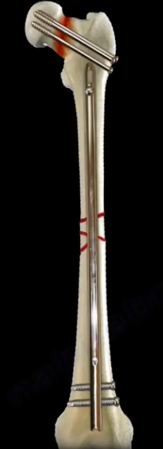
nerve stays lateral, distally. CTA around the knee can be done for dislocations
or severe fractures around the knee area. At this level of the distal femur,
you can see the popliteal artery. At the level of the proximal part of the leg,
you can see the three branches of the popliteal artery: anterior tibial artery,
posterior tibial artery, and peroneal artery. Be careful during placement. The anterior tibial artery arises just below the
popliteus muscle. The anterior tibial artery pierces the interosseous membrane
to enter into the extensor compartment or anterior compartment of the leg. The anterior
tibial artery gives the anterior and posterior tibial recurrent arteries. The anterior
tibial recurrent artery is the one that can be injured from tibial tubercle
fracture in children, which can cause compartment syndrome of the leg. The artery
then runs proximally between the tibialis anterior medially and the extensor
digitorum longus laterally. The extensor digitorum longus arises from the upper
3/4
Low back pain is a common condition. 90% of patients with
low back pain will improve without surgery. Usually they get better with
spontaneous resolution of the symptoms within 12 weeks. We usually advise the
patient for early return to activity and function as the symptoms and the pain
permits. The risk factors for development of low back pain are numerous, some
include: vibration exposure, poor physical fitness, smoking and obesity,
anxiety and depression, job dissatisfaction, or repetitive bending or
“stooping” on the job.In summary, if the patient has no
red flags and has a normal neurological exam, there is no reason to get early
radiological studies. Getting early x-rays and early MRIs leads to a better
patient satisfaction but does not give a better patient outcome. If there is no
specific pain pattern, then there is no need for further workup. MRIs are good
studies, but they give false positives. There is degeneration or a bulge of a
disc in 35% of all asymptomatic subjects between 25-39 years of age. In
patients 60 years old or older, the majority of the patients will have changes
in the MRI. MRI abnormalities are common and must be correlated with the age
and the clinical signs and symptoms of the patient. An MRI is good for
diagnosing the lumbar disc herniation, which is sometimes called a ruptured
disc, a slipped disc, or a herniated disc. The most common location of a disc
herniation is a posterolateral herniation involving one nerve root. A
foramninal L4-L5 herniation occurs in about 8%-10% of the cases. It involves
the exiting nerve. A central herniation involves multiple nerve roots. It
predominantly causes low back pain more than leg pain. It may cause bladder and
bowel symptoms. This type of disc herniation causes Cauda Equina Syndrome which
needs urgent diagnosis and surgical treatment. Clinical evaluation for a herniated
disc examines sensory and motor reflexes. The Straight Leg Raising Test is the
most important finding. It can be done in either the sitting or supine
position. The test is positive as indicated by pain in the leg when the
patient’s leg is raised to flex the hip with the knee extended. A positive
straight leg test means a tension sign, something is putting tension or stress
on the sciatic nerve. When the test is positive, it indicates possible disc
herniation.
Treatment is typically non-operative. First, reassure the patient.
Let the patient take some rest (no more than a few days), give the patient
anti-inflammatory medication, and instruct them to attend physical therapy.
Indications for surgery include progressive neurological deficits, Cauda Equina
Syndrome, the patient is not getting better with time and treatment or if the
symptoms are not getting better with conservative treatment, or the patient has
a positive tension sign with persistent sever pain. Patients with sciatica and
positive tension signs or patients with positive neurological findings on
clinical exam with positive MRI findings make ideal surgical candidates.
Surgery results in relief of leg pain in the majority of patients. Back pain
may persist in some patients. Surgery results in neurological improvement, 50 %
motor and sensory and 25% reflexes. In patients with discogenic back pain, they
may need fusion which is a major procedure.The worst pressure on the disc occurs with prolonged
sitting and bending over. This is the position that produces the highest
pressure on the disc. If a patient has back pain but no radiation, by the
patient’s history or physical examination and there are no red flags, then
there is no reason to get x-rays or MRI early in the treatment of the patient.
Red flags include a history of trauma, a tumor, infection, or Cauda Equina
Syndrome symptoms. To rule out a history of trauma you should rule out
fractures with x-rays, MRI, or CT scans. Tumors are a risk if the patient is
older than 50 years old, if the patient had weight loss, or if the patient has
pain at rest or at night. An infection may be present if the patient has fever
and chills, if the patient has a history of diabetes, or if the patient has a
history of IV drug abuse. Cauda Equina Symptoms may be present if the patient
has back pain more than leg pain or if the patient also has bladder and bowel
symptoms. Cauda Equina Syndrome needs to be diagnosed and surgically treated
early. An MRI needs to be ordered urgently in the course of treatment. The MRI
should be ordered STAT. There may need to be a wet read; a wet read is an early
preliminary read of the radiographs. A wet read needs to be communicated with
the physician and can be done while the patient is still on the table of the
MRI.
Femoral neck fracture nonunion has multiple facets and is
important to understand all aspects of this important problem.
Example:
40 year old patient had a displaced femoral neck fracture,
fixed with multiple cancellous screws about 9 months ago. The patient still has
persistent groin pain. The patient cannot bear full weight on the hip. The patient
has a painful limb, antalgic gait, and difficulty in walking. X-rays are not
clear and show a possible nonunion. CT scan shows the nonunion with some Varus
angulation. The treatment for this would be removal of the hardware and valgus
osteotomy. The scenario can be more complicated by adding a healed
femoral shaft fracture to the nonunion of the femoral neck. In this case, you
will do removal of the hardware from the femur and removal of the screws from
the femoral neck nonunion. You will do valgus osteotomy and fixation with a
plate, preferably a blade plate, to treat the nonunion of the femoral neck.
Intracapsular fractures of the proximal part of the femur
are not common in adults younger than 50 years old, but they are associated
with a high incidence of avascular necrosis and nonunion. About 10-30 % of
femoral neck fractures go to nonunion after ORIF. It is usually the vertical
fracture pattern, such as Type III in Pauwels Classification. These fractures
are more prone to nonunion due to shear stress, rather than compression forces
across the fracture site. In Garden Classification fracture Type IV, where the
fracture is completely displaced, the greater the displacement, the higher the
incidence of nonunion and reoperation rate after fixation of the femoral neck. The
inverted triangle pattern of fixation of femoral neck fractures is the one that
is commonly used with the inferior screw posterior to the midline and adjacent
to the calcar. Achieving and maintaining anatomic reduction is important for
femoral neck fracture fixation and healing. The femoral neck fractures are
intracapsular. There will be no abundant callus formation during the healing
(healing is intraosseous only). Sometimes it is difficult to know if the fracture
healed or not. There is no correlation between age, gender, and rate of
nonunion. Varus malreduction correlates with failure of fixation after
reduction and cannulated screw fixation. Posterior comminution of the fracture
does not allow stable fixation and can lead to nonunion. The comminution of the
femoral neck is usually posteriorly and inferiorly. Some recommend adding a
fourth screw in this situation. High energy fractures have a worse prognosis
for healing, especially in patients with metabolic bone disease and nutritional
deficiency. When you see a femoral neck nonunion after fixation, you need to
get blood work and rule out infection (get sedimentation rate and CRP).
For the
high angle femoral neck fracture, follow the patient up closely with clinical
exam and x-rays. There might be a Varus collapse on the x-rays. You may see a femoral
neck nonunion or a failed internal fixation. The patient walks with a limp, the
limb is shortened, and the patient may have rotational deformity of the
extremity. In the young patient with a femoral neck nonunion, arthroplasty is
not a desirable option. In a young patient with femoral neck fracture nonunion,
valgus intertrochanteric osteotomy with plate fixation produces a good result
in the majority of cases. Valgus intertrochanteric osteotomy with plate
fixation produces approximately 80% union rate and the procedure makes a
vertical fracture more horizontal, converting the shear forces into compressive
forces. It is done in a healthy, young patient with no joint arthritis and when
the femoral head is intact. This procedure also corrects the Varus
malalignment. Basically, the procedure changes the vertical fracture
orientation to a horizontal fracture to achieve compression. Other procedures
done in the young patient include revision ORIF with or without bone graft, but
this is rarely done. Other procedures done in the young patient also include
free vascularized fibular graft which is done in some patients especially in
the younger patient with a nonviable femoral head. Hemiarthroplasy is done in
patients with low physical demands. The articular cartilage of the patient is
preserved with no evidence of infection. Total hip arthroplasty is done in
patients that are older, in patients that have hip arthritis, if the femoral
head is not viable, or if the hardware is cut out. It can also be done in
younger patients that are active, when the femoral head is not viable and the
patient does not want a free vascularized fibular graft or if the patient had
collapse of the femoral head with nonunion. The problem with total hip
replacement in this situation is more dislocations of the hip postoperatively.
Cervical radiculopathy is caused by cervical nerve root
compression. The patient will have pain and/or progressive neurological deficit
that results from conditions such as disc herniation that irritates a nerve in
the cervical spine. Cervical radiculopathy is an irritation of the cervical
nerve root. Cervical spine and shoulder problems overlap. The condition is of
cervical spine etiology if the patient’s symptoms are relieved by shoulder
abduction, by placing the hand over the head. The relief of the symptoms occurs
due to decreased tension on the nerve roots. In cervical disc problems, be
aware of false positive MRIs especially if the patient is above the age of 40
years old. Nerve conduction studies are not useful; they have a high false
negative rate. EMG and nerve studies may differentiate radiculopathy from
peripheral nerve entrapment. Cervical disc problems usually affect the lower
numbered nerve root.
When you see the middle finger numbness, then this is C7.
When compression of the C7 nerve root, there will be middle finger numbness,
triceps weakness, and the triceps reflex will be affected. The cervical nerve
roots are horizontal in orientation. It does not matter if cervical disc
herniation is central or foraminal, it will compress the same nerve root. C7
nerve root runs above the pedicle of the C7 vertebra. C5-C6 is the most
commonly affected disc and that will compress the C6 nerve root. The patient
will come to the doctor with unilateral arm pain that is relieved by arm
elevation. The numbness and paresthesia will occur in specific dermatomes. The
patient may also have upper trapezius pain or interscapular pain. The patient
may complain of occipital headache. When you examine the patient, do
provocative tests such as the spurling’s test and the shoulder abduction test.
The Spurling’s test is done by extending and rotating the neck towards the
involved side. It reproduces the symptoms by narrowing the neuroforamen. The
Spurling’s test differentiates cervical radiculopathy from peripheral nerve
entrapment. Lifting the arm above the head relieves the symptoms if the
cervical nerve roots are irritated. The Shoulder Abduction test differentiates
cervical pathology from other causes of painful shoulder etiology. Make sure
that you do not have a double crush syndrome, one in the neck and one in the
peripheral nerve. Make sure that you differentiate radiculopathy from
myelopathy. Make sure that you exclude a coexisting myelopathy. Examine the
patient for upper motor neuron signs or cervical
myelopathy. Test the patient
for gait instability. Test the patient for Hoffman’s sign. Test the patient for
Babinski reflex. Test the patient for ankle Clonus. Check to see if the patient
has hyperflexia in the upper and lower extremities (triceps/quadriceps). Even
if there is a bad cervical spine disc problem on the MRI, treat it
conservatively for about 3 months. Give the patient therapy and nonsteroidal
anti-inflammatory medication (NSAIDS). 75% of the patients will improve with
nonoperative treatment. Cervical radiculopathy is generally treated
nonoperatively, in contrast to cervical myelopathy. Do surgery when there is
persistent, severe pain for 6-12 weeks and/or progressive neurological deficit
such as weakness or numbness. The procedure to treat cervical radiculopathy
surgically is usually done anteriorly with direct removal of the lesion that
causes the radiculopathy such as a herniated disc or spurs. When you place the
anterior bone graft or the allograft in the disc space, you open the
nueroforamen, and that will indirectly relieve the nerve. Then you will add the
anterior plate. Some surgeons prefer to do a posterior approach.
 The anterior tibial artery is a branch of the popliteal
artery (posterior aspect of knee), which divides into the anterior tibial
artery and the posterior tibial artery (posterior). The anterior tibial artery
is a branch of the popliteal artery (posterior aspect of the knee), which
divides into the anterior tibial artery and the posterior tibial artery
(posterior). Sometimes the two divisions are called the anterior tibial artery
and the tibio-fibular trunk. th area of the anterior fibula. Then the extensor hallucis longus
muscle appears, so the anterior tibial artery lies between the tibialis
anterior muscle and the extensor hallucis longus muscle. The extensor hallucis
longus muscle arises from the middle 2/4th area of the anterior
fibula. The extensor hallucis longus then crosses the leg medially to take a
position in the medial side. The big toe is definitely medial, so the extensor
hallucis longus will go towards the big toe and become medial. The other toes
are lateral, so the extensor digitorum longus will be inserted laterally and
the anterior tibial artery will then be between these two muscles in the distal
part of the leg and in front of the ankle. When the extensor hallucis longus
tendon crosses the leg to go medially, it then crosses the anterior tibial
artery. At this point, the anterior tibial artery is between the extensor
hallucis longus and the extensor digitorum longus tendons. At the level of the
ankle joint, this is how we remember the arrangement of the anterior ankle
structures. Tom Has a Very Nice Dog: Tibialis anterior,
extensor Hallucis longus, Vessels, Nerve, extensor Digitorum
longus. This is only good to remember the structures in the distal portion of the
tibia in front of the ankle. This does not work proximally, and this does not
work for the structures in the middle third of the tibia. Extensor hallicus
longus tendon is medial. Anterior tibial artery is lateral. After the anterior
tibial artery passes underneath the extensor retinaculum, the artery is then
called the dorsalis pedis, distally. The deep peroneal nerve pierces the intermuscular
septum to enter the anterior compartment and goes through the substance of the
extensor digitorum longus muscle. of retractors in the posterior part of the
proximal tibia to avoid damage to any of these branches of the popliteal artery
since the bifurcation of the popliteal artery is in this area.
The anterior tibial artery is a branch of the popliteal
artery (posterior aspect of knee), which divides into the anterior tibial
artery and the posterior tibial artery (posterior). The anterior tibial artery
is a branch of the popliteal artery (posterior aspect of the knee), which
divides into the anterior tibial artery and the posterior tibial artery
(posterior). Sometimes the two divisions are called the anterior tibial artery
and the tibio-fibular trunk. th area of the anterior fibula. Then the extensor hallucis longus
muscle appears, so the anterior tibial artery lies between the tibialis
anterior muscle and the extensor hallucis longus muscle. The extensor hallucis
longus muscle arises from the middle 2/4th area of the anterior
fibula. The extensor hallucis longus then crosses the leg medially to take a
position in the medial side. The big toe is definitely medial, so the extensor
hallucis longus will go towards the big toe and become medial. The other toes
are lateral, so the extensor digitorum longus will be inserted laterally and
the anterior tibial artery will then be between these two muscles in the distal
part of the leg and in front of the ankle. When the extensor hallucis longus
tendon crosses the leg to go medially, it then crosses the anterior tibial
artery. At this point, the anterior tibial artery is between the extensor
hallucis longus and the extensor digitorum longus tendons. At the level of the
ankle joint, this is how we remember the arrangement of the anterior ankle
structures. Tom Has a Very Nice Dog: Tibialis anterior,
extensor Hallucis longus, Vessels, Nerve, extensor Digitorum
longus. This is only good to remember the structures in the distal portion of the
tibia in front of the ankle. This does not work proximally, and this does not
work for the structures in the middle third of the tibia. Extensor hallicus
longus tendon is medial. Anterior tibial artery is lateral. After the anterior
tibial artery passes underneath the extensor retinaculum, the artery is then
called the dorsalis pedis, distally. The deep peroneal nerve pierces the intermuscular
septum to enter the anterior compartment and goes through the substance of the
extensor digitorum longus muscle. of retractors in the posterior part of the
proximal tibia to avoid damage to any of these branches of the popliteal artery
since the bifurcation of the popliteal artery is in this area.