Tuesday, August 28, 2018

Massive Rotator Cuff Tear


To view my Youtube video, 'Massive Rotator Cuff Tear- Classic', click here.


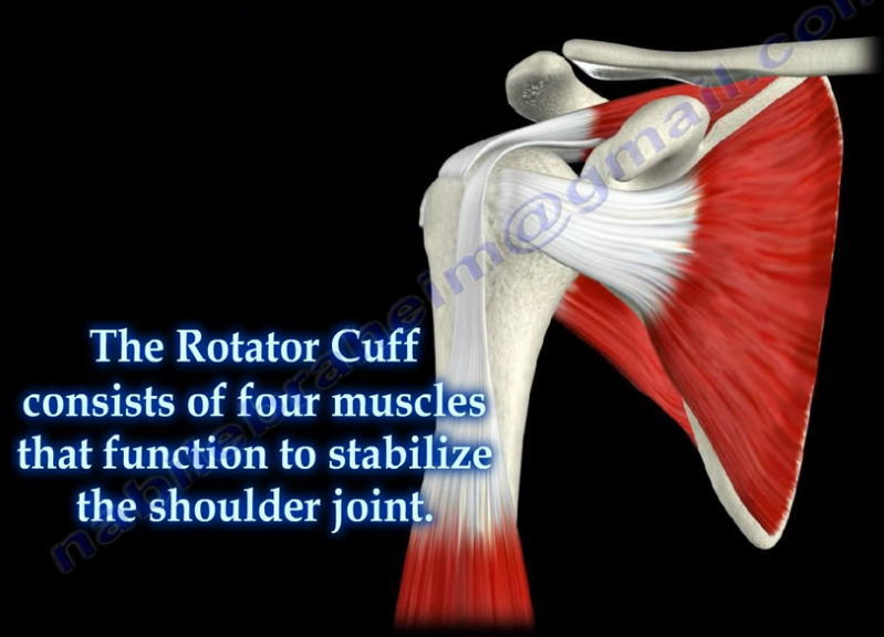
Massive rotator cuff tears are a disabling problem. These tears can cause pain, weakness, and sometimes swelling of the shoulder. The rotator cuff consists of four muscles that function to stabilize the shoulder joint: supraspinatus rotator cuff tendon, subscapularis tendon, infraspinatus rotator cuff tendon, and the teres minor rotator cuff tendon. The supraspinatus tendon is the most common of the rotator cuff tendons to become ruptured. Massive tears of the rotator cuff that are greater than 5cm usually involving both the supraspinatus and infraspinatus tendons.


Massive tears of the rotator cuff are defined as tears greater than 5cm, usually involving both the supraspinatus and infraspinatus tendons. Retraction of the rotator cuff tendons along with muscle atrophy and fatty infiltration can occur. This makes surgical reconstruction difficult with the surgical outcome being unpredictable and less than satisfactory.

Treatment varies from physiotherapy to replacement of the humeral head. Arthroscopic or open repair is usually the selected treatment. Reconstruction can be done in selected cases. A rotator cuff arthropathy is performed on massive cuff tears that are associated with superior migration of the humeral head as well as instability and arthritis of the shoulder. The patient will have pseudoparalysis and an x-ray will show shift of the humerus proximally. An MRI will show massive cuff tear with retraction at the level of the glenoid with atrophy of the muscle and fatty infiltration. A reverse shoulder is the treatment of choice for the elderly with rotator cuff arthropathy as it improves the pain and function. Hemiarthroplasty is the treatment for younger patients. A standard head or a big humeral head can be selected.


A patient with a massive tear of the cuff usually develops weakness of the shoulder and becomes unable to actively lift the arm without assistance. Fluid collection within the shoulder may occur with a massive tear of the rotator cuff.

Tuesday, August 21, 2018

Toe Deformities


Deformities of the toes are not uncommon and can occur from muscle imbalance, or other causes such as rheumatoid arthritis, diabetes, compartment syndrome, synovitis, or neurological disorders. Hammer toe occurs as flexion of the proximal interphalangeal (PIP) joint. Hammer toe is similar to the Boutonniere deformity of the finger. Claw Toe is a hyperextension deformity of the MTP joint and flexion of the PIP and DIP, resembling a pirate hook. Claw toe is similar to an intrinsic minus deformity of the hand, or “claw hand”. Mallet toe is similar in appearance to mallet finger, and is a flexion deformity of the DIP joint.

Tuesday, August 14, 2018

Triplane Fracture of the Distal Tibia


A triplane fracture of the distal tibia usually occurs during adolescence and occurs before complete closure of the distal tibial physis. The distal tibial physis (growth plate) is a weak area which closes from central to medial, with the lateral side being the last part to close. A Triplane fracture is a Salter-Harris Type IV Fracture, involving all three planes, the coronal (metaphysis), transverse (growth plate), and sagittal (epiphysis). The fracture has several variations and occurs due to external rotation forces. This fracture typically occurs in patients between 12-15 years of age.


Triplane fractures are complicated three-dimensional fractures. A two-part fracture is a Salter-Harris Type IV. A Three-part fracture is a combination of Salter-Harris Type III in an AP view and a Type II in a lateral view. CT scans are helpful.


An ORIF is necessary if there is displacement of the fragments of more than 2 mm.

Wednesday, August 8, 2018

Discoid Meniscus


The meniscus is a cushion structure made of cartilage which fits within the knee joint between the tibia and the femur. The medial meniscus is C-shaped and the lateral meniscus in the more circular. The meniscus is made up of type I collagen that provides shock absorption and stability to the knee joint. The meniscus helps to protect the knee joint, allowing the bones to slide freely on each other. Discoid meniscus is a rare variation of the meniscus that usually affects the lateral meniscus of the knee in less than 5% of the population and could be bilateral in about 25% of the cases.
Discoid meniscus is a large meniscus with abnormal attachment causing increased mobility of the meniscus. It causes a pop, click, or snapping with locking and pain. There will be loss of full knee extension with tenderness on the lateral joint space. Symptoms occur more during extension of the knee. The discoid meniscus occurs due to the abnormal development and increase in size of the meniscus. An x-ray could show increased widening of the joint space. An MRI will show the “bow tie” sign in three or more sagittal continuous cuts. The coronal MRI will show a thick and flat meniscus extending beyond the halfway point of the condyle.


Watanabe Classification of Discoid Lateral Meniscus


  • Type I: Block-shaped stable
  • Type II: Block-shaped, stable, partial meniscus (has good peripheral attachment)
  • Type III: Unstable meniscus with stability arising only form the ligament of Wrisberg. (no posterior meniscal tibial attachment).

Treatment


An asymptomatic patient will be treated with observation. A symptomatic patient may receive a partial meniscectomy and saucerization with repair of type III (no posterior tibial meniscal attachment)