The vastus lateralis muscle is a common site for
intramuscular injection. To inject, first locate the greater trochanter of the
femur. The top border of the vastus lateralis muscle can be found a hand width
below the groin. The bottom border of the vasus lateralis muscle is found a
hand above the knee. The side borders are from the mid anterior thigh to the
mid lateral thigh. Squeezing the muscle before injection increases the
thickness and depth of the muscle and avoids nerve injuries. Make sure you do
not advance the needle deep to touch the bone. Aspirate before you inject.
As the median nerve travels through the carpal tunnel, it
may become compressed. Carpal tunnel syndrome is the condition of pressure
being placed on the median nerve. The transverse carpal ligament is usually
thickened at the wrist and this causes the compression of the nerve. The condition
usually occurs due to an overuse injury such as repetitive hand grip movements.
The predisposing factors for carpal tunnel syndrome include trauma, fractures,
pregnancy, diabetes, rheumatoid arthritis, ganglion cyst, smoking, alcoholism,
advanced age, and obesity. As a result of this compression, the patient may
experience pain and paresthesia in the median nerve distribution as well as
weakness in the hand. The median nerve is inside the carpal canal. The transverse
carpal ligament covers the median nerve and if this ligament is thickened, it
can compress the median nerve. It is almost like a narrow tunnel, and the
median nerve is much like a truck passing through the narrow tunnel. o angle to the skin of the wrist. Next, direct the needle
towards the base of the thumb and advance the needle distally and slowly. You should
mark the site 4-5 cm proximal to the distal palmar crease between the palmaris
longus and the flexor carpi radialis tendons. For the proximal approach, you
should advance the needle distally towards the wrist at about a 20o
angle to the skin, keep the needle between the two tendons, be aware that the
nerve is in between the two tendons (the nerve is really superficial) and
adjust the needle as needed, and then inject the desired fluid.
We try to
make room for the nerve to past through. The tunnel is widened by doing carpal
tunnel surgery. Cutting the transverse carpal ligament, as seen in this
example. We can possibly make room for the median nerve with steroid injection,
because injection will decrease the inflammation. The typical patient with
carpal tunnel syndrome will have hand pain, numbness and tingling in the radial
3 ½ fingers (median nerve distribution). These symptoms may wake the patient up
at night. Treatment is usually a night splint or anti-inflammatory medication. Sometimes
we give some patients a steroid injection which can be helpful. In general, you
will know which patients will have a good prognosis from the treatment of
carpal tunnel, and these are the patients who will have night symptoms. Another
indication for a good prognosis is the response to steroid injection. If the
steroid injection helps the symptoms, then the patient will do well from the
carpal tunnel surgery. Failure to improve after steroid injection indicates a
less favorable outcome from the surgery. The injection is usually helpful when
it is not clear to the clinician where the symptoms are coming from. If you
inject the carpal tunnel and the patient improves, then you know the
predominant problem is the carpal tunnel, because all of the three conditions
can give the same symptoms. This is especially important in double crush
syndrome where the median nerve can be compressed at two different places along
its course, and one of these places can be the carpal tunnel. Injection also
has therapeutic value. Injection allows a period of relief in patients with
mild or moderate carpal tunnel symptoms. 80% of the patients will have some
transient improvement with injection and 20% of the patients will improve up to
1 year. The median nerve is located between the palmaris longus and flexor
carpi radialis tendons. You can give the injection by ultrasound or you can do
it blindly. When you do blind injection, you can do it by the usual approach or
by the proximal approach. With the usual approach, you should mark the
intersection of the palmaris longus tendon and the distal palmar crease. Next,
go 1 cm proximal and 1 cm ulnar to that site, this will be the point of the
injection.
Use a 25 gauge needle with the preferred brand and amount of
steroids and 1 mL of 1% lidocaine. The physician should warn the patient before
the injection that if any feeling of numbness, paresthesia, or sever pain
exists to let the physician know about it. If the symptoms exist, the doctor
should withdraw the needle or adjust it before injection. You may use local
anesthesia or a spray. You should use a sterile field, make sure that you have
a consent, make sure that you have a time-out sheet, and mark the site before
injection. To administer a usual approach injection, you should put the needle
at a 45
Low back pain is a common condition. 90% of patients with
low back pain will improve without surgery. Usually they get better with
spontaneous resolution of the symptoms within 12 weeks. We usually advise the
patient for early return to activity and function as the symptoms and the pain
permits. The risk factors for development of low back pain are numerous, some
include: vibration exposure, poor physical fitness, smoking and obesity,
anxiety and depression, job dissatisfaction, or repetitive bending or “stooping”
on the job. The worst pressure on the disc occurs with prolonged sitting and
bending over.In summary, if the patient has no red flags
and has a normal neurological exam, there is no reason to get early
radiological studies. Getting early x-rays and early MRIs leads to a better
patient satisfaction but does not give a better patient outcome. If there is no
specific pain pattern, then there is no need for further workup. MRIs are good
studies, but they give false positives. There is degeneration or a bulge of a
disc in 35% of all asymptomatic subjects between 25-39 years of age. In patients
60 years old or older, the majority of the patients will have changes in the
MRI. MRI abnormalities are common and must be correlated with the age and the
clinical signs and symptoms of the patient. An MRI is good for diagnosing the
lumbar disc herniation, which is sometimes called a ruptured disc, a slipped
disc, or a herniated disc. The most common location of a disc herniation is a
posterolateral herniation involving one nerve root. A foramninal L4-L5
herniation occurs in about 8%-10% of the cases. It involves the exiting nerve. A
central herniation involves multiple nerve roots. It predominantly causes low
back pain more than leg pain. It may cause bladder and bowel symptoms. This type
of disc herniation causes Cauda Equina Syndrome which needs urgent diagnosis
and surgical treatment. Clinical evaluation for a herniated disc examines
sensory and motor reflexes. The Straight Leg Raising Test is the most important
finding. It can be done in either the sitting or supine position. The test is
positive as indicated by pain in the leg when the patient’s leg is raised to
flex the hip with the knee extended. A positive straight leg test means a tension
sign, something is putting tension or stress on the sciatic nerve. When the
test is positive, it indicates possible disc herniation. Treatment is typically
non-operative. First, reassure the patient. Let the patient take some rest (no
more than a few days), give the patient anti-inflammatory medication, and
instruct them to attend physical therapy. Indications for surgery include
progressive neurological deficits, Cauda Equina Syndrome, the patient is not
getting better with time and treatment or if the symptoms are not getting better
with conservative treatment, or the patient has a positive tension sign with
persistent sever pain. Patients with sciatica and positive tension signs or
patients with positive neurological findings on clinical exam with positive MRI
findings make ideal surgical candidates. Surgery results in relief of leg pain
in the majority of patients. Back pain may persist in some patients.
Surgery results
in neurological improvement, 50 % motor and sensory and 25% reflexes. In patients
with discogenic back pain, they may need fusion which is a major procedure.This is the position that produces the highest pressure on the
disc. If a patient has back pain but no radiation, by the patient’s history or
physical examination and there are no red flags, then there is no reason to get
x-rays or MRI early in the treatment of the patient. Red flags include a
history of trauma, a tumor, infection, or Cauda Equina Syndrome symptoms. To rule
out a history of trauma you should rule out fractures with x-rays, MRI, or CT
scans. Tumors are a risk if the patient is older than 50 years old, if the
patient had weight loss, or if the patient has pain at rest or at night. An infection
may be present if the patient has fever and chills, if the patient has a
history of diabetes, or if the patient has a history of IV drug abuse. Cauda
Equina Symptoms may be present if the patient has back pain more than leg pain
or if the patient also has bladder and bowel symptoms. Cauda Equina Syndrome
needs to be diagnosed and surgically treated early. An MRI needs to be ordered
urgently in the course of treatment. The MRI should be ordered STAT. There may
need to be a wet read; a wet read is an early preliminary read of the
radiographs. A wet read needs to be communicated with the physician and can be
done while the patient is still on the table of the MRI.
The Extensor Digitorum Brevis originates from the anterior
part of the dorsal surface of the calcaneus. Four tendons insert into the
proximal phalanx of the big toe and long extensor tendons to toes 2, 3, and 4. The
medial part of the muscle is known as the extensor hallucis brevies and ends in
a tendon that is inserted into the dorsal surface of the base of the proximal
phalanx of the big toe.
The other three tendons insert into the lateral side of
the tendons of the extensor digitorum longus of toes number 2, 3, and 4. The extensor
digitorum brevis helps to extend the first four digits. The extensor digitorum
brevis is innervated by the deep peroneal nerve (predominantly L5 nerve root). The
EDB muscle has the same innervation as a disc herniation at L4-L5 which will
also involve the L5 nerve root. It is probably the only muscle of the foot that
is innervated by the deep peroneal nerve. The deep peroneal nerve supplies
sensation to the first web space. The EDB is the only muscle on the foot that
makes a fleshy enlargement anterior to the lateral malleolus. The extensor
digitorum brevis can cause pain on the top of the foot. Irreducible dislocation
of the medial subtalar joint can result from interposition of the extensor digitorum
brevis muscle. Irreducible dislocation of the lateral subtalar joint can result
from interposition of the tibialis posterior tendon.
25 Vitamin D, Tests Ortho
Surgeons Think About-Everything You Need To Know
Vitamin D 25 is the most appropriate study to assess and
monitor vitamin D status in the body. Vitamin D is important for proper
maturation and development of bone. Vitamin D is also important in immunity and
plays a role in other conditions. The main function of Vitamin D is absorption
of the calcium and phosphate from the intestine. Vitamin D comes from diet,
supplements, and exposure to the sun. Vitamin D is naturally found in fish.
Exposure to the sun for 15 minutes will give a person about 10,000 units of
Vitamin D. The average daily requirement of Vitamin D is approximately 400-800
International Units (IUs). Vitamin D gets activated metabolically in the liver
and in the kidney. The activation occurs by hydroxylation. Hydroxylation to 25
Vitamin D3 occurs in the liver. The big organ takes the big number-
25, so 25(OH)-Vitamin D3. Another hydroxylation occurs in the
kidneys. 2- Vitamin D3. This is the active form of Vitamin D
and works mainly on the intestines and bones. The activation of Vitamin D to 1,
25 hydroxyvitamin D is controlled by the parathyroid hormone. Any deficiency or
any problem in the process of activating Vitamin D3 to its active
form will lead to deficiency of Vitamin D in the body. Vitamin D deficiency is
very common and the majority of people are not aware of it. In fact, Vitamin D
deficiency symptoms are subtle and nonspecific. 25-hydroxyvitamin D has a long
half-life and a higher concentration. This is probably easier to measure and
obtain 25 hydroxyvitamin D than the active form, which is 1,25-dihydroxyvitamin
D. The half-life of 25-hydroxyvitamin D is 2-3 weeks. The half-life of
1,25-dihidroxyvitamin D is only 4-6 hours. The circulating levels of 25-
hydroxyvitamin D is 1000x more than 1,25 dihydroxyvitamin D. therefore,
25-hydroxyvitamin D test is the best study to determine the Vitamin D
deficiency in the body. A low level of 25- hydroxyvitamin D could mean that a
person is not getting enough exposure to the sun, is not getting enough dietary
Vitamin D, or there may be a problem with absorbing Vitamin D from the
intestines. The patient may be taking Dilantin, which interferes with
hydroxylation of Vitamin D in the liver. A low level of 1, 25-dihydroxyvitamin
D usually indicates kidney disease. 40% of the United States population have
Vitamin D deficiency.The small organ takes the small number-1. The result will be 1, 25
(OH)
Symptoms of Vitamin D deficiency may include:
·Fatigue and tiredness
·Not sleeping well
·Muscle weakness
·Bone pain
·Osteoporosis/ Osteomalacia
·Fractures
Elderly patients are vulnerable to Vitamin D deficiency
because they usually live indoors or in nursing homes with no sun exposure or
because these patients may not eat enough food containing Vitamin D or they may
not receive enough supplements. Vitamin D deficiency may impair or affect wound
healing. Vitamin D deficiency may cause bone loss and places the elderly
patient at risk of fractures. Deficiency may cause slow healing of fractures or
nonunion of the fractures. If you find a patient with fractures that are not
healing well, or a patient with fractures due to low energy trauma where you
find the bone mass is inadequate (osteoporosis), this is the time to get a 25-
hydroxyvitamin D blood test. The Endocrine Society defines Vitamin D deficiency
as 25 Vitamin D level below 20 ng/mL, and insufficiency as the level between
21-29 ng/mL. In general, a 25 Vitamin D level greater than 30 ng/mL is probably
adequate, but these numbers are controversial.
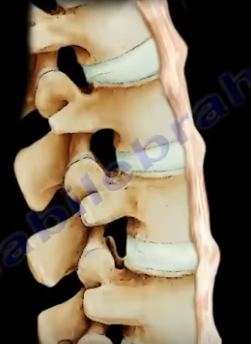
The DISH has flowing ossification along anterolateral aspect
of at least four continuous vertebrae. When you look at the x-ray, you find
ossification along the anterior aspect of the body but separate from the
vertebrae and the disc height is preserved. It occurs in older patients (50
years and above). It affects all of the spine (more in the thoracic spine),
especially on the right side, which is typical of DISH. The syndesmophytes are
equal on the right and left sides in the lumbar and cervical vertebrae.
There is
no involvement of the discs and there is no facet fusion or sacroiliac joint involvement.
The patient may have other comorbidities such as gout or diabetes, and you need
to get the hemoglobin A1c (HbA1c test) in these patients. Some patients may
have high cholesterol levels. The patient will complain of back pain and spinal
stiffness. DISH will have large syndesmophytes, and if the condition occurs in
the neck, it will cause dysphagia, hoarseness of the voice, and sleep apnea. Diagnosis
can be established by x-ray of the spine. On lateral x-ray of the cervical
spine, you will find anterior bony fragments and the discs are preserved. The fractures
in the spine are usually due to a hyperextension injury and can be occult,
resulting from minor trauma and may have major instability. There is an
increased mortality in c-spine trauma in DISH, high mortality especially in non-operative
treatment. If the patient has a history of sudden neck or back pain, then the
patient will be assumed to have an occult fracture, so try to get a CT scan or
an MRI even if the pain is minimal and even if the x-rays appear normal. Heterotopic
ossification after total hip arthroplasty is more in patients with DISH.
What is the difference between DISH and Ankylosing
Spondylitis?
DISH
-Flowing large syndesmophytes
-No bamboo spine
-Sacroiliac (SI) join will not be involved
-Occurs in older patients
-some patients may have diabetes, check hemoglobin A1c
Ankylosing Spondylitis
-Diffuse ossification of the disc space without large
osteophytes
Plantar fascia rupture is not a very common injury, and it
has the characteristic of acute pain in the arch of the foot. It occurs due to
a tear in the plantar fascia, and that tear is painful. Rupture is often
associated with long standing flat feet deformity or can occur from steroid
injections. Another predisposing factor for plantar fascia rupture is plantar
fasciitis.
Anatomy
The plantar fascia is formed by three bands: the medial, the
central, and the lateral. The plantar aponeurosis is the central part of the
plantar fascia. The plantar fascia is inserted into the medial tuberosity of
the
calcaneus and extends distally, becoming broader and thinner. The plantar
fascia acts as a bow string.
The rupture of the plantar fascia may be misdiagnosed as
plantar fasciitis. When the plantar fascia tears, the patient will describe a
tearing pain that usually occurs during athletic activity. The tear may be
complete or incomplete. Complete tear of the plantar fascia occurs from sudden
trauma or injury. The patient feels “popping” or “snapping” suddenly. Walking will
be very difficult with tenderness, swelling and significant bruising on the
sole of the foot (the condition is painful). Some patients may have a noticeable
tightness of the calf muscle (equinus contracture) in association with rupture
of the plantar fascia. Partial rupture is less common and occurs from overuse,
as in running. MRI will identify the rupture, and it can also identify if the
rupture is partial or incomplete. Rupture is often in the arch of the foot
opposed to where the plantar fascia inserts into the heel (calcaneus). Ultrasound
has the same accuracy as MRI for imaging the plantar fascia. Interpretation of
the plantar fascia rupture may be difficult. You may need dynamic maneuvers
with dorsal flexion of the forefoot to stretch the plantar fascia. Usually the
proximal part of the plantar aponeurosis is clearly visualized on ultrasound.
MRI is probably better in diagnosis plantar fascia rupture.
Treatment of Plantar Fascia Rupture
-Non-Weight Bearing for 2-3 Weeks
-Walking Boot
-Crutches
-Physical Therapy
-Surgical Treatment is the last resort.
-Could be used in some athletes who
continue to have pain despite a well conducted conservative treatment
-Surgery is done to release the
fascia and the excise the scar
Patients with rupture of the plantar fascia typically
achieve a favorable outcome with return to full activity.
Ganglion Cysts Pressure Motor Branch of Median Nerve
After passing through the carpal tunnel, the median nerve
gives a branch on the radial side called the recurrent motor branch. The recurrent
motor branch innervates the abductor pollicis brevis, the flexor pollicis
brevis (superficial head), and the opponens pollicis muscles.
The recurrent
motor branch of the median nerve has multiple variations of the nerve. 50% are
extraligamentous with recurrent innervation. 30% are subligamentous with
recurrent innervation. 20% are transligamentous with recurrent innervation. When
you release the carpal tunnel, it is important to cut the transverse carpal
ligament far ulnarly to avoid cutting the recurrent motor branch of the median
nerve. These are the patients that will get motor symptoms after you do carpal
tunnel release. There is another entity similar to this entity, and these are
the patients that have symptoms similar to carpal tunnel syndrome, but their presentation
is not classic. These are the patients that you may need to get an MRI or
ultrasound to check the carpal tunnel area. Pain symptoms of carpal tunnel
syndrome occur more at night. Self-administered hand diagram is extremely
helpful (most specific test for carpal tunnel syndrome). The patient should
highlight the areas where they are experiencing the symptoms. The patient may
complain of thenar atrophy, weakness, or clumsiness of the hand. The positive
compression test (Durkan’s test) is the most sensitive test. You can see the
Tinel’s Sign, do the Phalen’s test, or the Semmes Weinstein test. Some physicians
believe that EMG doesn’t really increase the diagnostic value of these tests
(if you have a combination of these test), and you will proceed with surgery
even if the EMG is normal. The problem is, that you will find a group of
patients that have weakness and atrophy of the thumb muscles, and the
provocative and sensory tests for carpal tunnel syndrome are negative. These are
the patients that you will get an MRI to rule out pressure on the motor branch
of the median nerve. These are the patients that you will probably find a
ganglion cyst pressuring the motor branch of the median nerve.
The ligaments of the ankle are complex. Injury to these
ligaments are called ankle sprains. Sprain of the ankle is usually a low ankle
sprain. Occasionally, it can be a high ankle sprain. Sprain of the ankle can be
confused with other conditions that can happen around the ankle such as:
·Osteochondral lesion
·Peroneal tendon subluxation
·Fracture of the lateral talar process
·Fracture of the anterior process of the
calcaneus
·High ankle sprain (syndesmotic injury)
Here are a few tests that are used to test for injury of
these ligaments:
Anterior Drawer Test
Squeeze Test
External Rotation Stress Test
Talar Tilt Test (Inversion Test)
If the patient cannot bear weight on the ankle, the patient
should get an x-ray. Injury of the deltoid ligament occurs at the medial side
of the ankle, and it is usually associated with ankle fractures. Sometimes injury
of the deltoid ligament is occult and the patient will need external rotation
stress x-rays to demonstrate injury of the deltoid ligament. Injury to the
lateral side ligaments is referred to as low ankle sprain. The anterior
talofibular ligament is the weakest ligament on the lateral side. The anterior
drawer test is done to test the competency of the anterior talofibular ligament.
It is done in 20 degrees of plantar flexion and compare it to the other side. A
shift of an absolute value of 9 mm on the lateral x-ray or 5 mm compared to the
other side is positive. The calcaneofibular ligament is usually injured after
the anterior talofibular ligament. The test used to diagnose injury of the
calcaneofibular ligament is called the talar tilt test or the inversion test. Less
than 5 degrees of tilt is usually normal. The final area of injury is called a
high ankle sprain or injury to the syndesmosis. Contrary to a low ankle sprain,
a high ankle sprain may require surgery. This is how injury to the syndesmosis
occurs. Always check the fibula proximally to avoid missing a Maisonneuve
fracture. The Maisonneuve fracture will have a proximal fibular fracture, a
syndesmotic injury, and a deltoid ligament injury. This will require surgery. The
tests used to diagnose high ankle sprains are the squeeze test and the external
rotation stress test. The squeeze test is performed by squeezing the tibia and
fibula at mid-calf. This will cause pain at the syndesmosis if a high ankle
sprain is present. The external rotation test is the other test used to
diagnose a high ankle sprain or an injury of the syndesmosis. The external
rotation test is performed by first placing the ankle into a neutral position. Then,
apply external rotation stress and finally, get a mortise view radiograph. There
is a positive result for syndesmotic injury if the tibiofibular clear space is
more than 5mm or if the medial clear space widening is more than 4mm.
Ganglion
cysts can occur anywhere. They usually occur at the wrist area, however,
ganglion cysts may occur at the foot (usually at the top of the foot). This mass
can change in size, and it may grow slowly. The patient notices a mass usually
on the top of the foot. The mass is usually asymptomatic. The patient may have
a burning sensation due to irritation of the nerve when the ganglion compresses
the nerve. The patient may have skin irritation and also may have difficulty
walking and wearing shoes. If the ganglion is pushing on a nerve and causing
irritation, aspiration or surgical removal of the cyst can help relieving the
symptoms. We need to differentiate ganglion cyst of the foot from plantar
fibromatosis. Plantar fibromatosis occurs at the bottom of the foot. Ganglion cysts
usually occur at the top of the foot. Ganglion cysts will transilluminate. Plantar
fibromatosis does not transilluminate
Ganglion Cyst of the Ankle (Tarsal Tunnel Syndrome)
What is
Tarsal Tunnel Syndrome?
Tarsal
tunnel syndrome is a compressive neuropathy which is caused by compression of
the tibial nerve around the ankle region. A ganglion cyst can be one of the
intrinsic causes of tibial nerve compression. In the tarsal tunnel, the patient
may have pain and burning sensation. An MRI is probably needed for the
diagnosis of a ganglion cyst in the tarsal tunnel. They found that they best
result after surgery occurs when there is a ganglion cyst compressing on the
nerve, and this cyst is removed.