Tuesday, July 31, 2018

Examination of the Acromioclavicular Joint


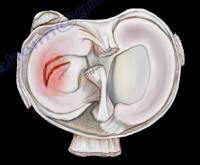
The acromioclavicular joint is located at the top of the shoulder, where the acromion of the scapula and the clavicle join together. The AC joint is a small synovial gliding joint that can be affected by arthritis and osteoarthritis. The oblique orientation of the joint’s articular surfaces may allow the acromion to be driven underneath the clavicle when the AC joint is injured. The condition could be subtle. Injuries of the acromioclavicular joint most commonly occur due to separation of the AC joint. Falling directly onto the shoulder can injure the ligaments that stabilize the AC joint. The AC ligament provides anterior-posterior stability of the AC joint. The posterior and superior AC ligaments are most important for stability. The coracoclavicular ligaments provide superior-inferior stability. Activity related pain with overhead activity and arm adduction.
During the physical examination, in order to test for injury to the AC joint, the physician will begin by palpating the AC joint. They should check to see if pain is present with direct palpation of the AC joint. If pressing down onto the AC joint causes pain, this is a sign of an AC joint problem such as distal clavicle osteolysis, arthritis, sprain of the AC ligament, or separation. Osteolysis of the distal clavicle is a localized area of inflammation, hyperemia, microfracture, bone resorption, and eventually arthritis of the AC joint. When pulling down on the shoulder, if there is a separation of the AC joint, the clavicle will rise and a bump will be seen in the area of the joint. Sometimes, this is demonstrated by adding weights and comparing both sides. The cross body adduction test can also be done by bringing the shoulder across the body. This squeezes the acromion and clavicle together, causing pain directly in the area of the joint if an AC joint separation or arthritis is present.

The acromioclavicular joint is best evaluated using the Zanca view radiograph. Using the Zanca view, the x-ray beam is directed with a cephalad angle of 15 degrees. Clavicular osteolysis can be assessed using the Zanca view. The acromion will be normal with the abnormality isolated to the distal clavicle. The Zanca view is also used for diagnosis of arthritis of the AC joint. It can show osteophytes and joint space narrowing. The patient’s symptoms may not correlate with the x-ray findings. An MRI will show an increased signal and edema in the AC joint.

Tuesday, July 24, 2018

Lumbosacral Plexus


The sciatic nerve is the key nerve of the lumbosacral plexus. It arises from the spinal nerves of L4 through S3. The sciatic nerve is the cornerstone of the lumbosacral plexus. Knowledge of the lumbosacral plexus starts with the sciatic nerve because it is the most important nerve. The lumbosacral plexus is comprised of the sciatic nerve and S4. The branches of the lumbosacral plexus are the superior gluteal nerve (L4-S1), the inferior gluteal nerve (L5-S2), the posterior cutaneous nerve of the thigh (S1-S3), and the pudendal nerve (S2-S4) (SIPP).

Tuesday, July 17, 2018

Intra-articular Extensile Approach for Tibial Plateau Fractures


Several types of tibial plateau fractures are a complex management problem. The knee joint may have a significant comminution and depression, and the physician may need to take an extensile approach for reduction and fixation of this fracture. Personally, I use the intra-articular extensile approach for tibial plateau fracture reduction and fixation. In general, fracture of the tibial plateau is a complicated problem.


A vascular evaluation is necessary. The ankle-brachial index (ABI) is needed in some types, such as in medial plateau fractures or in severe types, such as Schatzker Type V or Type VI. The ABI should be more than 0.9. Usually, medial tibial plateau fractures are considered to be a knee dislocation. A fasciotomy may be needed if compartment syndrome occurs. The soft tissue condition may be bad, and an external fixator may be initially used until the soft tissue condition improves.

The association between tibial plateau fractures and meniscal tear is not uncommon. A lateral plateau fracture will create a lateral meniscal tear, while the medial plateau fracture will cause a medial meniscal tear. A tear of the meniscus is usually peripheral. It should be recognized and dealt with. The physician may want to look at the x-ray and see if there is a depression or separation of more than 6mm, as this indicates a high chance of meniscal tear.

The posteromedial fragment is another problem with tibial plateau fractures which needs to be fixed separately. When an extensive comminuted displaced tibial plateau fracture occurs, the physician may need excellent exposure of the articular surface to allow for anatomic reduction of the joint and visualization and repair or debridement of the meniscus if it is torn. This extensile exposure is important, especially if the posterior part of the plateau is involved. The traditional way to see the articular cartilage of the tibial plateau is to use the submeniscal approach by cutting the coronary ligament, but the exposure is limited. Other extensile approaches are also developed; however, we use the extensile intra-articular approach for complex, comminuted tibial plateau fractures. This involves anterior detachment and retraction of the meniscus to improve visualization of the tibial articular surface. This approach can be utilized for lateral or medial tibial plateau fractures and it is especially helpful in diagnosing and repairing the torn meniscus. This allows for inspection of the meniscus pathology in fractures of the articular surface. This improves reduction of the fracture and the torn meniscus is repaired and reattached to the coronary ligament. Incision and reflection of the meniscus allows great exposure and inspection of the joint which is followed by reattachment and suturing of the anterior horn of the meniscus to its normal position which is followed by reattachment of the meniscotibial (coronary) ligament. The sutures are tied to the sides of the patellar tendon on the opposite side of the meniscus.

Tuesday, July 10, 2018

McMurray's Test- Meniscal Tear


Meniscal injuries are very common. The McMurray’s Test is a rotational maneuver of the knee that is frequently used to aid in the diagnosis of meniscal tears. With a meniscal tear, the patient usually complains of knee pain localized to the lateral or medial side of the knee joint. The patient will have locking, clicking, pain, or effusion.


During the physical examination, joint line tenderness is the most sensitive finding. Swelling of the knee and a possible extension lag (locked knee) is also a common finding. Pain at a higher level is usually associated with the medial collateral ligament. Pain at a lower level is usually associated with the pes anserine bursa.


What is the McMurrays test?             


The McMurray’s test is a knee examination test that provokes pain or a painful click as the knee is brought from flexion to extension with either internal or external rotation. The McMurray’s test uses the tibia to trap the meniscus between the femoral condyles of the femur and the tibia. When performing the test, the patient should be lying supine with the knee hyperflexed. The examiner then grasps the patient’s heel with one hand and places the other hand over the knee joint. To test the medial meniscus, the knee is fully flexed, and the examiner then passively externally rotates the tibia and places a valgus force. The knee is then extended in order to test the medial meniscus. To test the lateral meniscus, the examiner passively internally rotates the tibia and places a varus force. The knee is then extended in order to test the lateral meniscus. A positive test is indicated by pain, clicking or popping within the joint and may signal a tear of either the medial or lateral meniscus when the knee is brought from flexion to extension.


How reliable is the McMurray’s test?


There are mixed reviews for the validity of this test. An MRI is a very sensitive exam and makes the diagnosis easier, while excluding other associated injuries.


Tuesday, July 3, 2018

Hip Dislocation Following Total Hip Surgery


There are two types of hip joint dislocations: posterior and anterior. The position of the leg is important in determining the type of hip dislocation. When the hip is dislocated, the leg is usually shortened and it assumes a different position than the normal leg (the other leg). If the dislocation is posterior, the leg will be in adduction and internal rotation. If the dislocation is anterior, the leg will be in abduction and external rotation. Notice that the affected extremity is shortened and externally rotated. Leg shortening can also be seen in hip fractures and the leg will be shortened and externally rotated.


Dislocation of the hip following total hip surgery may require revision surgery, but it is rare. The majority of hip dislocations after total hip dislocations are posterior, and they are usually treated without surgery. Most occur within the first month of THA; 1-4% in primary, 16% in revision. There is more incidence of dislocation in revision hip replacement.


Causes & Risk Factors:

  • Posterior Approach (try to repair the capsule adequately)
  • Malposition of the component
    • Ideally, the normal cup component will be in 20° of anteversion and 40° of abduction
    • When the hip dislocates posterior, always check for retroversion of the cup.

  • Prior hip fracture surgery, especially in the elderly
  • Weakness of the abductor muscle—must achieve soft tissue tension and function
  • Alcohol abuse
  • Improper neck length—looseness of the hip

The patient should be careful to avoid all activities that cause dislocation after total hip surgery. The patient should use a pillow between the legs while sleeping on their back and they should be careful to not cross their legs in their sleep. Patients cannot sleep on their sides as well. The patient should not bend the body at the waist farther than 90°. When sitting, the patient must avoid chairs that make it difficult to stand up, and sit at more than a 90° angle. The patient must not sit with their legs crossed in the chair. The patient must be made aware that if the leg is changed from its usual position, or becomes shortened, then the hip is probably dislocated and their doctor should be consulted.


X-rays of the dislocated total hip should include AP and lateral views. Look for eccentric wear and look for the position of the prosthesis. CT scans may be needed before or after reduction of the dislocation to check the version of the components. Treatment is variable and depends on the situation. The treatment should be tailored for each case. The majority of these cases with early dislocations can be treated successfully with closed reduction and immobilization.

The treatment should start with closed reduction of the total hip and immobilization. Hip stability is checked after reduction of the dislocation. Immobilization can be done by a brace or a hip spica. Trochanteric osteotomy and advancement of the trochanter and tensioning the abductor muscle. Screws or wires can be used. The prosthesis must be in good alignment for this procedure to work. Constrained acetabular components are used when the abductor muscle is deficient and the component position is good. Revision total hip is done in recurrent dislocation with malposition of the component or polyethylene wear.