Showing posts with label hands. Show all posts

Monday, November 15, 2021

Nerve Injury Positions of the Hands and Fingers

Nerve Injury Positions of the Hands and Fingers

Written by: Alec Bryson with Dr. Nabil Ebraheim

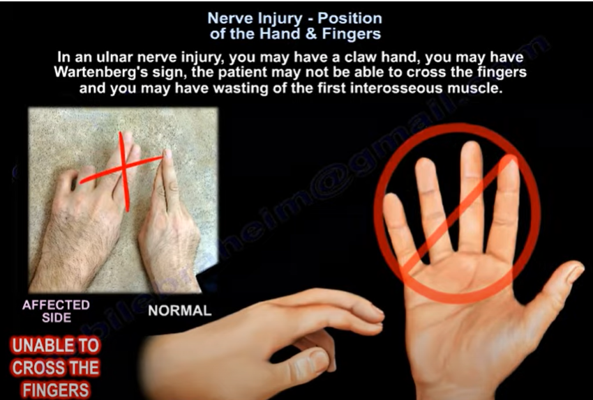
 The presentation of a patient’s hand may provide insight to which nerve is damaged and the approximate location of the damage. When presenting with ulnar n. damage, a patient may show a claw hand, Wartenberg’s sign, or Froment’s sign. Claw hand will present with clawing of the fourth and fifth digits due to the inability to extend the fingers specifically at the interphalangeal joints. This presentation is due to the lack of innervation to the intrinsic muscles of the hands, and the unopposed action of the flexor digitorum profundus m. (Moore et al., 2018). This indicates damage near the distal end of the ulnar n., below the elbow, and potentially near the wrist. Wartenberg’s sign will be seen as the inability to adduct the fifth digit when extended as well as an inability to cross the second and third digit. This is due to ulnar n. injury leading to wasting of the fifth interosseous m. This causes the fifth digit to rest in a more abducted position due to the unbalanced action of the extensor digiti minimi m. (Ebraheim, 2021). There will also be loss of function of the lumbrical m. in the fourth and fifth digits. A Froment’s test will detect palsy of the ulnar n. resulting from compression in the cubital tunnel. When asking the patient to pinch a piece of paper between their thumb and second digit, a positive Froment’s Sign will show as the patient flexing their thumb’s interphalangeal joint to grip the paper as the paper is pulled away (Attum, 2021). This will result from a weak adductor pollicis m. due to ulnar n. palsy. 

Depending on the location of a lesion to the median n., the patient will present with one of three signs. A positive Benedictine sign will be caused by proximal median n. damage. It will be seen as paralysis of the first and second digit, with weakness to the third digit. A proximal lesion would lead to paralysis of several muscles (FDS, FPL, FPB, and the radial half of FDP), leaving the ulnar half of the flexor digitorum profundus m. as the only remaining flexor (Ebraheim, 2021). When the patient is asked to make a fist, the hand will resemble the similar position taken during a blessing. A positive Benedict sign will also resemble the ulnar claw hand. However, the Benedict sign will present when the patient is flexing, not extending the fingers (Ebraheim, 2021). A median n. injury affecting the anterior interosseous n. branch will present as an inability to do the OK sign. This occurs due to paralysis of the flexor pollicus longus m. and the lateral part of the flexor digitorum m. (Moore et al., 2018). A positive Ape hand (Simian hand) is caused by paralysis of only the thenar m. from damage to the recurrent branch of the median n. to the thenar m. The thumb will be seen in the same plane as the other digits due the thumb being pulled more dorsal by the action of the adductor pollicis m., which is innervated by the ulnar n. (Moore et al., 2018). 
Finally, proximal radial n. damage will be seen as wrist drop. This is usually caused by fractures of the distal third of the humeral shaft (Holstein-Lewis Fracture) and caused paralysis of the wrist and fingers extensors (Ebraheim, 2010). Lower radial n. injury will present in the patient as the ability to extend the wrist, but the loss of finger extension. There will be no wrist drop, but the patient would not be able to make a hitchhiking sign.

Reference List

1. Attum B. Physical exam of the hand [Internet]. Orthobullets. Lineage Medical, Inc.; 2021 [cited 2021Oct21]. Available from: https://www.orthobullets.com/hand/6008/physical-exam-of-the-hand

2. Ebraheim N. Anterior Interosseous Nerve Injury - Everything You Need To Know - Dr. Nabil Ebraheim [Internet]. YouTube. 2021 [cited 2021Oct21]. Available from: https://www.youtube.com/watch?v=M9y-iDKLDPE

3. Ebraheim N. Claw Hand, Ulnar Claw Hand - Everything You Need To Know - Dr. Nabil Ebraheim [Internet]. YouTube. 2017 [cited 2021Oct21]. Available from: https://www.youtube.com/watch?v=GyqaKGg3HmM

4. Ebraheim N. Nerve Injury Position of the Hand & Fingers - Everything You Need To Know - Dr. Nabil Ebraheim [Internet]. YouTube. 2021 [cited 2021Oct21]. Available from: https://www.youtube.com/watch?v=nwd1h0Dfo5o

5. Ebraheim N. Radial Nerve Palsy, injury - WRIST DROP . Everything You Need To Know - Dr. Nabil Ebraheim [Internet]. YouTube. 2010 [cited 2021Oct21]. Available from: https://www.youtube.com/watch?v=_Cu6ttAhe8Y

6. Moore KL, Dalley AF, Agur A. Clinically Oriented Anatomy. 8th ed. Philadelphia, PA: Lippincott Williams and Wilkins; 2018.


Tuesday, October 9, 2018

Anterior Interosseous Nerve- The Benedictine and O.K. Sign


A patient with a complete anterior interosseous nerve injury or a high medial nerve injury should be asked to make a fist. The first and second digits will have difficulty in flexing, while the other digits will flex. The third digit will be weak, while the fourth and fifth digits are normal. This position of the hand is similar to the position taken during a hand blessing. The Benedictine sign is different from an “ulnar claw hand”. Ulnar claw hand refers to damage to the ULNAR nerve and is seen when attempting to extend all the digits (leaving the 4th and 5th digits flexed). The O.K. sign is used to check for paralysis of the anterior interosseous nerve due to entrapment or compression injury. A patient with paralysis of the anterior interosseous nerve will be unable to make the O.K. sign. This is due to weakness of the flexor pollicis longus and flexor digitorum profundus muscles. A typical pinch attitude is associated with anterior interosseous nerve injury.

The anterior interosseous nerve arises from the median nerve about 4-6cm distal to the elbow, which is about 1/3 of the way down the forearm. It exits from the anterolateral aspect of the median nerve and it runs between the radius and the ulna on the interosseous membrane between and below the muscles of the flexor digitorum profundus and the flexor pollicis longus.
The anterior interosseous nerve supplies the flexor digitorum profundus muscle for the index and long fingers. It also supplies the flexor pollicis longus and the pronator quadratus muscles. The flexor digitorum profundus muscle for the index and long fingers is supplied by the anterior interosseous nerve. The medial part of the FDP is supplied by the ulnar nerve (FDP has dual innervation). The anterior interosseous nerve passes dorsal to the pronator quadratus with the anterior interosseous artery and provides innervation to the volar wrist capsule. The terminal branch of the anterior interosseous nerve innervates the carpal joint capsule.


In patients with Martin-Gruber Connection, the median nerve, or anterior interosseous nerve to the ulnar nerve in the forearm may present with intrinsic muscle weakness. It may be differentiated also from Parsonage-Turner Synrome (acute brachial plexus neuritis) and patient may have pain in the affected extremity. In anterior interosseous nerve entrapment, the median nerve conduction study result will be normal, however the needle EMG of the anterior interosseous innervated muscles will be abnormal.

Friday, January 12, 2018

PIP Dorsal Fracture Dislocation


Proximal interphalangeal (PIP) dorsal fracture dislocations can be challenging in management. In PIP dorsal fracture dislocations, there is an involvement of the articular surfaces of the joint. These injuries are the most disabling PIP joint injuries. A reduction of the middle phalanx on the condyles of the proximal phalanx is the primary goal of treatment. The patient frequently presents with a chronic dorsal subluxation due to delay in seeking treatment or from failed treatment.


85% of motion for grasping objects occurs at the PIP joint. The PIP joint has the largest arc of motion (120 degrees) of the three joints in each digit.

There are three different types of injury:

  1. Hyperextension
  2. Impact Shear
  3. Pylon


In cases of Hyperextension injuries, the size of the fragment can range from a small piece of bone, up to 30% of the joint surface with minimal comminution (usually stable).  In order to provide accurate treatment, the physician will want to test the stability of the fracture and apply a dorsal blocking splint. It is important to observe the lateral view for any subluxation.


Impaction/Shear injuries are produced by an axial load applied to a slightly extended or flexed PIP joint. The middle phalanx is driven over the head of the proximal phalanx with comminution and impaction of the base of the middle phalanx. These injuries are characterized by the loss of the volar plate tether. A 50% loss of the palmar of the middle phalanx base will make the PIP joint unstable. The extensor tendon and superficialis tendon will aggravate the dorsal subluxation. Splints will be inadequate if the injury is not stable. Treatment will include skeletal traction and early range of motion. An open reduction and internal fixation may be necessary if the fragment is large enough. A Palmar Plate Arthroplasty may be considered to advance the palmar plate into the defect. This procedure will support the palmar plate by filling the defect behind it with fracture chips, bone graft, or by a superficialis slip. Chronic impaction/shear cases are a difficult problem and can be treated with an arthrodesis or volar plate arthroplasty.


A pylon fracture results from an axial force that fractures the volar and dorsal articular surfaces with impaction on the central part. A pylon fracture is not a true fracture dislocation and will not have a good outcome. Dynamic external traction should be done in order to help in ligamentotaxis. Early range of motion is important for remodeling of the joint.


Classification of a PIP Dorsal Fracture Dislocation will depend on the degree of involvement of the articular surface.

  • Type I (stable) <30%
  • Type II (tenuous)= 30-50%
  • Type III (unstable) > 50%

Treatment will consist of a Dorsal Extension Blocking Splint if the PIP can be reduced in less than 30% of flexion. If less than 40%, the joint involved and stable. For Type I and Type II fractures, decrease the flexion gradually every week. Type III fractures will require ORIF, volar plate arthroplasty, or hemi-hamate graft (if >40% joint involved and unstable).


With a Hemi-Hamate Graft, the damaged palmar lip of the middle phalanx with a size matched portion of the hamate bone obtained from its distal dorsal articular surface between the 4th and 5th metacarpals. It restores both articular congruity and osseous stability. Extension block pinning is probably a helpful technique. Treatment must provide stable reduction to allow for early mobilization.


Stability of the reduction depends on the size of the avulsed fragment and the amount of ligament remaining attached to the middle phalanx. If less than 40% of the articular segment is avulsed, some of the collateral ligament will be intact. This will keep the reduction stable. If more than 40% of the articular segment has avulsed, only very little or none of the ligament will remain attached to the base of the middle phalanx, rendering the reduction unstable.

Recognizing Subluxation

AP and lateral x-rays are necessary for diagnosis. An AP view will help to recognize impaction fractures.  Subluxation can be missed on x-rays. The physician will want to look for the characteristic “V” sign of diverging joint surfaces, which indicates injury and subluxation.


If a reduction of the avulsion fracture is achieved with less than a 30 degree bend, nonoperative treatment is a good choice. However, if it takes more than 30 degrees of flexion to reduce the fragment, this may be an indication for surgery.