Tuesday, December 19, 2017

Chondroblastoma


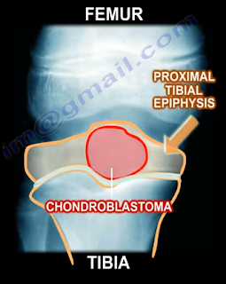
Chondroblastoma is a benign, aggressive cartilage tumor with an epiphyseal location. Chondroblastoma occurs more often in males, in younger patients between the ages of 10-25 years

of age, and in patients that may be skeletally immature. Chondroblastoma occurs commonly at the distal femur and the proximal tibia. 30% of chondroblastoma occur around the knee, followed by the proximal humerus, proximal femur, and the calcaneus.


Other epiphyseal lesions include:
  • Clear Cell Chondrosarcoma
    • Occurs in older age groups
    • Has a more aggressive histological pattern
    • Large cells with central nuclei
    • Occurs in the proximal humerus and proximal femur
  • Giant Cell Tumor
    • Occurs in older age groups
    • Has uniform cells and the nuclei of the stroma are similar to the nuclei of the giant cells
  • Osteomyelitis
    • Brodie’s abscess


During the clinical examination, the patient may complain of pain due to the tumor. The tumor abuts the joint which may cause joint symptoms and may cross the physis. 1% of these tumor metastasize to the lung. Radiologic images will show a lytic epiphyseal lesion with a sclerotic bony rim and sharp, well-defined borders. There may be calcification in the matrix. An MRI will show extensive surrounding edema.


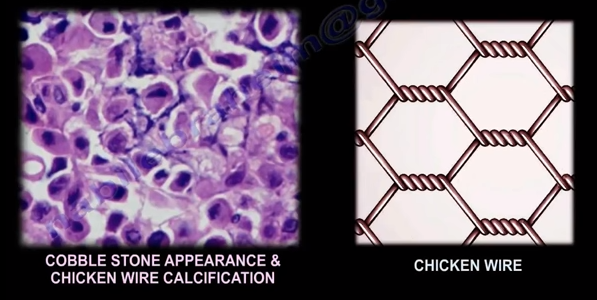
You may find scattered giant cells and a cobblestone appearance of the cells with chicken wire calcifications. One-third of the lesions may have an aneurysmal bone cyst (ABC). The lesion is chondroid with polygonal cells. The lesion will have a defined cytoplasmic border and oval shaped nuclei with a prominent longitudinal groove (coffee bean appearance of the nuclei). Chondroblasts can be distinguied from giant cell tumors by staining for the S100 protein as the chondroblastoma will be reactive.
Chondroblastomas can be treated with an intralesional curettage and bone graft. The recurrence rate is less than 10%. Some surgeons may use adjuvants such as phenol or liquid nitrogen.

Tuesday, December 12, 2017

Wrist Pain Part I


There are eight carpal bones of the wrist that fit into a shallow socket formed by the bones of the forearm. Carpal Tunnel Syndrome occurs when there is pressure placed on the median nerve due to thickening of the transverse carpal ligament.
Thenar atrophy is commonly associated with severe carpal tunnel syndrome. Carpal tunnel syndrome can occur during pregnancy due to swelling, which compresses the median nerve in the hand, causing the fingers to feel numb. These symptoms typically go away after the delivery of the baby. Common causes of Carpal Tunnel syndrome are: obesity, hypothyroidism, arthritis, diabetes, trauma, and repetitive work (typing and lifting).  Carpal Tunnel Syndrome can lead to numbness, tingling, or weakness in the hand and fingers. The numbness usually occurs at night because we tend to sleep with our wrists flexed.
The Tinel’s test is used to determine symptoms of carpal tunnel syndrome. The test is considered positive if symptoms of tingling worsen while tapping on the median nerve at the wrist. The Phalen’s maneuver is a diagnostic test performed to determine if the patient has carpal tunnel syndrome. During this test, the back of the hands are pressed together, compressing the nerve, which may cause the symptoms of carpal tunnel syndrome.


Treatment for Carpal Tunnel Syndrome includes: anti-inflammatory medications, a splint, therapy, injections, and carpal tunnel release surgery. A carpal tunnel release is performed by cutting through the transverse carpal ligament. A carpal tunnel release allows the median nerve to pass freely through the tunnel to receive sensations from the thumb, index, and middle fingers of the hand. The idea is similar to a tunnel being widened so a large truck can pass through.

Tuesday, December 5, 2017

Pelvic Fractures with Bleeding


Pelvic fractures may cause significant bleeding. The superior gluteal artery is responsible for the majority of bleeding in pelvic fractures with an arterial injury. Most of the bleeding in pelvic fractures is from the veins and the fracture itself. Hemorrhage is the most life-threatening complication associated with pelvic injuries and will typically occur at the Superior Gluteal Artery. Hemorrhage of the Superior Gluteal Artery can be fatal. Approximately 10% of patients will have severe bleeding. Severe bleeding usually occurs in fracture patterns that are highly unstable to both rotational and vertical forces.


APC III (open book like type) is the complete disruption of anterior SI, sacrotuberous, and sacrospinous ligaments; disrupted posterior SI ligaments.
Vertical Shear Fractures are very bad fractures as they cause the complete disruption of the anterior and posterior ligaments; cephaloposterior displacement. Anteroposterior compression or vertical shear injuries are consistently associated with a higher risk of mortality from bleeding. The mortality rate is directly related to the amount of shock the patient is in at the time of presentation.
When treating patients with pelvic fractures and massive bleeding, it is important to remember that the patient will lose approximately 35% of their blood volume with acute hemorrhage before a sustained decrease in systolic blood pressure occurs. Immediate application of a pneumatic anti-shock garment is absolutely contraindicated in patients with a rupture of the diaphragm. Ringer’s lactate is the preferred initial fluid replacement used to resuscitate hypovolemic trauma patients in shock. A hypotensive blunt trauma patient will be given an initial fluid push with 2,000mL of Ringer’s lactate. A patient with bleeding and in shock will probably require O negative blood. If the patient is given 4 units of blood but remains hemodynamically unstable, then angiography and embolization is needed. Immediate application of an external fixator is another method to control bleeding, especially if the pelvis is unstable in external rotation. An abdominal and pelvic CT scan will clearly define the bony injury as well as the extent and source of the bleeding.
The best treatment for pelvic fractures with bleeding is a blood transfusion with correction of hypothermia and coagulopathy.      

Thursday, November 30, 2017

Barlow & Ortolani Signs—DDH, Congenital Hip Dislocation


In order to remember these tests, remember that “B” comes before “O” in the alphabet and will go in this order for these procedures as well. The “B” stands for Barlow and “O” for Ortolani. When reading the word “barlow” think: “We are going out tonight!”, because the hip can be popped out of the acetabulum with this maneuver. To remember Ortolani, think: “Once we have been out, now it is time to go home!”—during the Ortolani maneuver, the femoral head is reduced back into the acetabulum.
The Barlow maneuver identifies the unstable hip that is in a reduced position that the clinician can passively dislocate. When performing the Barlow Test, the examiner will flex the hip and knees to 90 degrees. The maneuver is performed by bringing the thigh towards the midline (adducting the hip). Mild pressure is then placed on the knee while directing the force posteriorly. The femoral head will be pushed out of the socket. The Barlow Test is considered positive if the hip can be popped out of socket with this maneuver. The dislocation will be palpable.


The Ortolani Test is used to confirm the findings of the Barlow test. The Ortolani maneuver is performed following the Barlow test to determine if the hip is actually dislocated. The Ortolani test is performed by the examiner flexing the hips and knees to 90 degrees. Reduction is done by abduction of the hip and pushing the thigh anteriorly. The test is positive is a palpable and audible clunk is heard from the hip being reduced. A hip click is a nonspecific finding.
In summary, The Barlow Test is performed when the hip is reduced and is used to dislocate the hip. The Ortolani Test is performed only after the Barlow Test has been performed and the hip has been dislocated. The Ortolani Test will reduce the hip.
Both of these tests are used for screening newborns during the neonatal period. The hips are examined one at a time and usually the hips are flexed during these maneuvers. Early diagnosis by these tests and preferably with ultrasound is essential to detect hip instability and dislocation in the neonatal period.

Treatment is directed at stabilizing the hip that has positive Barlow and Ortolani Signs. The first born female with a breach presentation and a positive family history are at risk of developing developmental dysplasia of the hip (DDH). If the hip remains dislocated for weeks, these two tests are usually not reliable. Barlow and Ortolani are not positive after 3 months due to the soft tissue contracture around the hip region. Limitation of abduction becomes the most consistent clinical findings. Children older than 12 months will have other findings which include asymmetry of hip abduction, a positive Trendelenburg gait and a positive Galeazzi sign.

Tuesday, November 21, 2017

Froment's Sign


The Froment’s sign occurs due to weakness of the adductor pollicis muscle in ulnar nerve palsy. The adductor pollicis muscle has two heads:

  1. Transverse Head
    1. Originiates from the anterior body of the third metacarpal
  2. Oblique Head
    1. Originates from the base of the second and the third metacarpals as well as the trapezoid and capitate bones


The two heads of the adductor pollicis muscle then insert into the base of the proximal phalanx of the thumb and the ulnar sesamoid bones. The muscle is innervated by the deep branch of the ulnar nerve. The function of the adductor pollicis muscle is to adduct the thumb. It is important in pinch strength. When the ulnar nerve is injured, the adductor pollicis function is lost and thumb adduction will not occur.

The Foment’s Sign is used to test the function of the adductor pollicis muscle. When pinching a piece of paper between the thumb and index finger against resistance, the thumb IP joint will flex if the adductor pollicis muscle is weak. The flexion of the thumb occurs by the flexor pollicis longus, which is innervated by the median nerve. The flexor pollicis longus, which is innervated by the median nerve, substitutes the function of the adductor pollicis which is innervated by the ulnar nerve.

Monday, November 6, 2017

Bulbocavernosus Reflex and Spinal Shock


The bulbocavernosus reflex indicates the absence or presence of spinal shock. Spinal shock usually occurs between 24-72 hours after a spinal injury. Spinal shock is manifested by the absence of the bulbocavernosus reflex, hypotension, bradycardia, and complete loss of motor sensation and reflexes. When the reflex is absent, this means that the patient is in spinal shock because the anal sphincter will not contract when the reflex is absent.

When the reflex is present, this signals the end of spinal shock; the anal sphincter will contract when the reflex is present. The reflex is check by monitoring anal sphincter contraction in response to squeezing of the penis of clitoris, or by pulling on an indwelling Foley catheter. It is a polysynaptic response mediated by S2-S4.

What is Spinal Shock?

Spinal shock is the loss of sensation and motor power following a spinal cord injury. Spinal shock is the loss of sensation and motor power following a spinal cord injury. After an injury to the spine, if the patient has no motor or sensory below the level of the lesion, the physician must determine if the patient is in spinal shock by checking the bulbocavernosus reflex.
If there is no anal contraction (absent bulbocavernosus reflex), this indicates that the patient is still in shock and the prognosis cannot be determined. If anal contraction is present (positive bulbocavernosis reflex), this indicates the end of spinal shock. The patient’s condition and prognosis at this point can be determined by examining sacral sparing (positive sacral sparing indicates an incomplete lesion).

Loss of sensation and motor power below the level of injury indicates complete spinal cord injury. Once the diagnosis of neurogenic shock is established, the blood pressure should be managed with vasopressors to prevent fluid overload. With the end of spinal shock, the prognosis can be determined. Examine the patient thoroughly, including sacral sparing. The patient may have normal, partial, or complete indications. 

Tuesday, October 31, 2017

Platelet Rich Plasma Therapy


Platelet Rich Plasma, or PRP, is a volume of the plasma of autologous blood having a platelet concentration above the baseline. Platelets facilitate healing by stimulating the release of different growth factors. The growth factors recruit stem cells that assist with healing, repair, or regeneration of the injured tissue. PRP is injected directly into the injured tissue, stimulating a healing response in a more powerful form.
Red Blood Cell
In order to understand PRP, you must know that platelets are components of blood cells. The development of PRP focuses on the concentration of the patient’s own blood platelets to create an active mixture of growth factors. The other components of blood are the red and white blood cells. Red blood cells carry oxygen from the lungs and deliver it to the body tissues. White blood cells form our immune system and help the body to fight against infection and foreign materials.


Active Platelet
Platelets aid in hemostasis (stop the bleeding) and in building new tissues—they act as a scaffold for tissue regeneration. Platelets aid in the attraction and binding of stem cells. Platelets act as the directors while stem cells work. Platelets divide, multiply and differentiate to become the healing cells for injured tissue. The platelets become activated by thrombin and other factors which cause a change in their morphology and the release of multiple growth factors. These growth factors bind themselves to the receptors of the cell causing intracellular changes down to the nucleus and affecting its DNA.  The result is a change in the performance and function of the cell.
In order to produce PRP, 30-60mLs of the patient’s own venous blood is drawn from the antecubital vein. The blood is then placed into a device to be centrifuged which separates the blood into platelet poor plasma (PPP), red blood cells (RBC), and platelet rich plasma (PRP). The blood is then placed in a centrifuge for 15 minutes at 3,200 rpm. The centrifuge spins and separates the platelets from the rest of the blood components and increases the concentration of platelets and growth factors. The more platelet concentration, the greater the healing power. After the centrifuge process is complete, the plasma has been separated from the blood producing the PRP and the platelet poor plasma is withdrawn to be discarded. Platelet rich plasma is withdrawn for injection. Sodium Bicarbonate is used to neutralize the acidity of the sample. The more platelet concentration, the more growth factors and healing power the sample has.
Ultrasounds help deliver a concentrated sample of platelets to the injured tissue. Ultrasound increases the accuracy of delivering the sample to the injured tissue. Preferably, injections should be performed with the aid of ultrasound imaging. Needling induced injury releases thrombin which activates the platelets. Platelets help in hemostasis and produce growth factors as well as chemotactic factors. Platelets act as a scaffold for mesenchymal stem cells which start the process of tissue regeneration. Patients will typically experience minimal to moderate discomfort which may last up to one week following the injection. Avoid the use of anti-inflammatory medications for 6 weeks after the injection.

Tuesday, October 24, 2017

Hangman's Fracture


The hangman’s fracture refers to the bilateral fracture of the pars interarticularis. When this fracture occurs, the spinal canal is widened and there is a low risk for spinal cord injury. This fracture usually occurs due to motor vehicle accidents.

Hyperextension will fracture the pars interarticularis with secondary flexion, injuring the disc and posterior ligament. The patient may have other associated spine fractures.


Hangman’s fractures are identified by types using the Levine and Edwards Classification.
Type I


Type I:


  • Stable fracture with less than 3mm displacement
  • No angulation
  • Treatment: Cervical Orthosis
Type II

Type II:

  • Most common type
  • Significant translation and some angulation
  • Unstable fracture
  • Treatment: Cervical traction to improve the displacement and immobilization in halo vest
Type IIa

Type IIa:

  • Slight translation but severe anglulation seen in flexion distraction injuries with tearing of the posterior longitudinal ligament and the disc.
  • The fracture is unstable
  • Treatment: reduction in extension and compression in a halo
  • Do NOT use traction when there is severe angulation of the fracture  

 
Type III

Type III:

  • C2-C3 facet dislocation
  • Rare fracture that results from initial anterior facet dislocation of C2 on C3, followed by an extension injury fracturing the neural arch.
  • Results in translation with unilateral or bilateral facet dislocation of C2-C3.
  • Unstable fracture
  • Treatment: surgery for reduction of the facet dislocation and stabilization of the injury
 

Typical and Atypical fractures

A typical hangman’s fracture displaces the vertebral body anteriorly and its posterior element posteriorly. This creates increased space for the spinal cord.

Atypical Hangman's Fracture
An atypical hangman’s fracture line leave the canal circumferentially intact, which puts the spinal cord at risk of injury if displacement occurs.


Wednesday, October 18, 2017

Total Hip Acetabular Screw Placement


Utilizing acetabular screws in total hip arthroplasty is controversial. Screws can compress the cup and close any gap. Screws could be helpful in osteoporotic bone and when the bone coverage is not sufficient. Screws and its holes can be conduit for migration of polyethylene debris that can cause osteolysis (The surgeon should insert the least number of screws).
Many surgeons prefer to use screws in the acetabular cup. The quadrant technique is a safe technique of screw placement. It provides the surgeon with a simple, basic guide during surgery for safe placement of the screws during the primary and revision surgery.


In order to find the safe zone for the placement of acetabular screws and avoid the danger zone, you will need to draw a line from the Anterior Superior Iliac Spine (ASIS). Then you will draw a second line, perpendicular to the first line. Drawing these lines will divide the acetabulum into four quarters. The posterior-superior quarter is considered the safe zone. There is minimal risk to the sciatic nerve, superior gluteal nerve, and vessels. It is important to direct the screws away from the notch. The anterior-superior and anterior-inferior quarters are considered to be the danger zone due to the external iliac artery and vein as well as the obturator nerve, artery, and vein are at risk. The posterior-inferior quarter is considered the caution zone. Placing screws here can be safe if the screws are less than 20mm. There are still several risks when placing screws in this area and these risks include: the sciatic nerve, inferior gluteal nerve and vessels, internal pudendal nerve and vessels.
A retroverted vertical cup will be at most risk for injury to neurovascular structures. The structures can be at risk in a high hip center, which is reamed superiorly ½ the native acetabular diameter. The new hip will have different anatomic zones than what the surgeon might think. The antero-superior and the antero-inferior quadrants are positioned in the postero-superior quadrant (Be aware of the shift in the zones). The insertion of the screws as guided by the quadrant method is based on a study done by Dr. Ray Wasielewski and his colleagues. When in doubt, aim the screws anteriorly to the greater sciatic notch. The bone will be stronger and thicker. Screws longer than 35mm are usually not needed. Avoid drilling eccentrically because it may lift up the liner. The screw must be fully seated and the head of the screw should not be proud. If the screw is proud, the liner may not be seated properly.


Vascular injuries occur more in females and more often in the left hip.

Tuesday, October 10, 2017

Tibial Plateau Fracture Balloon Osteoplasty


A tibial plateau depression fracture occurs when axial forces from the femoral condyles against the articular surface of the tibia cause the injury. The compression fracture may also be associated with other fractures. Several techniques are used for the treatment of this fracture. Balloon osteoplasty is proposed as a useful tool for reduction and elevation of the tibial plateau depression fracture.

The technique of balloon osteoplasty can be done with or without the supplementation of screws and plate fixation. Trajectory lines are drawn on the medial side of the knee for access to the fracture area. A small incision is made at the point where the lines cross. A cannula and trocar tip stylet is used to access the fractured area under radiographic guidance. The cannula and trocar is advanced under the area of depression using a mallet or drill when needed. Once inserted, the trocar is then removed from the cannula.
 The precision drill is then inserted into the cannula and advanced until it passes under the depressed area of bone. Then, the precision drill is removed from the cannula and the inflatable bone tamp is inserted through the cannula to the area of the depressed fragments. The inflatable bone tamp is centered under the area of depression and then inflated to reduce the depression fracture. Once the depression has been adequately reduced, the inflatable bone tamp can be deflated and removed from the cannula.
The void that has been left by the inflatable bone tamp is now ready to be replaced with bone filler. The device loaded with injectable material is inserted and used until the void is completely filled. Finally, you will remove the injectable material device and the cannula. Plate fixation may be added before or after the injectable material is inserted.

Tuesday, September 19, 2017

Lactic Acid and Base Deficit in Trauma Patients


Lactic acid is a byproduct of anaerobic metabolism (Metabolism without oxygen). Normally, the cells use anaerobic metabolism and it breaks down the glucose to form energy. Normally, the cells use oxygen available to breakdown the glucose and produce ATP. This occurs when you have oxygen. If the body does not have the oxygen, it then converts the metabolism to an anaerobic metabolism. The end product is lactic acid. If the body is acidic, and the pH drops, this means that there is a tissue ischemia. If we take the glucose (CCCCCC), and break it down, you will have two pyruvates—each attached to a CCC. That’s not a lot of energy and the pyruvate is a mild acid.

There are different ways to get more energy, such as going to the mitochondria to use the oxygen and produce more ATP. When we break down the glucose to produce energy in the presence of oxygen, so the glucose will split into two separate three carbon molecules called the pyruvate and produces ATP. If you don’t have oxygen, then the pyruvate will be attached to hydrogen atoms, which is H+ (proton that is an acid). If you are acidotic and a sick patient, you will have a lot of floating hydrogen atoms and a lot of floating protons.

The pyruvate will attach to the free hydrogen protons. The H+ pyruvate is called lactic acid. It is a pyruvate that is holding onto hydrogen. The lactic acid is the end product of anaerobic metabolism.

Now, with a base deficit, as the number of protons goes up, which is hydrogen, then the pH will go down and the patient will become acidotic. The body uses bicarbonate as a buffer if the pH goes down. There are a lot of protons within the body and the bicarbonate will be exhausted in this situation. The bicarbonate goes down because we are combining it with the protons. When the bicarbonate goes down, this becomes metabolic acidosis.

When you combine the bicarbonate with the protons, this will make carbonic acid (H2CO3), rather than HCO3-, which is bicarbonate. The carbonic acid can give water (H2O), and CO2. The lactic acid will give out the protons and be buffered by the bicarbonate, leaving lactic acid. When this occurs, the bicarbonate will go down, and this will be the base deficit.

When checking if a patient has been resuscitated, you can check in several ways

The two ways this is asked on exams:
  1. Base deficit from -2 to +2
  2. Serum lactate level (normal is less than 2.5, some sources use normal less than 2)

Friday, September 8, 2017

Orthopaedic Emergencies Part IV


In our final blog post regarding Orthopaedic Emergencies, we will review:

  1. Transverse Atlantal Ligament Rupture
  2. Bilateral Cervical Facet Dislocation
  3. Spinal Cord Compression
  4. Cauda Equina Syndrome


Transverse Atlantal Ligament Rupture


The normal Atlanto-Dental Interval is less than 3mm. An A.D.I measuring greater than 3mm will be translationally unstable in the sagittal plane due to transverse atlantal ligament rupture. This is usually apparent on x-rays or CT scan. If the condition is not diagnosed, it can result in spinal cord compression, respiratory arrest, and a catastrophic outcome. Treatment typically requires a posterior atlanto-axial arthrodesis.


Bilateral Cervical Facet Dislocation


Facet dislocations of the cervical spine:

  1. Unilateral Facet Dislocation
    1. Displacement is less than 50% of the vertebral body width
    2. May need surgery
  2. Bilateral Facet Dislocation
    1. Displacement greater than 50% of the vertebral body width
    2. Usually needs surgery
    3. Exclude disc herniation

Obtain a preoperative MRI to rule our disc herniation associated with facet dislocations.


Spinal Cord Compression


Spinal cord compression is more common with cervical spine injuries and thoracic spine injuries. Neurogenic shock resulting from spinal cord injury may complicate resuscitation of the patient and should be differentiated from hypovolemic shock. It is important to look for hypotension and bradycardia as well as thoracolumbar fractures which could be missed. Treatment consists of emergency management involving resuscitation and hemodynamic stabilization with concurrent neurologic examination. Protocol requires steroids given early. Definitive treatment consists of stabilization of unstable spinal injuries.


Cauda Equina Syndrome


Central disc herniation compressing the cauda equine. It results from injury to the lumbosacral nerve roots within the spinal canal. This syndrome presents with involvement of the bladder, bowel, and lower limbs and usually results from central disc herniation or fractures. Central disc herniation or bony fragments results in the compression of the nerve roots. Early diagnosis is imperative to find the cause of the compression on the nerve roots. Urgent decompression by the removal of the central disc herniation or stabilization of the fracture is necessary for treatment.

Tuesday, August 29, 2017

Measles— Everything You Need to Know


Measles, also known as Rubeola, is an extremely contagious viral infection caused by a Paramyxovirus. It usually occurs in children under the age of 5 years and the reservoir for this virus is the human respiratory tract. Transmission occurs through inhalation of infected droplets produced by infected individuals by sneezing, coughing, or even talking.


Risk factors to contracting this viral infection include:
  •          Lack of vaccination
  •          Travelling to endemic areas
  •          Vitamin A deficiency
  •          Immunocompromised individuals


The measles virus has an incubation period of 10-14 days during which the patient has no signs or symptoms. After the 14 days of the incubation period, the patient will start developing the following signs and symptoms—fever, cough, coryza (runny nose), and conjunctivitis (pink eye).  A characteristic rash also develops which is a red maculopapular rash appearing first on the face- behind the ears, and then spreads downwards towards the neck, trunk, arms, legs, and feet. Disease specific Koplik Spots may develop, which are tiny white spots that appear on the buccal mucosa.
A measles patient is infective for a total duration of 8 days. Infectivity starts four days before the appearance of the rash and stops around the fourth day of having the rash. Thus, the patient may appear to be well and still infect other people.
A diagnosis is usually achieved by thorough history taking and examination to identify disease specific features such as the unique rash patter and the characteristic koplik spots. Blood tests could be ordered to confirm the presence of measles IgM antibodies. Furthermore, respiratory specimens may be obtained to isolate the virus.
Complications of Measles include:
  •          Otitis media
  •         Pneumonia
  •         Laryngotracheitis
  •          Subacute sclerosing panencephalitis

Vaccination is quite important and is considered the most widely used method of prevention. Widespread vaccination leads to herd immunity, which helps to contain the disease and prevent outbreaks. When only a few individuals are vaccinated, the disease may spread easily through the population. The measles vaccine is a live attenuated vaccine that is given to children as part of the MMR (Measles, Mumps, and Rubella) vaccine. The vaccine is typically administered by the age of 1 year, followed by a booster dose at the age of 5. The vaccine helps the individual develop lymphocytes and anti-bodies to attack and eliminate the virus upon exposure. It is important to remember not to vaccinate immunocompromised individuals because this is a live attenuated virus vaccine.


Treatment is largely supportive including fluids, fever reduction, vitamin A, and in some cases, antibiotics may be given to prevent superinfection.

It is worth noting that a controversy remains regarding the use of the measles vaccine.