Simple Tests for Diagnosis of Orthopaedic
Conditions
Dr.Ebraheim Examination/Evaluation educational videos
Part 1
Before studies are done or medications are prescribed, an orthopaedist
begins with a thorough physical examination of the patient. During this
process, the physician investigates the patient’s body with a series of tests
to locate areas of pain or concern. Once the tests are completed, imaging is
often used to confirm physical examination findings. Many tests are used to
diagnose conditions of the hand and wrist, including Finkelstein and Phalen
tests, Froment’s sign, “ok” sign and wrist drop.
Finkelstein’s Test (Figure 1)
 |
| Figure 1 |
Used to diagnose DeQuervain’s syndrome, an inflammation of the tunnel
or sheath that surrounds two tendons controlling movement of the
thumb, often caused by repeated motion of the wrist and hand. To perform the
test, a patient is asked to make a fist with the thumb far enough inside to touch the
little finger.
Next, the patient is instructed to move the wrist in the
direction of the little finger. If a patient experiences pain during this movement, DeQuervain’s tenosynovitis may be the cause.
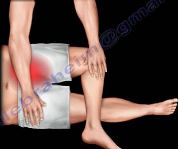
Faber's Test (Figure 2)
 |
| Figure 2 |
An acronym for flexion, abduction, external rotation, this test examines
the sacroiliac joint and evaluates back pain. During this test, a
physician forces external rotation of the affected hip in the supine position
which causes pain in the sacroiliac joint. In addition, there would
be tenderness over the sacroiliac joint.
Phalen's Test (Figure 3)
An alternative test used to identify carpal tunnel syndrome.
Here, the physician bends the patient’s
wrists downward and pushes the backs of the hands together for approximately one minute.
 |
| Figure 3 |
A positive test is indicated by numbness, pain or tingling along the
median nerve.
This test increases the pressure in the carpal tunnel and has
the affect of pinching the median nerve between the proximal edge of the transverse
carpal tunnel ligament and the anterior border of the distal end of the radius.
Froment’s Sign (Figure 4)
 |
| Figure 4 |
Another exam of the wrist and hand is the Froment’s sign.
Patients are asked to hold an object (usually a piece of paper) between the thumb
and palm (which is flat); the object is then pulled away.
A patient with a negative Froment’s sign will be able to maintain a hold
on the object without problem. A patient with a positive test, however,
will have trouble holding the paper and will compensate by flexing the
flexor pollicis longus of the thumb.
This test is used to identify ulnar nerve palsy --
paralysis caused by damage or compression of the ulnar nerve.
 |
| Figure 5 |
“OK” Sign (Figure 5)
Patients with an injury to the anterior interosseus nerve cannot
perform the “ok” sign, opposing the second finger and thumb. The anterior interosseous
nerve is a branch of the median nerve that supplies the deep muscles
of the front of the forearm
Wrist Drop (Figure 6)
 |
| Figure 6 |
A patient with a wrist drop cannot extend or raise the wrist. It
is typically caused by damage to the radial nerve, which stimulates the muscles in the
forearm.
Adson's Test (Figure 7)
Another test used to evaluate upper extremity region, specifically for
thoracic outlet
 |
| Figure 7 |
Lift-Off Test
The Lift-off test is used to diagnose tears of the subscapularis
tendon. The patient places the back of the hand on his or her back with the arm
in internal rotation. The patient is then asked to lift the hand away from the
back. The physician will push the hand toward the back to test the strength of
the subscapularis if the patient is able to take the hand away from the back.
If the patient is unable to lift the hand against the physician’s resistance, a
tendon rupture or injury to the subscapularis is present.
Straight Leg Raise Test (Figure 8)
 |
| Figure 8 |
Another test used to evaluate back pain is the straight leg
raise test. Which can determine whether a patient with low back pain has a herniated
disk. The physician will place one hand under the ankle and the other hand on the knee. The physician
then lifts the ankle and flexes the thigh relative to the pelvis.
A positive test will yield reproducible pain in the patient’s leg and lower
back.
Thompson's Test (Figure 9)
 |
| Figure 9 |
The Thompson test is used to examine the Achilles tendon. Here, the
patient is asked to lie prone on the examination table with the foot extended
beyond the end of the table. The physician will then squeeze the calf. A
non-injured response to this maneuver is a slight plantar flexion (movement
which increases the angle between the foot and the leg). Lack of movement can
indicate a rupture of the Achilles tendon.
