Showing posts with label evaluation. Show all posts

Tuesday, July 3, 2018

Hip Dislocation Following Total Hip Surgery


There are two types of hip joint dislocations: posterior and anterior. The position of the leg is important in determining the type of hip dislocation. When the hip is dislocated, the leg is usually shortened and it assumes a different position than the normal leg (the other leg). If the dislocation is posterior, the leg will be in adduction and internal rotation. If the dislocation is anterior, the leg will be in abduction and external rotation. Notice that the affected extremity is shortened and externally rotated. Leg shortening can also be seen in hip fractures and the leg will be shortened and externally rotated.


Dislocation of the hip following total hip surgery may require revision surgery, but it is rare. The majority of hip dislocations after total hip dislocations are posterior, and they are usually treated without surgery. Most occur within the first month of THA; 1-4% in primary, 16% in revision. There is more incidence of dislocation in revision hip replacement.


Causes & Risk Factors:

  • Posterior Approach (try to repair the capsule adequately)
  • Malposition of the component
    • Ideally, the normal cup component will be in 20° of anteversion and 40° of abduction
    • When the hip dislocates posterior, always check for retroversion of the cup.

  • Prior hip fracture surgery, especially in the elderly
  • Weakness of the abductor muscle—must achieve soft tissue tension and function
  • Alcohol abuse
  • Improper neck length—looseness of the hip

The patient should be careful to avoid all activities that cause dislocation after total hip surgery. The patient should use a pillow between the legs while sleeping on their back and they should be careful to not cross their legs in their sleep. Patients cannot sleep on their sides as well. The patient should not bend the body at the waist farther than 90°. When sitting, the patient must avoid chairs that make it difficult to stand up, and sit at more than a 90° angle. The patient must not sit with their legs crossed in the chair. The patient must be made aware that if the leg is changed from its usual position, or becomes shortened, then the hip is probably dislocated and their doctor should be consulted.


X-rays of the dislocated total hip should include AP and lateral views. Look for eccentric wear and look for the position of the prosthesis. CT scans may be needed before or after reduction of the dislocation to check the version of the components. Treatment is variable and depends on the situation. The treatment should be tailored for each case. The majority of these cases with early dislocations can be treated successfully with closed reduction and immobilization.

The treatment should start with closed reduction of the total hip and immobilization. Hip stability is checked after reduction of the dislocation. Immobilization can be done by a brace or a hip spica. Trochanteric osteotomy and advancement of the trochanter and tensioning the abductor muscle. Screws or wires can be used. The prosthesis must be in good alignment for this procedure to work. Constrained acetabular components are used when the abductor muscle is deficient and the component position is good. Revision total hip is done in recurrent dislocation with malposition of the component or polyethylene wear.

Tuesday, February 13, 2018

Pronator Teres Syndrome


The nerve that is involved in pronator teres syndrome is the median nerve. Pronator Teres Syndrome is caused by a compression of the median nerve at the level of the elbow which occurs more in women. In the forearm, the median nerve runs between the two heads of the pronator teres muscle and then it lies between the flexor digitorum superficialis and flexor digitorum profundus muscles. This syndrome may be associated with medial epicondylitis. The principle symptoms of numbness in the radial 3 ½ fingers as well as thenar weakness which may be mistakenly attributed to carpal tunnel syndrome.
The most common cause of entrapment is due to compression of the median nerve between the two heads of the pronator teres muscle. This commonly occurs in people who perform repetitive forceful pronation of the forearm. Compression may be due to the thickening of the bicipital aponeurosis. The aponeurosis crosses from lateral to medial over the antecubital fossa and may irritate the median nerve. Compression of the nerve may also occur due to the fibrous arch of the origin of the flexor digitorum superficialis (FDS).


The median nerve runs down the medial side of the arm and passes 2 ½ to 4 cm below the level of the medial epicondyle before it enters between the two heads of the pronator teres. About 1% of
patients have a medial supracondylar humeral spur about 5cm proximally to the medial epicondyle. The ligament of Struthers is attached to this bony projection which connects the process to the medial epicondyle. The bony process points towards the elbow joint and the median nerve can become compressed by the supracondylar spur. The median nerve can also become trapped by the ligament of Struthers that extends from the supracondylar process to the medial epicondyle. The ligament of Struthers is different from the arcade of Struthers, which deals with the compression of the ulnar nerve around the elbow.


Paresthesia in these lateral 3 ½ fingers may occur with the compression of the median nerve at the elbow region or at the carpal tunnel region. These symptoms are similar to carpal tunnel syndrome but the symptoms are worse with rotation of the forearm. The patient will complain of dull aching pain over the proximal forearm with no nighttime symptoms. The pain is usually worsened by repetitive or forceful pronation. Tenderness of palpation to the pronator teres muscle will be detected. The median nerve gives off a palmar cutaneous branch before entering the carpal tunnel. Sensory disturbances over the palm of the hand occur due to involvement of the palmar cutaneous branch of the medial nerve and this occurs proximal to the carpal tunnel. Sensory disturbances in this area indicates median nerve problems proximal to the carpal tunnel. This differentiates between carpal tunnel syndrome and pronator teres syndrome.

There are specific provocative tests that produce the pain and distal paresthesia that are used to localize the site of compression. The Tinel’s sign at the wrist and the Phalen’s test will be negative. The Median nerve compression tests are negative at the carpal tunnel; however, there will be a positive Tinel’s sign at the proximal forearm. There will be abnormal sensation in the “palmar triangle”. When compression of the nerve involves the supracondylar process, the test is considered positive if symptoms of tingling worsen while tapping on the spur.
Occassionally, the spur can be felt. The pronator teres muscle can be assessed as the cause of the median nerve compression in different ways. Resisted forearm pronation with elbow flexion will test for compression at the two heads of the pronator teres muscle. During this test, the patient’s forearm is held in resisted pronation and flexion. While remaining in a pronated position, the forearm is gradually extended. Compression of the median nerve may also be tested by: resisted elbow flexion with forearm supination (compression at the bicipital aponeurosis) and resisted contraction of the FDS to the middle finger (compression at the FDS arch).


Differential Diagnosis

C6/C7 Radiculopathy occurs due to involvement of the nerves at these levels which will cause numbness of the thumb, index, and long fingers, as well as weakness of the muscles of the forearm that are innervated by the median nerve. The radial nerve part of C6-C7 will show normal function of the wrist extensors and the triceps.

X-rays, imaging and nerve conduction studies may be helpful in the diagnosis.

Treatment typically consists of rest, splints, and NSAIDs. Surgical decompression of the median nerve through all 4 or 5 possible sites of compression when non-operative management fails for 3-6 months. The results of surgery are variable. Full recovery is not always seen in all patients as only about 80% of patients improve from surgery. The skin incision may leave an unsatisfactory scar.

Tuesday, November 21, 2017

Froment's Sign


The Froment’s sign occurs due to weakness of the adductor pollicis muscle in ulnar nerve palsy. The adductor pollicis muscle has two heads:

  1. Transverse Head
    1. Originiates from the anterior body of the third metacarpal
  2. Oblique Head
    1. Originates from the base of the second and the third metacarpals as well as the trapezoid and capitate bones


The two heads of the adductor pollicis muscle then insert into the base of the proximal phalanx of the thumb and the ulnar sesamoid bones. The muscle is innervated by the deep branch of the ulnar nerve. The function of the adductor pollicis muscle is to adduct the thumb. It is important in pinch strength. When the ulnar nerve is injured, the adductor pollicis function is lost and thumb adduction will not occur.

The Foment’s Sign is used to test the function of the adductor pollicis muscle. When pinching a piece of paper between the thumb and index finger against resistance, the thumb IP joint will flex if the adductor pollicis muscle is weak. The flexion of the thumb occurs by the flexor pollicis longus, which is innervated by the median nerve. The flexor pollicis longus, which is innervated by the median nerve, substitutes the function of the adductor pollicis which is innervated by the ulnar nerve.

Tuesday, August 29, 2017

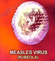
Measles— Everything You Need to Know


Measles, also known as Rubeola, is an extremely contagious viral infection caused by a Paramyxovirus. It usually occurs in children under the age of 5 years and the reservoir for this virus is the human respiratory tract. Transmission occurs through inhalation of infected droplets produced by infected individuals by sneezing, coughing, or even talking.


Risk factors to contracting this viral infection include:
  •          Lack of vaccination
  •          Travelling to endemic areas
  •          Vitamin A deficiency
  •          Immunocompromised individuals


The measles virus has an incubation period of 10-14 days during which the patient has no signs or symptoms. After the 14 days of the incubation period, the patient will start developing the following signs and symptoms—fever, cough, coryza (runny nose), and conjunctivitis (pink eye).  A characteristic rash also develops which is a red maculopapular rash appearing first on the face- behind the ears, and then spreads downwards towards the neck, trunk, arms, legs, and feet. Disease specific Koplik Spots may develop, which are tiny white spots that appear on the buccal mucosa.
A measles patient is infective for a total duration of 8 days. Infectivity starts four days before the appearance of the rash and stops around the fourth day of having the rash. Thus, the patient may appear to be well and still infect other people.
A diagnosis is usually achieved by thorough history taking and examination to identify disease specific features such as the unique rash patter and the characteristic koplik spots. Blood tests could be ordered to confirm the presence of measles IgM antibodies. Furthermore, respiratory specimens may be obtained to isolate the virus.
Complications of Measles include:
  •          Otitis media
  •         Pneumonia
  •         Laryngotracheitis
  •          Subacute sclerosing panencephalitis

Vaccination is quite important and is considered the most widely used method of prevention. Widespread vaccination leads to herd immunity, which helps to contain the disease and prevent outbreaks. When only a few individuals are vaccinated, the disease may spread easily through the population. The measles vaccine is a live attenuated vaccine that is given to children as part of the MMR (Measles, Mumps, and Rubella) vaccine. The vaccine is typically administered by the age of 1 year, followed by a booster dose at the age of 5. The vaccine helps the individual develop lymphocytes and anti-bodies to attack and eliminate the virus upon exposure. It is important to remember not to vaccinate immunocompromised individuals because this is a live attenuated virus vaccine.


Treatment is largely supportive including fluids, fever reduction, vitamin A, and in some cases, antibiotics may be given to prevent superinfection.

It is worth noting that a controversy remains regarding the use of the measles vaccine. 

Friday, August 25, 2017

Supracondylar Fracture Humerus & Circulation


The neurovascular status must be examined in patients with supracondylar fractures. Avoid treating the patient with a cast that may cause hyperflexion of the elbow. Bending the elbow too much may affect the brachial artery. It may not be acceptable to reduce the fracture at 90° of elbow flexion. In these cases, choose a different alternative to casting, such as pinning (closed or open technique).
It is important to remember that Volkmann’s ischemic contracture may occur due to injury to the brachial artery. You have to make sure that you restore the circulation.


A few scenarios to go over
1.       The Patient has good circulation with no radial pulse (hand perfused)

a.    In this case, you would do a closed reduction and pinning as well as in-patient monitoring for 24-48 hours in order to assess the circulation of the extremity

2.       Cold Cyanotic Hand (no perfusion or you may have underperfusion)
a.       This may occur before or after attempting reduction
b.      The patient must immediately go to the operating room for closed or open reduction and pinning (No matter if the hand is underperfused or perfused at all)
c.       Monitor the circulation for anticipation of improvement
d.      If there is no immediate improvement, explore the antecubital fossa in order to explore the brachial artery
e.      Have the help of a vascular surgeon
f.        Assess the circulation

You want to think of this scenario like a knee dislocation. If you have pulses or no pulses with a knee dislocation, then you reduce the knee dislocation. It is the same with supracondylar fractures: pulses or no pulses, pink or not pink hand, cold cyanotic hand—do closed reduction and pinning. It is a more urgent condition if there are perfusion problems.


3.       Perfusion Disappears During Reduction or Monitoring
a.       If the perfusion gets worse after reduction of the fracture, then you need to find out what has happened.
b.      You will need to perform an open exploration and without an arteriogram

4.       Circulation Disappears After Closed Reduction and Pinning
a.       Reduction caused harm to the patient
b.      The pins need to be removed, the fracture needs to be unreduced and check the circulation.

c.       Check to make sure that the neurovascular bundle does not become trapped in the fracture gap after closed reduction and pinning. 

Monday, August 14, 2017

Orthopaedic Emergencies Part II


Knee Dislocations
Dislocations at the knee occur as a result of a violent trauma. For example, a Posterior Dislocation—the dashboard injury, is the most common mechanism of injury which includes exaggerated hyperextension of the knee and dashboard (posteriorly directed force with the knee flexed at 90 degrees). Posterior dislocation is associated with a high incidence of popliteal artery injury. With an established popliteal artery injury and resultant ischemia, blood flow must be restored within 6 hours. Posterior tibialis and dorsalis pedis pulses should be carefully evaluated in any patient with a knee dislocation. Look for any evidence of ischemia, diminished blood flow, or compartment syndrome. Incidence of nerve injury range from 14 percent to 35 percent. Be cautious of spontaneously reduced knee dislocations and its associated pathology.
Urgent reduction of the knee dislocation is mandatory. Once the reduction is complete, it is important to reevaluate circulation. If the circulation is normal, serial follow-up up to 48 hours with clinical examination and non-invasive studies (ABI). If the circulation is abnormal, an arteriography should be performed. If no pulses are palpable, immediate exploration will need to be initiated. The arterial injury is treated, circulation restored, and prophylactic fasciotomy may be necessary.

Posterior Sternoclavicular Joint Dislocation
A Posterior Sternoclavicular Joint Dislocation typically results from either a direct force applied to the front of the medial clavicle or an indirect force applied to the posterolateral aspect of the shoulder. Posterior dislocation of the sternoclavicular joint could be missed. It is imperative to look for compression of the trachea, esophagus, or great vessels of the neck. A posterior dislocation is difficult to diagnose by x-ray so a CT scan is the preferred method for diagnosing the dislocation and any associated complications. An urgent reduction is mandatory in order to assure that a closed reduction is successful and stable. Open reduction may be performed if a closed reduction is unsuccessful. If an open reduction is decided, during the operation, a cardiac surgeon will be waiting standby.


Scapulothoracic Dissociation
Scapulothoracic Dissociation is a rare entity that consists of disruption of the scapula-thoracic articulation. It is a closed avulsion of the scapula with associated clavicular fracture or disruption of its articulations and severe soft tissue injury. This injury has been described as a closed, traumatic fore-quarter amputation. It is a traumatic lateral displacement of the scapula with intact skin. It is associated with upper extremity fractures such as fractures of the scapula, clavicle, and humerus. Most often, there are varying degrees of injury to the brachial plexus and the subclavian artery, resulting in a flail and pulseless upper extremity. An arteriogram should be performed to diagnose a vascular injury. A chest x-ray shows significant lateral displacement of the scapula; however, the injury can be missed!
First method of treatment consists of advanced trauma life support (airway breathing, circulation), followed by an arteriogram for evaluation of the vascular injury and repair of the arterial injury, if possible.


Fat Embolism
Fat embolism syndrome is a clinical diagnosis with non-specific or insensitive diagnostic tests. This occurs in trauma patients with multiple long bone fractures or pelvic fractures. Suspect fat embolism syndrome with the appropriate signs and underlying risk factors. The clinical signs usually develop within 24-72 hours of the injury. A fat embolism will develop earlier than a pulmonary embolism. Early stabilization of the fractures decreases the rate of incidence of this complication.
Major signs of a fat embolism include: confusion, agitation, petechial rash—axillae, conjunctivae, palate, and shortness of breath. Minor signs are listed as: tachycardia, fever, anemia, thrombocytopenia, and fat in the urine. For a diagnosis of a fat embolism, there must be one major sign and four minor signs, as mentioned above. Treatment of the fat embolism consists of diagnostic tests—however these are non-specific and insensitive, supportive treatment—such as intubation and oxygenation, and prevention (stabilization of long bone fractures).

Femoral Fracture in the Multiply Injured Patient

In a multiply injured patient, early skeletal stabilization of a femoral fracture within 24 hours results in decreased incidence of pulmonary complications and fat embolisms. The effect of reamed intramedullary nailing for femoral fractures on the incidence of pulmonary complications in a multiply injured patient or patients with concomitant chest injury is controversial. Multiple studies have shown that reamed intramedullary nailing for the acute stabilization of femoral fractures in the multiply injured patient with a thoracic injury did not increase the occurrence of pulmonary complications. External fixation is indicated for early stabilization of femoral fractures in severely injured patients as a form of damage control in orthopedics and as a temporary bridge to femoral nailing. External fixation is also indicated in the presence of an associated vascular injury requiring stabilization before repair and in the presence of severe soft tissue injuries with extensive contamination.

Hip fractures in an elderly patient
Nonoperative treatment in elderly patients with hip fractures results a high complication rate including pneumonia, thromboembolism, urinary tract infection, and decubitus ulcers, resulting in a high mortality rate.

The mortality rate is 25% in the first year following the fracture. Early surgery within 48 hours of an injury has been shown to be associated with a decreased one-year mortality rate. 

Wednesday, July 26, 2017

Gout, Arthritis and Joint Pain


The most common joint affected by gout is the 1st metatarsophalangeal joint. The most common joint affected by pesudogout is the knee joint (Figure 1). Gout and pseudogout are similar problems with different causes.

Gout is caused by the buildup of uric acid and the deposit of uric acid crystals inside a joint. The best test to diagnose gout is with a joint fluid analysis. Elevated uric acid is not a good criteria. 90% of patients suffering from gout are men between the ages of 40-60 years. Gout crystals are needle shaped and negatively birefringent. When placed under polarized light they will be yellow (Figure 2).


Uric acid builds up the body by two main mechanisms. These two mechanisms are excessive urate production and diminished urate clearance. Uric acid is produced from the breakdown of proteins inside the body and from the proteins of food that is eaten.


Precipitating Factors:
The sudden attack of gout can be brought on by anything that increases the level of uric acid in the blood such as dehydration, increased consumption of alcohol, eating large amount of meat or seafood, and trauma/surgery.


Diagnostic Testing:
Aspiration and analysis of the joint fluid is the best method for diagnosis (Figure 3). There are blood tests such as white blood cell count, C-reactive protein, erythrocyte sedimentation rate, and uric acid level that are helpful in supporting the diagnosis if elevated, but if normal, it cannot definitively rule out gout or pseudogout.


Pesudogout or chondrocalcinosis is the deposition of calcium pyrophosphate dehydrate crystals in the hyaline cartilage or fibrocartilage (CPPD). Pseudogout is a metabolic disease where calcium pyrophosphate dehydrate crystals (CPPD) are formed within the joint space. It most often affects the knee and occurs more in older patients. It is a calcification of fibrocartilage (chondrocalcinosis). Pseudogout crystals are rhomboid shaped and positively birefringent. Crystals will be blue when placed under polarized light (Figure 4). Associated conditions are hyperparathyroidism, rheumatoid arthritis and gout.


Gout and pseudogout both show a sudden onset of pain, redness and swelling typically affecting a single joint in 80% of the cases. Gout symptoms include joint pain, swelling and arthritis. Patients with gout have periarticular erosions along with the formation of uric acid soft tissue masses in and around the joint which can be seen on x-ray. Soft tissue tophus deposition with periarticular erosions called “punch-out” lesions (Figure 5).


X-rays in pseudogout will show this calcification in the articular cartilage or menisci, with involvement of the patellofemoral joint (Figure 6). Calcifications of the synovium, tendon, and ligaments can also be seen.


Treatment of Gout and Pseudogout:
Acute gout can be treated with indocine and colchicine (be aware of peptic ulcer). In cases of chronic gout the patient will be treated with allopurinol (xanthine oxidase inhibitor) and colchicine. Uricosuric drugs such as Probenecid may increase uric acid excretion by the kidneys may be helpful. Pseudogout is treated with NSAIDs and intra-articular injections.

Wednesday, July 19, 2017

Precious Blood Supply of Bones


There are five major bones with previous blood supply. Fractures in these bones can interrupt this peculiar blood supply, causing a threat of death of the bone and nonunion of the fracture. Fractures in these areas usually occur as a result of trauma or stress related injuries.

These areas are the proximal humerus, scaphoid, proximal femur, talus, and fifth metatarsal. Interruption of the blood supply causes death of the bone and nonunion.


There are three types of fractures at the fifth metatarsal; avulsion fracture, Jones fracture, and mid-shaft fractures. The avulsion and mid-shaft fractures have good healing due to a sufficient blood supply. The Jones fracture compromises the blood supply which leads to nonunion of the fracture. Treatment can be achieved by non-weight bearing immobilization or may require intramedullary screw fixation in athletes and active individuals.