Showing posts with label Dr. Nabil Ebraheim. Show all posts

Monday, October 4, 2021

Fractures of the Calcaneus: Everything You Need to Know

Fractures of the Calcaneus: Everything You Need to Know

Written by Dominic Ruwe and Dr. Nabil Ebraheim

Fractures of the calcaneus can be open or closed.1 Open fractures are more serious than closed fractures.1 The primary fracture line is caused by an axial load injury.1 The primary fracture line goes from anterolateral to posteromedial.1 The primary fracture line divides the calcaneus into two main fragments: the superomedial fragment which is also called the constant or sustentacular (SAS) fragment and the superolateral or tuberosity fragment.1 The superomedial fragment includes the sustentaculum tali and is stabilized to the talus by ligaments. So, the talus is attached to the constant fragment.1 The sustentacular fragment is a useful reference point for fracture reduction.2 The flexor hallucis longus tendon lies underneath the sustentaculum. If screw placement to the sustentacular fragment is too long, the flexor hallucis longus tendon could be affected, causing fixed flexion of the big toe.3

                The Essex-Lopresti classification system is a useful way to differentiate between different joint fractures. There are two types of Essex-Lopresti fractures: a tongue-type fracture and a joint depression type fracture.1 In the tongue-type, the posterior facet is attached to the tuberosity. In the joint depression type, the posterior facet is not attached to the tuberosity.4 In the tongue-type, the primary fracture line exits anterolaterally and posteromedially.5 The secondary fracture line appears beneath the posterior facet and exits posteriorly through the tuberosity.5 The superolateral fragment and posterior facet are attached to the tuberosity. The tongue-type fracture can be treated with open reduction and internal fixation.6

                In the joint depression type, the primary fracture line splits the calcaneus obliquely through the posterior facet and exits anterolaterally and posteromedially.1 The secondary fracture line exits superiorly just behind the posterior facet.1 The posterior facet is a free fragment. The lateral portion of the posterior facet is usually involved and depressed.4

The Sander’s classification of calcaneal fractures is used to guide the treatment and predict the outcome of the treatment. This classification system is based on the number of posterior facet fracture fragments seen on a coronal CT scan.7 Type I is a nondisplaced fracture which requires nonoperative treatment.7 Type II is a two-part fracture of the posterior facet.7 Type III is a three-part fracture of the posterior facet.7 Type II and III calcaneal fractures benefit from surgery of reduction and fixation.1 Type III fractures normally result in more arthritis because it has more fracture fragments and may end by fusion.8 Type IV fractures are highly comminuted.9 They may require primary subtalar arthrodesis.1

Calcaneal avulsion fractures are typically serious. These types of fractures require urgent reduction and internal fixation to prevent skin complications.10 In joint depression fractures of the calcaneus, the swelling must go down before surgery. Avulsion fractures of the calcaneus are emergencies, so emergency surgery is performed before the swelling goes down. Open reduction and internal fixation of the calcaneus is generally delayed for 1-2 weeks to allow for improvement of the soft tissue swelling, except with avulsion fractures.1 Avulsion fractures can cause skin tenting and urgent reduction is recommended.10

There are many associated conditions with calcaneal fractures. Ten percent are associated with spinal fractures.11 Ten percent are associated with compartment syndrome of the foot.12 If this is neglected, it will lead to claw toes due to contracture of the intrinsic flexor muscles.12 Approximately ten percent are associated with bilateral fractures.13 Sixty percent are associated with calcaneocuboid joint fractures.14 Calcaneal fractures may also be associated with peroneal tendon subluxation. Peroneal tendon subluxation may be detected on axial CT scans or it may be seen as an avulsion fracture of the fibula on x-rays.15

                Complication rates for calcaneal fractures are high. Factors associated with poor outcomes are age greater than 50, smoking, early surgery, history of a fall, heavy manual labor, males, bilateral injury, workman’s compensation, and peripheral vascular disease.1,16,17 Men do worse with calcaneal fractures than women. Calcaneal fractures in men are normally associated with workman’s compensation, heavy labor, and a 0˚ Bohler angle.1 These fractures typically need subtalar fusion.18 Calcaneal fractures in females have a simple fracture pattern. Since calcaneal fractures in males are usually more severe, it follows that better outcomes are seen in females with calcaneal fractures.19

                The Bohler angle is measured on lateral x-rays.1 This angle is normally between 20˚-40˚.1 The Bohler angle is formed by a line drawn from the highest point of the anterior process of the calcaneus to the highest point of the posterior facet and a line drawn tangential to the superior edge of the tuberosity.1 A decrease in this angle indicates a collapse of the posterior facet.1 When viewing calcaneal fractures with the Harris view, the calcaneus appears to be shortened and widened with varus.1 When viewing calcaneal fractures through CT scans, the axial cut shows the calcaneocuboid joint and peroneal tendon subluxation.1,20 The sagittal view shows the subtalar joint and its depression.21 The coronal view shows the displacement of the posterior facet.22 Coronal CT scans can also show the number of the joint fracture fragments.1 The surgical outcome of calcaneal fractures correlate with the number of the joint fracture fragments and the quality of reduction.1 MR imaging shows stress fractures of the calcaneus and the integrity of the peroneal tendons.23,24

Stress fractures of the calcaneus may be misdiagnosed as plantar fasciitis.25 Stress fractures usually occur in female runners.26 It is characterized by swelling and tenderness with medial and lateral compression of the hindfoot during the squeeze test.27 If the X-ray is negative, an MRI should be obtained. The fracture will be seen in T1 MR imaging as a linear streak or a band of low signal intensity in the posterior calcaneal tuberosity.28 In T2 imaging, the signal will be increased.28

                There are several complications with calcaneal fractures. Wound-related complications are the most common complication.29 Wound-related complications occur more in smokers, diabetics, and patients with open fractures.1 Open fractures of the calcaneus is another common complication. Open fractures of the calcaneus can lead to amputation.30 There is also a high risk of infection with open fractures.30 Grade I and Grade II open fractures have wounds that open medially. Open reduction and internal fixation (ORIF) can be done to treat this complication.30 Open reduction and internal fixation should not be done in Grade III medial wounds and in most lateral wounds.30 Another complication is malunion of the calcaneus.31 This is characterized by widening of the heel, varus deformity, and loss of height.31 The talus is dorsiflexed, limiting dorsiflexion of the ankle.31 Peroneal tendon irritation and impingement from the lateral wall is another complication.32

                Surgery on the calcaneus decreases the risk of post-traumatic arthritis.33 Tongue-type and joint depression type fractures may benefit from open reduction and internal fixation.6 Subtalar distraction arthrodesis is a good operation to treat calcaneal fractures associated with loss of height and limited dorsiflexion of the ankle.31 This operation improves talar inclination and decreases anterior ankle impingement.31 Additionally, it takes care of arthritis in the subtalar joint.31 Another surgical approach is extensile lateral approach. The lateral calcaneal artery provides blood supply to the lateral flap associated with the calcaneal extensile approach.34 It is important to be aware that the Sural nerve is in the vicinity of the surgical area.35 Delayed wound healing is a common complication in the extensile lateral approach.35


References:

1. Trompeter A, Razik A, Harris M. Calcaneal fractures: Where are we now? Strategies in Trauma and Limb Reconstruction. 2017;13(1):1–11.

2. Berberian W, Sood A, Karanfilian B, Najarian R, Lin S, Liporace F. Displacement of the SUSTENTACULAR fragment in INTRA-ARTICULAR CALCANEAL FRACTURES. Journal of Bone and Joint Surgery. 2013;95(11):995–1000.

3. Carr JB. Complications of CALCANEUS fractures entrapment of the Flexor hallucis longus. Journal of Orthopaedic Trauma. 1990;4(2):166–8.

4. Rothberg DL, Yoo BJ. Posterior facet cartilage injury in OPERATIVELY Treated Intra-articular CALCANEUS FRACTURES. Foot & Ankle International. 2014;35(10):970–4.

5. White EA, Skalski MR, Matcuk GR, Heckmann N, Tomasian A, Gross JS, et al. Intra-articular tongue-type fractures of the calcaneus: Anatomy, injury patterns, and an approach to management. Emergency Radiology. 2018;26(1):67–74.

6. Chhabra N, Sherman SC, Szatkowski JP. Tongue-type calcaneus fractures: a threat to skin. The American Journal of Emergency Medicine. 2013;31(7).

7. Jiménez-Almonte JH, King JD, Luo TD, Aneja A, Moghadamian E. Classifications in Brief: Sanders classification OF INTRAARTICULAR fractures of the calcaneus. Clinical Orthopaedics & Related Research. 2018;477(2):467–71.

8. Rammelt S, Marx C. Managing severely malunited calcaneal fractures and fracture-dislocations. Foot and Ankle Clinics. 2020;25(2):239–56.

9. Piovesana LG, Lopes HC, Pacca DM, Ninomiya AF, Dinato MC, Pagnano RG. Assessment of reproducibility of sanders classification for calcaneal fractures. Acta Ortopédica Brasileira. 2016;24(2):90–3.

10. Berringer R. Avulsion fracture of the calcaneus. Canadian Medical Association Journal. 2018;190(45).

11. Rowe CR. Fractures of the os calcis. JAMA. 1963;184(12):920.

12. Myerson Mark, Manoli Arthur. Compartment syndromes of the foot after calcaneal fractures. Clinical Orthopaedics and Related Research. 1993;&NA;(290).

13. Popelka V. Súčasné trendy v liečbe intraartikulárnych zlomenín pätovej kosti [Current Concepts in the Treatment of Intra-Articular Calcaneal Fractures]. Acta Chir Orthop Traumatol Cech. 2019;86(1):58-64. Slovak. PMID: 30843515.14.

14. Kinner B, Schieder S, Müller F, Pannek A, Roll C. Calcaneocuboid joint involvement IN CALCANEAL FRACTURES. Journal of Trauma: Injury, Infection & Critical Care. 2010;68(5):1192–9.

15. Park C-H, Gwak H-C, Kim J-H, Lee C-R, Kim D-H, Park C-S. Peroneal tendon Subluxation and dislocation In CALCANEUS FRACTURES. The Journal of Foot and Ankle Surgery. 2021;60(2):233–6.

16. Su J, Cao X. Can operations achieve good outcomes in elderly patients with SANDERS II–III calcaneal fractures? Medicine. 2017;96(29).

17. Clare MP, Crawford WS. Managing complications of CALCANEUS FRACTURES. Foot and Ankle Clinics. 2017;22(1):105–16.

18. Csizy M, Buckley R, Tough S, Leighton R, Smith J, McCormack R, et al. Displaced Intra-articular CALCANEAL FRACTURES. Journal of Orthopaedic Trauma. 2003;17(2):106–12.

19. Barla J, Buckley R, McCormack R, Pate G, Leighton R, Petrie D, et al. Displaced intraarticular calcaneal fractures: Long-term outcome in women. Foot & Ankle International. 2004;25(12):853–6.

20. Toussaint RJ, Lin D, Ehrlichman LK, Ellington JK, Strasser N, Kwon JY. Peroneal tendon DISPLACEMENT Accompanying INTRA-ARTICULAR CALCANEAL FRACTURES. Journal of Bone and Joint Surgery. 2014;96(4):310–5.

21. Badillo K, Pacheco JA, Padua SO, Gomez AA, Colon E, Vidal JA. Multidetector CT evaluation Of CALCANEAL FRACTURES. RadioGraphics. 2011;31(1):81–92.

22. Buckley R. Displaced fracture of the calcaneus body [Internet]. AO Foundation Surgery Reference. [cited 2021Sep29]. Available from: https://surgeryreference.aofoundation.org/orthopedic-trauma/adult-trauma/calcaneous/displaced-body-fractures/definition

23. Kato M, Warashina H, Kataoka A, Ando T, Mitamura S. Calcaneal insufficiency fractures following ipsilateral total knee arthroplasty. Injury. 2021;52(7):1978–84.

24. Park HJ, Cha SD, Kim HS, Chung ST, Park NH, Yoo JH, et al. Reliability of MRI findings OF PERONEAL Tendinopathy in patients with LATERAL CHRONIC Ankle Instability. Clinics in Orthopedic Surgery. 2010Nov5;2(4):237.

25. Weber JM, Vidt LG, Gehl RS, Montgomery T. Calcaneal stress fractures. Clinics in Podiatric Medicine and Surgery. 2005;22(1):45–54.

26. Labronici P, Pires RE, Amorim L. Calcaneal stress fractures in civilian patients. Journal of the Foot & Ankle. 2021;15(1):54–9.

27. Kiel J, Kaiser K. Stress Reaction and Fractures. 2021 Aug 4. In: StatPearls [Internet]. Treasure Island (FL): StatPearls Publishing; 2021 Jan–. PMID: 29939612.

28. Lawrence DA, Rolen MF, Morshed KA, Moukaddam H. MRI of heel pain. American Journal of Roentgenology. 2013Apr18;200(4):845–55.

29. Ding L, He Z, Xiao H, Chai L, Xue F. Risk factors for postoperative wound complications of calcaneal fractures following plate fixation. Foot & Ankle International. 2013;34(9):1238–44.

30. Heier KA, Infante AF, Walling AK, Sanders RW. Open fractures of THE Calcaneus: Soft-tissue Injury DETERMINES OUTCOME. The Journal of Bone and Joint Surgery-American Volume. 2004;86(11):2569.

31. Guang-Rong Y, Xiao Y. Surgical management Of Calcaneal Malunion. Journal of Orthopaedics, Trauma and Rehabilitation. 2013;17(1):2–8.

32. Davis D, Seaman TJ, Newton EJ. Calcaneus Fractures. 2021 Aug 9. In: StatPearls [Internet]. Treasure Island (FL): StatPearls Publishing; 2021 Jan–. PMID: 28613611.

33. Vilá-Rico J, Ojeda-Thies C, Mellado-Romero MÁ, Sánchez-Morata EJ, Ramos-Pascua LR. Arthroscopic posterior subtalar arthrodesis for salvage of posttraumatic arthritis following calcaneal fractures. Injury. 2018;49.

34. Mehta CR, An VV, Phan K, Sivakumar B, Kanawati AJ, Suthersan M. Extensile lateral versus sinus Tarsi approach For displaced, intra-articular Calcaneal Fractures: A meta-analysis. Journal of Orthopaedic Surgery and Research. 2018;13(1).

35. Buckley R. Extended lateral approach to the calcaneus [Internet]. AO Foundation Surgery Reference. [cited 2021Sep29]. Available from: https://surgeryreference.aofoundation.org/orthopedic-trauma/adult-trauma/calcaneous/approach/extended-lateral-approach-to-the-calcaneus


Tuesday, October 2, 2018

Tests Orthopaedic Surgeons Should Think About


There are some important tests that every Orthopaedic Surgeon should think about. This doesn’t mean the tests are needed for every patient. It just means that the physician needs to think about these tests to see if it will benefit the patient or not. Some tests commonly ordered are hemoglobin A1C (HbA1c), Vitamin D25, and C-reactive protein (CRP) & sedimentation rate.


Hemoglobin A1C test are ordered for diabetic patients. HbA1c is a good test for monitoring long-term glucose (sugar) control on patients with diabetes. HbA1c is a percentage of the glycanated hemoglobin relative to the total hemoglobin in the blood. The normal range of HbA1c is 4-6%. More than 7% is high. Another test, the 25-Hydroxy Vitamin D blood test is ordered for patients with osteoporosis, nonunions, fragility fractures, and occasionally in patients with infections. If infection is suspected and the physician needs to monitor the progress of treatment, C-reactive protein (CRP) & sedimentation rate tests should be ordered. A Methicillin-resistant staphylococcus aureus (MRSA) screening should be ordered for patients who could be carriers. Nutritional assessments may be necessary for other patients.

Joints should be aspirated prior to injecting of the joint. The physician should additionally be sure that there is no infection when injecting the joint. A fluid analysis from the joint should be completed. Important vascular studies that can be ordered include: A.B.I., CTA, or a Doppler. Some radiological studies are performed with a dye injection. For example, an MRI of the spine will require gadolinium, while an MRI arthrogram may be used for the hip or shoulder. Tests rarely ordered include: alpha-defensin test (infection); Nicotine/Cotinine test (smoking); Protein S, Protein C, or Factor 5 leiden tests. There are some special tests and precautions that must be taken for patients with epilepsy. It is important that the physician does not perform a procedure if the epilepsy is not controlled. It is important to know that anti-epileptic medication can interfere with vitamin D metabolism in the liver.
Patients on anticoagulation medications should be monitored, especially patients with atrial fibrillation, which makes the orthopaedic procedure more complicated. You want to give the patient anticoagulation, but not encroaching on the management of atrial fibrillation. Patients with a short or thick neck, or a history of sleep apnea, may need additional sleep studies before surgery and may need special precautions after surgery. Sleep apnea will affect the post-operative care of the patient. The physician should avoid ordering unnecessary tests and focus on ordering the most important tests. Orthopaedics deal with concepts and every condition will have a reasonable way of diagnosing it and a reasonable way of treating it.  

Wednesday, September 26, 2018

Adhesive Capsulitis, Frozen Shoulder


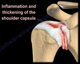
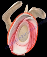
Adhesive Capsulitis, or frozen shoulder, is a painful progressive loss of shoulder motion. It affects both active and passive movement of the shoulder joint. The shoulder will be stiff and painful and occurs due to inflammation, fibrosis, scarring, and contraction of the capsule. A normal shoulder joint capsule is elastic and allows great range of motion. Inflammation and thickening of the shoulder capsule and may lead to adhesive capsulitis. Frozen shoulder may occur without any specific cause, however it may be triggered by a mild trauma to the shoulder.


This condition develops slowly and goes through three phases:

  1. Pain and freezing
  2. Stiffness or frozen
  3. Resolution

During the pain and freezing phase, the pain is worse at night and increases with any movement. This phase will last several months. During the second phase, range of motion is limited as pain is diminishing. This may last up to one year. The resolution phase may begin overtime and may last up to three years.


Conditions associated with frozen shoulder include:

  • Diabetes
  • Thyroid problems
  • Auto immune disease
  • Stroke
  • Rheumatoid arthritis
  • Trauma or post-surgery

A patient with frozen shoulder will have loss of both active (movement without assistance) and passive (movement with assistance) motion. External rotation of the shoulder is very limited and the condition is self-limiting and may resolve on its own. X-rays are needed to rule out degenerative arthritis.  An MRI or arthrogram will show small fluid in joint cavity. Rotator cuff may be normal and synovitis and narrowing of the rotator cuff interval is usually seen.

Treatment consists of anti-inflammatory medications, physical therapy, injections, and manipulation under anesthesia. Surgery will be done in the form of a release of the capsule when nonoperative methods fail. The physician should always check the patient for diabetes.  

Tuesday, September 18, 2018

Ganglion Cyst of the Shoulder


Ganglion cysts can be important when they are located around the shoulder, especially when they are located in the suprascapular notch and the spinoglenoid notch. The suprascapular nerve passes under the transverse scapular ligament at the suprascapular notch. The transverse scapular artery runs above the transverse scapular ligament. The artery and nerve joint and then pass through the spinoglenoid notch under the inferior scapular ligament. The suprascapular nerve gives branches to the supraspinatus muscle and branches to the infraspinatus muscle.

Nerve compression from a ganglion cyst at the suprascapular notch affects both the supraspinatus and infraspinatus muscles, causing a decrease in abduction and loss of external rotation of the shoulder. Nerve compression at the spinoglenoid notch affects only infraspinatus muscle, causing loss of external rotation of the shoulder with the arm to the side. Spinoglenoid notch compression is usually associated with cysts and ganglia. In addition to compression of the suprascapular nerve, these patients may also have associated posterior labral tears.

Tuesday, September 11, 2018

Anatomy of the Posterior Cutaneous Nerve of the Thigh


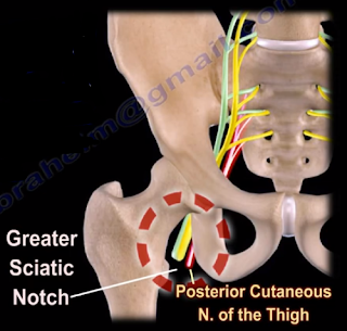
The posterior cutaneous nerve of the thigh (small sciatic nerve) arises from the sacral plexus from S1-S3. The posterior cutaneous nerve of the thigh exits from the pelvis through the greater sciatic notch below the piriformis muscle. The nerve descends below the gluteus maximus muscle along with the inferior gluteal artery. It runs into the back of the thigh beneath the fascia lata and over the long head of the biceps femoris muscle to the back of the knee. The nerve then pierces the deep fascia and accompanies the short saphenous vein to the middle of the back of the leg. The posterior cutaneous nerve of the thigh innervates the distal part of the gluteal region, the skin of the perineum and the posterior part of the thigh.
The nerve can become compressed when passing through the tunnel below the piriformis muscle and under the gluteus maximus muscle. This may result in sensitivity disturbances of the innervation area of the nerve. Causes of the syndrome may be hypertrophy or abnormality of the piriformis muscle such as entrapment below the piriformis which compresses the nerve. Compression of the nerve can also occur due to prolonged sitting. During the clinical examination, pain and sensitivity will be evident. Pain and sensitivity disturbances are characteristic of the nerve distribution site in the posterior part of the thigh down the knee. This disturbance can be from hyperesthesia to hypoesthesia or burning sensation similar to meralgia paresthetica of the lateral cutaneous nerve of the thigh.

Differential diagnosis include piriformis syndrome. The patient should avoid sitting for long periods of time, especially on a hard base. Treatment consists of physical therapy, massage, and injection. Surgery is rarely needed.

Tuesday, September 4, 2018

SLAP Tear- Symptoms, Diagnosis, and Treatment


A SLAP tear is a tear that occurs where the biceps tendon inserts into the superior labrum. A SLAP tear is different from a Bankart lesion. SLAP tears are not common and can be hard to diagnose. Symptoms of a SLAP tear include: pain deep within the shoulder or in the back of the shoulder, as well as catching, popping, or clicking sensations. The patient may also experience pain when throwing a ball with a decrease in velocity and the feeling of having a dead arm after pitching. Patients will also experience pain with overhead activity which mimics impingement syndrome. This typically affects throwing athletes. When the biceps tendon is involved, pain may also be located at the front of the shoulder. A SLAP tear can be an isolated lesion or it can be associated with internal impingement, articular sided cuff tear, or instability.
A SLAP tear is diagnosed with a clinical examination and testing. The O’Brien’s test is the most commonly used test. Multiple tests are usually used including the anterior slide test and the clunk test. An MRI with contrast is the best imaging technique. When performing the O’Brien’s test, the patient is standing or sitting with the arm at 90° of flexion, 10° of adduction, and full internal rotation with the forearm pronated.  The examiner applies pressure to the forearm and instructs the patient to resist the applied downward force. Pain at the shoulder joint suggests a SLAP lesion. Decrease in pain of the shoulder joint on supination of the arm is suggestive of a SLAP tear.
Treatment consists of physical therapy, anti-inflammatory medications, injections, and surgery (when conservative treatment fails). If surgery is necessary, a labral debridement will be performed for minor tearing and fraying. Biceps Tenodesis is becoming popular, as it is a procedure that cuts the biceps tendon where it attaches to the labrum and reinserts it in another area, usually in front of the shoulder. A biceps tenotomy is a procedure that cuts the biceps tendon from the glenoid, releasing the long head of the biceps tendon from its attachment allowing it to fall into the upper arm out of the shoulder joint. A biceps tenotomy is probably best suitable for some elderly patients. A SLAP repair is a procedure which uses sutures to anchor the torn labrum to the glenoid. This repair is usually done for athletes and patients under the age of 40 years.

Tuesday, August 28, 2018

Massive Rotator Cuff Tear


To view my Youtube video, 'Massive Rotator Cuff Tear- Classic', click here.


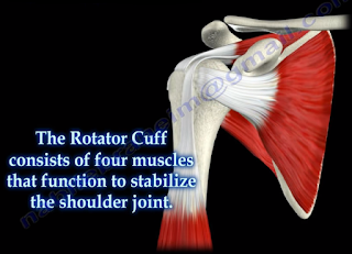
Massive rotator cuff tears are a disabling problem. These tears can cause pain, weakness, and sometimes swelling of the shoulder. The rotator cuff consists of four muscles that function to stabilize the shoulder joint: supraspinatus rotator cuff tendon, subscapularis tendon, infraspinatus rotator cuff tendon, and the teres minor rotator cuff tendon. The supraspinatus tendon is the most common of the rotator cuff tendons to become ruptured. Massive tears of the rotator cuff that are greater than 5cm usually involving both the supraspinatus and infraspinatus tendons.


Massive tears of the rotator cuff are defined as tears greater than 5cm, usually involving both the supraspinatus and infraspinatus tendons. Retraction of the rotator cuff tendons along with muscle atrophy and fatty infiltration can occur. This makes surgical reconstruction difficult with the surgical outcome being unpredictable and less than satisfactory.

Treatment varies from physiotherapy to replacement of the humeral head. Arthroscopic or open repair is usually the selected treatment. Reconstruction can be done in selected cases. A rotator cuff arthropathy is performed on massive cuff tears that are associated with superior migration of the humeral head as well as instability and arthritis of the shoulder. The patient will have pseudoparalysis and an x-ray will show shift of the humerus proximally. An MRI will show massive cuff tear with retraction at the level of the glenoid with atrophy of the muscle and fatty infiltration. A reverse shoulder is the treatment of choice for the elderly with rotator cuff arthropathy as it improves the pain and function. Hemiarthroplasty is the treatment for younger patients. A standard head or a big humeral head can be selected.


A patient with a massive tear of the cuff usually develops weakness of the shoulder and becomes unable to actively lift the arm without assistance. Fluid collection within the shoulder may occur with a massive tear of the rotator cuff.

Tuesday, August 21, 2018

Toe Deformities


Deformities of the toes are not uncommon and can occur from muscle imbalance, or other causes such as rheumatoid arthritis, diabetes, compartment syndrome, synovitis, or neurological disorders. Hammer toe occurs as flexion of the proximal interphalangeal (PIP) joint. Hammer toe is similar to the Boutonniere deformity of the finger. Claw Toe is a hyperextension deformity of the MTP joint and flexion of the PIP and DIP, resembling a pirate hook. Claw toe is similar to an intrinsic minus deformity of the hand, or “claw hand”. Mallet toe is similar in appearance to mallet finger, and is a flexion deformity of the DIP joint.

Wednesday, August 8, 2018

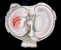
Discoid Meniscus


The meniscus is a cushion structure made of cartilage which fits within the knee joint between the tibia and the femur. The medial meniscus is C-shaped and the lateral meniscus in the more circular. The meniscus is made up of type I collagen that provides shock absorption and stability to the knee joint. The meniscus helps to protect the knee joint, allowing the bones to slide freely on each other. Discoid meniscus is a rare variation of the meniscus that usually affects the lateral meniscus of the knee in less than 5% of the population and could be bilateral in about 25% of the cases.
Discoid meniscus is a large meniscus with abnormal attachment causing increased mobility of the meniscus. It causes a pop, click, or snapping with locking and pain. There will be loss of full knee extension with tenderness on the lateral joint space. Symptoms occur more during extension of the knee. The discoid meniscus occurs due to the abnormal development and increase in size of the meniscus. An x-ray could show increased widening of the joint space. An MRI will show the “bow tie” sign in three or more sagittal continuous cuts. The coronal MRI will show a thick and flat meniscus extending beyond the halfway point of the condyle.


Watanabe Classification of Discoid Lateral Meniscus


  • Type I: Block-shaped stable
  • Type II: Block-shaped, stable, partial meniscus (has good peripheral attachment)
  • Type III: Unstable meniscus with stability arising only form the ligament of Wrisberg. (no posterior meniscal tibial attachment).

Treatment


An asymptomatic patient will be treated with observation. A symptomatic patient may receive a partial meniscectomy and saucerization with repair of type III (no posterior tibial meniscal attachment)

Tuesday, July 10, 2018

McMurray's Test- Meniscal Tear


Meniscal injuries are very common. The McMurray’s Test is a rotational maneuver of the knee that is frequently used to aid in the diagnosis of meniscal tears. With a meniscal tear, the patient usually complains of knee pain localized to the lateral or medial side of the knee joint. The patient will have locking, clicking, pain, or effusion.


During the physical examination, joint line tenderness is the most sensitive finding. Swelling of the knee and a possible extension lag (locked knee) is also a common finding. Pain at a higher level is usually associated with the medial collateral ligament. Pain at a lower level is usually associated with the pes anserine bursa.


What is the McMurrays test?             


The McMurray’s test is a knee examination test that provokes pain or a painful click as the knee is brought from flexion to extension with either internal or external rotation. The McMurray’s test uses the tibia to trap the meniscus between the femoral condyles of the femur and the tibia. When performing the test, the patient should be lying supine with the knee hyperflexed. The examiner then grasps the patient’s heel with one hand and places the other hand over the knee joint. To test the medial meniscus, the knee is fully flexed, and the examiner then passively externally rotates the tibia and places a valgus force. The knee is then extended in order to test the medial meniscus. To test the lateral meniscus, the examiner passively internally rotates the tibia and places a varus force. The knee is then extended in order to test the lateral meniscus. A positive test is indicated by pain, clicking or popping within the joint and may signal a tear of either the medial or lateral meniscus when the knee is brought from flexion to extension.


How reliable is the McMurray’s test?


There are mixed reviews for the validity of this test. An MRI is a very sensitive exam and makes the diagnosis easier, while excluding other associated injuries.


Tuesday, July 3, 2018

Hip Dislocation Following Total Hip Surgery


There are two types of hip joint dislocations: posterior and anterior. The position of the leg is important in determining the type of hip dislocation. When the hip is dislocated, the leg is usually shortened and it assumes a different position than the normal leg (the other leg). If the dislocation is posterior, the leg will be in adduction and internal rotation. If the dislocation is anterior, the leg will be in abduction and external rotation. Notice that the affected extremity is shortened and externally rotated. Leg shortening can also be seen in hip fractures and the leg will be shortened and externally rotated.


Dislocation of the hip following total hip surgery may require revision surgery, but it is rare. The majority of hip dislocations after total hip dislocations are posterior, and they are usually treated without surgery. Most occur within the first month of THA; 1-4% in primary, 16% in revision. There is more incidence of dislocation in revision hip replacement.


Causes & Risk Factors:

  • Posterior Approach (try to repair the capsule adequately)
  • Malposition of the component
    • Ideally, the normal cup component will be in 20° of anteversion and 40° of abduction
    • When the hip dislocates posterior, always check for retroversion of the cup.

  • Prior hip fracture surgery, especially in the elderly
  • Weakness of the abductor muscle—must achieve soft tissue tension and function
  • Alcohol abuse
  • Improper neck length—looseness of the hip

The patient should be careful to avoid all activities that cause dislocation after total hip surgery. The patient should use a pillow between the legs while sleeping on their back and they should be careful to not cross their legs in their sleep. Patients cannot sleep on their sides as well. The patient should not bend the body at the waist farther than 90°. When sitting, the patient must avoid chairs that make it difficult to stand up, and sit at more than a 90° angle. The patient must not sit with their legs crossed in the chair. The patient must be made aware that if the leg is changed from its usual position, or becomes shortened, then the hip is probably dislocated and their doctor should be consulted.


X-rays of the dislocated total hip should include AP and lateral views. Look for eccentric wear and look for the position of the prosthesis. CT scans may be needed before or after reduction of the dislocation to check the version of the components. Treatment is variable and depends on the situation. The treatment should be tailored for each case. The majority of these cases with early dislocations can be treated successfully with closed reduction and immobilization.

The treatment should start with closed reduction of the total hip and immobilization. Hip stability is checked after reduction of the dislocation. Immobilization can be done by a brace or a hip spica. Trochanteric osteotomy and advancement of the trochanter and tensioning the abductor muscle. Screws or wires can be used. The prosthesis must be in good alignment for this procedure to work. Constrained acetabular components are used when the abductor muscle is deficient and the component position is good. Revision total hip is done in recurrent dislocation with malposition of the component or polyethylene wear.

Tuesday, June 19, 2018

Isolated Fibular Fractures


Fibular fractures are usually associated with a complex injury, however they can be an isolated fracture. Complex injuries where a fibula fracture can occur include: fracture of the fibula and tibia, ankle fracture, pilon fracture, and Maisonneuve fractures.

Maisonneuve fractures involve a fracture of the proximal fibula associated with an occult injury of the ankle. Isolated fibular fractures are rare and usually the result of direct trauma. The fibula carries about 15% of the axial load and is the site of muscle attachment for the peroneus muscles and the flexor hallucis longus muscle. Check the patient who has a fibular fracture and no other fracture involving the tibia to rule out a possible Maisonneuve fracture, especially if there is no history of direct trauma to the leg. A high index of suspicion is necessary to diagnose and treat this injury. For high fibular fractures, the physician should look for signs of syndesmotic injury. Syndesmotic injury may include an unexplained increase in the medial clear space or the tibiofibular clear space is widened (should be less than 5mm). The x-ray will show the fracture to be rotational or oblique. Maisonneuve fractures require surgery to fix the syndesmosis.


Treatment will consist of reduction and fixation. It is important to determine if the injury is a Maisonneuve fracture or an isolated fibular fracture. An isolated fibular fracture will not need surgery.

Tuesday, June 12, 2018

Congenital Dislocation of the Knee


Congenital Dislocation of the knee is rare and may occur due to a contracture of the quadriceps. This condition usually occurs in patients with myelo, arthrogryposis, or Larsen’s syndrome. The patient with a congenital dislocation of the knee may have developmental dysplasia of the hip (DDH) and club foot. On examination, the patient will have a hyperextended knee at birth. They may have their foot placed against their face and there will be limited flexion at the knee. The patient may have a dimple or skin crease at the anterior aspect of the knee. You must examine the hip to rule out ipsilateral hip dislocation. 50% or more patients will have hip dysplasia. The etiology is not known; however, it could be due to fetal positioning or congenital absence of the cruciate.
There are grades, or a spectrum, for this deformity. Grade I deformities are referred to as Severe Genu Recurvatum, and the knee is hyperextended. If the range of passive flexion is more than 90°, it is considered to be a simple recurvatum. Grade II deformities are identified by subluxation with a range of 30-90° in passive flexion. Grade III deformities are complete dislocations with a range of passive flexion being less than 30°.

Congenital dislocation of the knee will take priority over treatment of hip dysplasia or club foot. The Pavlik harness and club foot cast will require knee flexion, so the physician will need to treat the knee dislocation first. With Grade I deformities, the initial treatment will be stretching of the knee and serial casting with the knee in flexion. In serial stretching and casting, the goal is to obtain at least 90° of flexion and reduction of the deformity over the course of several weeks. The physician should avoid pseudo-correction through an iatrogenic fracture of the proximal tibial physis. The prognosis is usually good if reduction is achieved without surgery. With Grade II deformities, if the infant is less than 1 month old, you will do serial casting first followed by percutaneous quadriceps recession, especially if the flexion is less than 90°. In Grade III deformities, a V-Y quadricepsplasty with above the knee cast is done in Grade III (frank dislocation), especially if nonsurgical treatment fails to reduce the tibia on the femur.   The result of open surgery is better when it is done in children younger than 6 months. In general, open reduction is reserved for children who did not respond to stretching and cast immobilization. It is important that the hip dysplasia is recognized and the knee dislocation is corrected early. This will help in early reduction of the hip.

Tuesday, June 5, 2018

Martin-Gruber Anastomosis


Martin-Gruber Anastomosis is median to ulner anastomosis in the forearm. It occurs through a communicating nerve branch between the median nerve and the ulnar nerve in the forearm. This connection carries motor nerve fibers. It can be confusing clinically and also on an EMG. It has a clinical significance for understanding the median nerve lesions and carpal tunnel syndrome. The axons will leave the median nerve or the anterior interosseous nerve crossing through the forearm to join the main trunk of the ulnar nerve, innervating the intrinsic muscles of the hand. The lesion above the communicating branch will affect the median nerve muscles. A lesion below the anastomosis (connecting branch) will not affect the median nerve muscles, it will spare the thenar motor intrinsic muscles of the hand. An isolated ulnar nerve lesion at the elbow will produce an unusual pattern for intrinsic muscle paralysis. Martin-Gruber Anastomosis is the most common anastomosis anomaly between the two nerves. In cases of nerve lesions of the median or ulnar nerve, this anastomosis serves as a conduit or an alternative innervation of parts of the hand and the forearm (it is really a detour). This can be a good explanation of difficult challenges, especially in the differential diagnosis. Incidence is high (about 15%). The physician should factor Martin-Gruber anastomosis into the differential diagnosis and the diagnosis.


If the communicating nerve arises from the anterior interosseous nerve, then a patient with anterior interosseous nerve palsy may present with hand intrinsic weakness, normally supplied by the ulnar nerve. Damage of the ulnar nerve at the wrist will lead to severe deficit of the intrinsic hand function greater than expected. There are other anastomoses available and reported as well as many variations that are possible.
There are three common anastomoses:

  1. Ulnar to median anastomosis in the forearm-reverse of Martin-Gruber (Marinacci anastomosis)
  2. Ulnar to median anastomosis in the hand (Riche-Cannieu anastomosis)
    1. Connection between the deep branch of the ulnar nerve and the recurrent branch of the median nerve
    2. It carries motor fibers and this anastomosis usually occurs in the region of the thenar and adductor pollicis muscles.
  3. Berrettini Anastomosis
    1. Communication between the digital nerves (sensory nerves) arising from the ulnar and median nerves in the hand
    2. Most common nerve anastomosis pattern

When the examination does not make sense and it is confusing, you can consider Martin-Gruber anastomosis.

Tuesday, May 1, 2018

Patellar Tendon Rupture


A patellar tendon rupture is a rupture of the tendon that connects the patella to the tibia. Rupture often occurs at the lower pole insertion site of the patella and it could be associated with degenerative changes. Rupture most often occurs in patients younger than 40 years of age. When the tendon is ruptured, the quadriceps muscle pulls the patella upward. One way to measure the height of the patella is by measuring the Blumensaat’s line. The knee needs to be flexed at least 30 degrees, then a line can be drawn through the roof of the intercondylar notch and usually touches the tip of the patella. The patella moves upward with the patellar tendon rupture (patella alta).

Associated Risk Factors


  • Rheumatoid Arthritis
  • Diabetes
  • Chronic Renal Failure
  • Systemic Corticosteroid Therapy
  • Chronic Patellar Tendonitis
  • Degenerative Changes


During the radiographic evaluation, an AP and Lateral x-ray is necessary. The patella alta is seen on the lateral view (*patella superior to Blumensaat’s line). An MRI is effective in assessing the patellar tendon, especially if other intraarticular or soft tissue injuries are suspected.
Treatment consists of a surgical reattachment of the tendon. The patient will need to keep their knee in extension and in a knee immobilizer for about 4-6 weeks.