25% of tibial shaft fractures can be open. Open fractures
can lead to complications including wound problems, osteomyelitis, nonunions,
and infected nonunions. The treatment of open fractures of the tibia can be
challenging. A lot of the concepts are not black and white; they may be in the
grey zone. We don’t know the best time for debridement. We don’t know what the
optimal irrigation solution is and what the optimal pressure for the fluid is.
We don’t know for sure the ideal duration of giving antibiotic prophylaxis, but
we know that it is important to give the appropriate antibiotics early and do
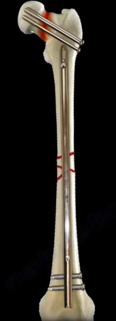
meticulous debridement. We know that the IM rod is better than the plate
fixation or external fixator, and the result of the reamed IM rod or unreamed
IM rod is the same. We need to close or cover the wound before 1 week and the
vac can be used provisionally when we cannot close the wound, primarily at the
optimal time. A grade I fracture is less than 1 cm. a grade II fracture is
1-10cm. A grade III fracture is more than 10 cm, and there is contamination.
Grade III fractures are divided into three types. Grade IIIa fractures require
adequate tissue for closure (or skin graft). Grade IIIb fractures require
extensive periosteal stripping and the patient will need a flap (rotational or
free flap). Grade IIIc fractures have a vascular injury that requires repair or
amputation. The relative indication for amputation is warm ischemia for more
than 6 hours, absent plantar sensation and severe ipsilateral foot trauma.
The
most predictive factor for amputation is the severity of the soft tissue injury
in the ipsilateral extremity. When comparing limb salvage versus amputation,
the patient’s outcome is generally the same at 1-5 years. Lack of plantar
sensation does not predict poor outcome after limb salvage. Segmental fractures
are Grade III fractures, even if the open fracture is 1 cm. The ideal
irrigation solution and the pressure used to controversial. Timing of the
initial debridement is controversial. Irrigation and debridement within 6 hours
was the gold standard in the past. Debridement is performed as a priority
procedure no later than the morning after admission. There is no difference in
infection rate for a patient who has the initial surgery before or after 6
hours, including patients with Type III open fractures. More than 40% of the
patients usually wait longer than 6 hours for their initial surgery after
arrival at the hospital. Delayed surgery for less severe fractures is
acceptable as long as the debridement is done as a priority the following day.
Unless there is a gross contamination, evidence is not clear as to when is the
best time for the debridement. It seems like giving the patient antibiotics
promptly is more important than the time of debridement. The preferred solution
is normal saline and low pressure irrigation. Low pressure lavage may reduce
reoperation rates due to infection, nonunions, and wound healing problems.
Normally the tradition is to use 3, 6, and 9 liters of solution for Type I,
Type II, and Type III open fractures (just recommendations). There is increased
risk of wound healing with antibiotic solution. Meticulous irrigation and
debridement of open fractures is important in decreasing the infection risk.
Prophylaxis should be started as soon as possible. All patients with open
fractures should receive first generation Cephalosporin’s that will cover
gram-positive bacteria. You can give penicillin for farm injuries and
clostridia prone wounds. You will give clindamycin if there is a penicillin
allergy. In Type III open fractures, add aminoglycoside, such as gentamicin. It
was found that local antibiotics delivery at the site of injury decreased the
infection risk, such as cement beaded loaded with antibiotics. Antibiotic
should be given within 3 hours of the time of injury (preferably given as soon
as possible). There is reduction of 59% of acute infection in patients with
open fractures treated with antibiotics. The infection rate is 1.6 times
greater if antibiotics are given after 3 hours. Type I and Type II open
fractures require antibiotic coverage for 24 hours after wound closure. For
Type III open fractures, antibiotic administration should be given for a period
of 72 hours after the injury and no more than 24 hours after wound closure. After
the initial debridement, the patient will need staged debridement within 24-48
hours. There is a reduction infection rate, acute and chronic, for Type III
open fractures with the use of systemic antibiotics and aminoglycoside cement
beads compared with antibiotics alone. This combination of antibiotics lowers
the infection rate for any open tibial fracture that is treated with an IM rod.
Its affect is more noticed in Type III injuries. Plating of open fractures may
cause chronic infection and infected nonunion. The healing time is doubled with
plated open fractures. The IM rod resulted in a better alignment and lower
reoperation rate than using external fixator. Also, no difference in the
infection rate between the IM rod and the external fixator. You can use a
reamed nail or an unreamed nail. They both have a comparable result and no
difference in the outcome.
When reaming, you can use a bigger rod that provides
better stability. Reamed nailing is superior in closed tibial fractures, but it
is not superior in open tibial fractures. Reaming can cause increased pressure
and disruption of the endosteal blood supply, can cause thermal necrosis and
fat embolism with increased intramedullary risk of infection. The unreamed rod
uses smaller nails and results in less stability but preserves the endosteal
blood supply. Unreamed IM tibial rod appears to have a shorter time to union
and fewer incidence of knee contractures when compared with circular wire
external fixator. Nowadays, more and more orthopedic surgeons are using reamed
nails for open fractures of the tibia. If you have a spiral fracture of the
distal 1/3 of the tibia, you will need to get a CT scan of the ankle to
identify a posterior malleolar fracture, which should be fixed before insertion
of the IM rod. There is a lack of evidence to support the value of external
fixator over the IM rod in open factures of the tibia. Due to patient
discomfort, the high incidence of pin tract infection and loss of alignment,
external fixator should not be used as a definitive fixation. Use external
fixator temporarily (less than 4 weeks) and replace it with a rod in about 14
days. External fixator may be utilized for severely contaminated open
fractures. Tibial fractures treated with a shorter duration of external fixator
has reduction of the infection risk by 80%. When there is a shorter interval
between removal of the external fixator and insertion of the IM rod of the
tibia, there is a reduction of the risk of infection by 85%. In less severe
soft tissue injuries, you do primary closure without tension. In cases of
delayed closure, soft tissue coverage should be done within 7 days. Soft tissue
coverage beyond 7 days will increase the infection. There is no difference in
the incidence of infection in patients who had primary closure and delayed
closure of the wound. It is recommended to do primary closure for Type I, Type
II, and Type IIIa fractures with tension free closure and after timely
antibiotic prophylaxis and adequate debridement. Intraoperative culture after debridement
has no value. It does not predict future infection. In the upper 1/3 of the
tibia, you can treat it by a medial gastrocnemius flap. In the middle 1/3 of
the tibia, you can treat it by a soleus rotational flap. The use of a free flap
for soft tissue coverage was less likely to have wound complications than the
use of a rotational flap. The zone of injury may be larger than expected, and
it may include the rotated muscle flap. The negative pressure wound therapy
(the vac) is used frequently. The vac provides provisional coverage for wounds
where the physician cannot do primary closure. There is decreased infection
rate when using the vac. The vac is used for coverage after the initial
debridement of the open fracture until the definitive coverage is done. It is a
good temporizing dressing and can also be used in fasciotomy wounds. The vac
promotes local wound healing. Bone morphogenetic protein (BMP2) decreases the
need for secondary surgery and is used in acute open tibial fractures treated
with IM rod.
The lumbosacral plexus starts from L4 to S4. The lumbar
plexus starts from L1 to L4. It has 6 branches. To remember this, think “I
Twice Got Lunch on Friday”. Knee extension is done by muscles that are
innervated by the L2, L3, and L4 nerve roots via the femoral nerve. The femoral
nerve innervates the muscles of the anterior compartment of the thigh
(quadriceps). th
lumbar nerve root. All nerves except the genitofemoral nerve and the obturator
nerve emerge from the lateral aspect of the psoas major muscle. The
genitofemoral nerve emerges anteriorly, and the obturator nerve emerges
medially in relationship to the psoas major muscle. L2, L3, and L4 will give
the obturator and femoral nerves. The obturator nerve and the femoral nerve are
equal.
The femoral nerve also provides sensation to this area. These
muscles cause hip adduction: adductor brevis, adductor longus, and adductor
magnus. They are innervated by the L2, L3, and L4 nerve roots via the obturator
nerve. This is the area of sensation supplied by the obturator nerve. With its
origin coming from the nerve roots of L2 and L3, the lateral femoral cutaneous
nerve of the thigh innervates the skin on the lateral part of the thigh. Risk
of injury to this nerve can occur with bone graft, with hip or acetabular
procedures, and with external fixator of the pelvis. The genitofemoral nerve
comes from the L1 and L2 nerve roots. Six branches come from the lumbar plexus.
Two branches come from one nerve root. Two branches come from three nerve
roots. Two branches come from two nerve roots. The two nerves that arise from
one nerve root are: iliohypogastric and iliolinguinal. Both of these nerves arise
from L1 nerve root. The iliohypogastric nerve is above the iliolinguinal nerve.
The two nerves that arise from two nerve roots are the genitofemoral (L1, L2)
and the lateral femoral cutaneous nerve of the thigh (L2, L3). The two nerves
that arise from three nerve roots are the obturator and the femoral. Both arise
from the L2, L3, and L4 nerve roots. There is also contribution from T12. The
fourth lumbar nerve root also passes a branch to the lumbosacral trunk at the 5
The ulnar nerve arises from the medial cord of the brachial
plexus. After the ulnar nerve passes through the arm, it runs from the medial
epicondyle to the pisiform bone in the wrist in a direct trajectory. Two carpal
bones are important in relation to the ulnar nerve in the wrist: the pisiform
and the hamate. Both the ulnar nerve and the ulnar artery enter the Guyon’s
canal. The Guyon’s canal is approximately 4 cm long. The ulnar nerve enters the
wrist medial to the ulnar artery. The Guyon’s canal has one proximal entrance
and two distal exits, one superficial and one deep. The Guyon’s canal contains
the ulnar nerve with its superficial sensory and deep motor branches. th
lumbricals. The deep branch of the ulnar nerve also innervates the hypothenar
muscles, the adductor pollicis muscle, and the deep head of the flexor pollicis
brevis muscle. The superficial branch of the ulnar nerve is mainly sensory. It
gives supply to the digital nerves of the 4th and 5th
fingers and a motor branch to the palmaris brevis muscle. Based on the location
of compression in the Guyon’s canal, the affected area of the nerve may be
purely motor, purely sensory, or a mixture of motor and sensory. Pain and
paresthesia in the ulnar 1 ½ digits or clawing of the 4th and 5th
fingers can be symptoms. Another symptom can be loss of function of the
intrinsics (it normally flexes the MCP and extends the IP joints). In low ulnar
nerve injury, the flexor digitorum profundus is working (functional). It flexes
the 4th and 5th fingers and causes clawing (unopposed by
the intrinsic muscles). Ulnar nerve palsy results in paralysis of the intrinsic
muscles; test the first dorsal interosseous muscle and check for atrophy. 70%
of pinch is lost due to loss of adductor muscles.th and 5th
fingers and causing clawing of these two fingers. High ulnar nerve palsy has
less clawing of the 4th and 5th fingers and sensory
deficit to dorsum of the hand. In cubital tunnel syndrome (high ulnar nerve
involvement) you can also find Tinel’s sign at the elbow and positive elbow
flexion test. The dorsal cutaneous branch arises before the Guyon’s canal. If
you have high ulnar nerve palsy, there will be sensory deficit on the dorsum of
the hand, because that nerve will be affected. If you have a low ulnar nerve
palsy, that nerve will not be affected because it already branched out or came
off from the ulnar nerve and escaped (sensation on that part of the dorsum of
the hand). Nonoperative treatments include activity modification, NSAIDS, and
splinting. Surgical treatment includes release of the carpal tunnel .In
patients diagnosed with both carpal tunnel and ulnar tunnel syndrome, the
Guyon’s canal is adequately decompressed by the release of the carpal tunnel. Local
decompression can be done especially if nonoperative treatment fails.
Decompression of the ulnar nerve by addressing the cause; success of surgery
depends on finding a cause. To determine the cause of compression, explore and
release of all three zones in the Guyon’s canal, vascular treatment of ulnar
artery thrombosis, hook of hamate excision, decompress ganglion cysts, and
release hypothenar muscle origin. All of these things are possible causes that
need to be addressed. Tendon transfer can be a treatment in late cases; it will
correct claw fingers, restore power pinch, and improve Wartenberg’s sign.
The Froment’s test is
positive. When pinching a piece of paper between the thumb and index finger,
the thumb IP joint will flex if the adductor pollicis muscle is weak due to
ulnar nerve palsy. Symptoms also include weak grasp due to intrinsic weakness,
Wartenberg’s Sign, and the Allen’s test. Carpal tunnel view x-rays and CT scans
are useful to evaluate hook of the hamate fractures and nonunions. MRI is
useful to evaluate ganglion cysts. Ultrasound is useful to check vascular
status of the hand and to diagnose ulnar artery thrombosis. EMG and nerve
studies are helpful. High or low ulnar nerve injury is a differential
diagnosis. In low ulnar nerve injury or compression, the flexor digitorum
profundus muscle is working; flexing the 4
The
Guyon’s canal contains the ulnar nerve with its superficial sensory and deep
motor branches. Ulnar nerve compression neuropathy can occur in the Guyon’s
canal. The most common causes include volar ganglion, hook of hamate fracture,
repetitive trauma, ulnar artery thrombosis, palmaris brevis muscle hypertrophy,
or pisiform fracture or dislocation. Volar ganglion cysts may protrude or grow
into the canal, which could compress the ulnar nerve. Hook of hamate fracture
can lead to neuropathy of the nerve in the tunnel. Compression and inflammation
also can result from repetitive trauma such as using a hammer. Trauma over the
hook of hamate, where the superficial branch of the palmar artery lies, leads
to vascular insufficiency of the ulnar side of the hand. Ulnar nerve
compression may occur as a result of pisiform dislocation or fracture. The
Guyon’s canal is bound on the floor by the transverse carpal ligament, on the
roof by the volar carpal ligament, on the ulnar border by the pisiform and
pisohamate ligament, and on the radial border by the hook of hamate. There are
three zones of Guyon’s canal compression. Zone I is located proximal
bifurcation of the nerve; it is characterized by mixed motor and sensory
symptoms, and it is caused by ganglia and hook of hamate fractures. Zone II is
located in the deep motor branch; it is characterized by motor symptoms only,
and it is caused by ganglia and hook of hamate fractures. Zone III is located
by superficial sensory branch; it is characterized by sensory symptoms only,
and it is caused by ulnar artery thrombosis or aneurysm. The deep branch of the
ulnar nerve innervates all of the interosseous muscles and the 34d and 4
The reverse pivot shift test helps to diagnose acute or
chronic posterolateral instability of the knee. A significantly positive
reverse pivot shift test suggests that the PCL, the LCL, the arcuate complex,
and the popliteal fibular ligament are all torn. The reverse pivot shift test
begins with the patient supine with the knee in 900 flexion. Valgus
stress is then applied to the knee with an external rotation force. Bring the
knee from 90o of flexion to full extension. The tibia reduces from a
posterior subluxed position at about 20o of flexion. A shift and
reduction of the lateral tibial plateau can be felt as it moves anteriorly from
a posteriorly subluxed position. This is called the reverse pivot shift because
shift of the lateral tibial plateau occurs in the opposite direction of the
true pivot shift (seen in ACL tears). If the tibia is posterolaterally
subluxed, the iliotibial band will reduce the knee as the IT band transitions
from a flexor to extensor of the knee. It is very important to compare this
test to the contralateral knee. The pivot shift test indicates an ACL tear. The
reverse pivot shift test indicates posterolateral instability of the knee.
Posterolateral corner injury includes the LCL, popliteal fibular ligament,
arcuate complex, and the lateral capsule.
The anterior cruciate ligament is located in the front of
the knee. Rupture of the anterior cruciate ligament (ACL) is a condition
commonly seen in sports usually due to a non-contact pivoting injury. The Pivot
Shift test is a specific test for ACL deficient knee (ACL injury). Pivot shift
is pathognomonic for an ACL tear and is best demonstrated in a chronic setting.
Lachman’s test is the most sensitive examination test for ACL injury. o of flexion. The anteromedial bundle is
tight in flexion, and it increases anterior translation at 90o of
flexion. The Lachman’s test is the most sensitive test especially in acute
settings, and the examiner will find no end point with anterior translation of
the tibia. In an acute setting, physical examination can be difficult or
limited due to pain. With the Pivot shift test, the patient must be completely
relaxed, and the test is helpful in chronic situations especially if the
patient complains of the knee giving way.
In the Pivot shift, the knee subluxes
in extension and reduces at 20-30 degrees of flexion. The Pivot shift
correlates closely with patient satisfaction of their reconstructed knee. It is
a measure of functional instability following ACL reconstruction. Vertical
femoral tunnel placement will cause rotational instability seen as a positive
pivot shift, and the malposition of the bone tunnel will be seen in an AP view
x-ray of the knee. The 9 or 10 o’clock position is better than the 12 o’clock
position; the vertical position is bad. The patient with an ACL injury usually
has a non-contact pivoting injury event with an awkward landing, feeling a
“pop” sensation, or immediate swelling. Aspiration usually shows blood in the
knee which proves a 75% chance of ACL tear when you aspirate blood from the
knee. Patients will also exhibit a positive Lachman’s test which may be hard to
examine because of the pain. Aspiration of the knee may make the examination
easier. MRI of the knee joint will show the hematoma, and it may show bone
lesions or bruising in the typical location which is characteristic with tears
of the ACL. These injuries are typically located at the middle of the femoral
condyle and posterior part of the tibia laterally. You may find a triple injury
within the MRI (O’Donoghue’s Unhappy Triad). The O’Donoghue’s Unhappy Triad
include an anterior cruciate ligament (ACL) injury, a medial cruciate ligament
(MCL) injury, and a lateral meniscus injury. In chronic ACL tears, the posterior
horn of the medial meniscus is the most commonly injured structure. In acute
ACL tear, send the patient for therapy for range of motion, brace the patient
and allow the MCL to heal and reconstruct the ACL later if needed. Patients
should do stress hamstring therapy in ACL tears. The patient will probably
complain of instability immediately or later on.The tibia can be pulled forward more than
normal (anterior translation). The examiner will have a sense of increased
movement and lack of a solid end point. For the pivot shift test, the patient
should be lying supine and totally relaxed. With pivot shift, the knee is in
the subluxed position when the knee is in full extension. The pivot shift
starts with extension of the knee and you can feel a “clunk” at 20-30 degrees
of flexion. To perform, hold the knee in full extension then add valgus force
plus internal rotation of the tibia to increase the rotational instability of
the knee. Then take the knee into flexion. A palpable clunk is very specific of
an ACL tear. The iliotibial band will reduce the tibia and create the clunk on
the outside of the knee. Always compare with the other side. The ACL prevents
anterior translation of the tibia. It is a secondary restraint to tibial
rotation and varus and valgus stress. The ACL consists of two bundles: the posterolateral
bundle and the anteromedial bundle. The posterolateral bundle prevents pivot
shift, contributes to rotational stability, prevents internal rotation of the
tibia with the knee in near extension, and increases the anterior translation
and tibial rotation at 30
The ACL
keeps the tibia from sliding out in front of the femur and provides rotational
stability to the knee. Rupture of the ACL causes anterolateral rotatory
instability. The tibia moves anterolaterally in extension; however, when you
flex the knee, the IT band becomes a flexor of the knee. The IT band pulls back
and reduces the tibia. The pivot shift test goes from extension (tibia
subluxed) to flexion, with the tibia reduced by the iliotibial band. Both the
Lachman’s tests and the Pivot shift test are associated with 20-30 degrees of
knee flexion. The Lachman’s test starts at 20-30 degrees of flexion. With the
Pivot shift test, you feel the clunk at 20-30 degrees of flexion. 20-30 degrees
of flexion is important for examination of the ACL. For the Lachman’s test, the
femur is stabilized with one hand and the other hand pulls the tibia anteriorly
and posteriorly against the femur.
Herpetic whitlow occurs from the herpes simplex virus. It is
a self-limited disease, and it often involves the tip of the fingers. It occurs
due to contact with oral or tracheal secretions and from self-inoculation.
Herpetic whitlow is seen in dentists, respiratory therapists,
anesthesiologists, and toddlers (children who suck their thumb). The symptoms
of herpetic whitlow include, burning, inflammation, and a clear fluid (not
purulent). Vesicles on the finger which can be grouped together with inflammation
and redness at the base. The gram stain will usually be negative. Diagnosis can
be done with the Tzanck test. Giant cells can be seen. Treatment is acyclovir,
no surgery. Surgery can make the situation worse.
Galeazzi fracture is a fracture of the distal 1/3 of the
radius with disruption of the distal radioulnar joint (DRUJ). The fracture is
always located above the proximal border of the pronator quadratus. The
pronator quadratus rotates the distal fragment towards the ulna and pulls it
proximally. We usually fix the fractured radius, and we then evaluate DRUF for
instability after we fix the distal radius. If you have instability, make sure
that the joint is reduced, then you will do percutaneous fixation of that
joint. If you don’t have instability, you will do nothing or maybe give a long
arm splint in supination if you think the patient needs the splint.
Basically,
you will need intraoperative evaluation of the DRUJ. Not all distal radial
fractures will be associated with distal radioulnar joint instability. They
found that if the radius fracture is less than 7.5 cm from the join, then the
distal radioulnar joint can be unstable. If the fracture radius is more than
7.5cm from the joint, then the distal radioulnar joint will be rarely unstable.
So the closer the fracture of the radius is to the joint, the more likely that
the distal radioulnar joint is involved, and we need to work diligently to find
the problem and address it. The problem can be instability of the DRUJ. You may
find an ulnar styloid fracture or you find that the radius is short (about 5 mm
or more). In the AP view of the wrist, you may find widening of the joint or in
the lateral view, you find that the ulna goes dorsally or volarly. The distal
radioulnar joint has ligaments, volar and dorsal, that stabilize the joint, and
that joint is usually stable in supination. Sometimes in old, complicated or
difficult cases, you can’t really evaluate the distal radioulnar joint without
getting a CT scan of both wrists (make sure that you position the wrist in the
same position. Anatomic reduction and fixation of the radius with a volar
plate. Then you assess the stability of the distal radioulnar joint. If the
distal radial ulnar joint remains unstable, supination of the wrist may reduce
that joint. If not, either a closed reduction or open reduction with pinning of
the joint is done. If after anatomic restoration and plate fixation of the
radius, the distal radioulnar joint remains irreducible, then the structure
that is most likely obstructing the reduction is the extensor carpi ulnaris. It
is imperative to recognize the problem of Galeazzi fracture, which is the
distal radioulnar joint injury. The treatment of the problem acutely is better
than late reconstruction. When you fix the radius, make sure that the radial
bow is restored. The reduction of the joint is done by supination of the
forearm, and you do immobilization in supination if the distal radioulnar joint
is stable following open reduction of the distal radius. So there is an obvious
injury that you will see, and you will test that injury and see if the joint is
stable in supination. If it is, keep the forearm in supination. You will do pin
fixation if the joint is reducible, but is unstable. The pin fixation will be
done by cross pinning from the ulna to the radius and leave the pins for about
4 weeks. You can do open reduction of the joint if the joint is not reduced and
something is blocking the reduction, such as the extensor carpi ulnaris tendon.
If there is a large ulnar styloid process fracture, you probably need to open
that fracture after you fix the radius, and then do open reduction and internal
fixation of the large ulnar styloid fragment and immobilize the forearm in
supination. It might be difficult to evaluate the stability of the distal
radial ulnar joint. In general, the DRUJ is stable in most cases after anatomic
reconstruction of the radius.
Mycobacterium marinum is the most common atypical
mycobacterium that can cause infection in humans. It is found in salt and fresh
water. It is an acid-fast bacilli. The wrist and the hand are affected in about
50% of the cases. It may cause skin and soft tissue infections after skin
abrasion. The patients are exposed to aquatic environments such as aquariums
and swimming pools. The disease often occurs following the cleaning of fish
tanks. The bacilli enter the body through scratches and abrasions, causing
lesions in the tissue. The diagnosis is usually delayed because the condition
is rare, and the history of aquatic exposure is usually not obtained. The hand
and wrist are commonly involved. There will be painful swelling of the hand.
Subcutaneous granules, masses, nodules, ulcers, and noncaseating granulomas are
present. It may present as chronic tenosynovitis of the hand. It affects the
extensors more than the flexors. It can cause a TB like disease in fish. The
chronic skin lesion is sometimes called a “swimming pool granuloma” or “fish
tank granuloma” in humans. The bacteria grows in a low temperature culture at
30o centigrade. The bacteria grows on Lowenstein-Jensen medium. It
requires lower temperature and a longer period of the incubation (up to 6 weeks
or more). It can be treated with oral antibiotics antimicrobial therapy.
Ethambutol and Rifampin if diagnosed early. Minocycline and Clarithromycin has
been described. Surgery is done in late stages and in deep infection. Surgery
entails synovectomy and debridement in addition to oral antibiotics for
approximately 3 months. Mycobacterium avium-intracellulare occurs in terminal
AIDS patients, or it also can occur in a non HIV patient.
Monteggia fracture is a fracture of the proximal ulna and
radial head subluxation. It is imperative that you restore the length and the
proper alignment of the ulna so that the radial head can be reduced. If we
malalign the ulna, then the radial head will remain subluxed. There are some
cases where the fracture of the ulna is so comminuted that we will be unable to
restore the length of the ulna. We will not even know if we restored the length
of the ulna.
There is a technique that I use in the reconstruction of the ulna
in cases where the ulna is too comminuted. I will open the fracture ulna, and I
will approach the radial head. I will reduce the radial head to the capitellum
and reduce the ulna to the radius and make sure that the proximal radioulnar
joint is anatomic. Once that joint is anatomic, I will pin it with either one
or two K wires. I transfix the ulna to the radial head. We know that the radial
head is reduced, now the ulna will be reduced because the radioulnar joint is
reduced. We are temporarily transfixing the ulna to the radial head, and that
will help to restore the proper length of the ulna. Once the proper length of
the ulna is defined, then reconstruction of the ulna is simplified utilizing a
dorsal ulnar plate. Next, the K wires are removed and the radioulnar joint is
tested for stability. Occasionally, the K wires may be left in place for a few
weeks if needed to provide additional stability, then removed later on.
Monteggia fracture is not a simple fracture. It is a
fracture of the proximal ulna with dislocation of the radial head. Monteggia
fracture can happen in children and in adults. It is one of the most common
injuries that is missed in the emergency room in children. The radial head may
be dislocated or subluxed, and this problem may not be clear on x-rays. If this
injury is missed, then the child will probably need a big surgery to deal with
this big problem. Treatment of this fracture depends on the age of the patient.
In general, in pediatric patients, you will do closed reduction of the ulna and
closed reduction of the radial head. In adult patients, you will do open
reduction with internal fixation of the ulna with dorsal plate and closed
reduction of the radial head. A line drawn from the proximal radius should
bisect the capitellum in all x-ray views. If you are in doubt and not sure, get
x-rays of the other side and compare. Always examine the patient for posterior
interosseous nerve injury.
The most common type is anterior Monteggia. That
means that the apex of the fracture is anteriorly and the radial head goes
anteriorly. Just make it a practice, when you have a fracture of the proximal
ulna, look at the radius and the radial head, and see the position of the
radial head in relationship to the capitellum. Anterior Monteggia is more
common in children. Posterior Monteggia constitutes 70-80% of Monteggia
fractures in adults. There are four types of Monteggia: Type I, Type II, Type
III, and Type IV. Monteggia fracture is classified according to the direction
of displacement of the radial head. The radial head has two relations: relation
with the capitellum and relation with the proximal radioulnar joint. When the
radial head subluxes or dislocates, it subluxes or dislocates from these two
joints. The radial head becomes free. This means that the radius is not
connected to the capitellum or to the superior radioulnar joint. A Type I
fracture is of the middle or the proximal third of the ulna with anterior
dislocation of the radial head, and it has the characteristic that the apex of
the ulnar fracture is anteriorly. Type I fracture is the most common of all
types (especially in children). Type I occurs in about 60% of fractures. In
children, reduce the fractured ulna and reduce the dislocation of the radial
head and immobilize the elbow in flexion and supination. When you flex the
elbow, especially more than 90 degrees, you will relax the biceps (watch the
circulation). A Type II fracture is a posterior type fracture. Posterior
Monteggia is the most common type in adults. It is associated with a higher
complication rate and carries the worst prognosis. 15% of Monteggia fractures
are Type II. It is a fracture of the middle or proximal third of the ulna with
posterior dislocation of the radial head. You should immobilize the elbow in
extension. Type III is a lateral Monteggia. About 20% of Monteggia fractures
are Type III. It is a fracture of the proximal ulna with lateral dislocation of
the radial head. 5% of Monteggia fractures are Type IV; it is very rare. It is
a fracture of the proximal ulna with anterior dislocation of the radial head
and fracture of the proximal third of the radius below the bicipital
tuberosity. The patient will need surgery, even in children. In this case, the
radial head is dislocated, and you also have fractures of the radius and the
ulna. The posterior interosseous nerve is adjacent to the radial neck, placing it
at risk for a traction injury with dislocation of the proximal radius.
You
should do a neurovascular examination. A nerve injury which involves the
posterior interosseous nerve is not uncommon. Ask the patient to “hitchhike”
and extend their fingers. Make sure the wrist is in dorsiflexion when you ask
the patient to extend the fingers. In posterior interosseous nerve injury, the
finger extensors will not be working. If the posterior interosseous nerve is
injured, observe the patient. In case of posterior interosseous nerve injury in
Monteggia fracture, you will reduce and stabilize the fracture and reduce the
radial head dislocation. Observe the nerve; do not explore the nerve. Typically
the nerve injury is a neuropraxia. It can be expected to resolve itself with
observation in 6-12 weeks. If it does not resolve, you will do EMG and nerve
studies after that period of observation. Any time that you have an ulnar shaft
fracture or any fracture of the proximal ulna, check the radial head position.
Make sure that the radial head is reduced to the capitellum (be aware that the
subluxation may be subtle). Recognition of Monteggia fracture in children is
important. Early appropriate treatment is much easier than treating a missed
radial head dislocation. To treat a Monteggia fracture in adult patients, do
open reduction internal fixation (ORIF) of the ulna. When the ulna is properly
aligned and fixed, the radial head will reduce by itself. After fixation of the
ulnar fracture, if the radial head is still not reduced, then assess the ulnar
reduction. Check for malalignment or malreduction of the ulna. It is imperative
that you restore the length and the proper alignment of the ulna, so that the
radial head can be reduced. If we malalign the ulna, then the radial head will
remain subluxed. Radial head instability may be caused by nonanatomic reduction
of the ulna or by interposition of the annular ligament. Fracture of the ulna
may be too comminuted, and it may not be reduced properly. The fracture may
also need bone graft later on for healing. A Monteggia variant associated with
radial head fracture, in addition to dislocation of the radial head fracture,
in addition to dislocation of the radial head and fracture of the ulna can be a
problem. The radial head fracture is usually fixed or replaced, a prosthesis is
used to replace the radial head in the elderly, especially if the fracture is
comminuted. The subluxation of the radial head is reduced, and the fractured
ulna is fixed as usual. Treatment is different in pediatric patients. The
radial head ossifies around four years of age. In Type I, Type II, and in Type
III Monteggia fractures, you will do closed reduction of the ulna to restore
the length of the ulna, and you will do closed reduction of the radial head.
Closed reduction is much more successful in young children. In anterior
Monteggia, you will immobilize the elbow in flexion and supination. In
posterior Monteggia, you will immobilize the elbow in extension. Ulnar fixation
with a rod or a plate is needed in older patients with unstable fractures. Type
IV fractures require surgery. Surgery is also done in cases where we are unable
to restore the proper length of the ulna, we are unable to reduce the ulna, and
we are unable to reduce the radial head. In this situation, we can use IM rod
or a plate. Dislocation of the radial head with fracture of both the radius and
ulnar shaft. Do closed reduction of the radial head with intramedullary pin
fixation of the radius and the ulnar shaft fractures. The radius and ulnar
shaft fractures are stabilized surgically to give a lever arm for reduction of
the radial head. In this type of fracture, the radial head subluxation may be
missed or unappreciated, because the focus is usually on the forearm fractures.
To treat a missed or neglected Monteggia fracture in children, do osteotomy of the
ulna and lengthening with correction of the angulation, and reduction of the
radial head in addition to plating of the ulna. The patient may need open
reduction of the radial head.
Lisfranc injury is an important topic. If Lisfranc injury is
not diagnosed and treated properly, it can lead to an altered gait, midfoot
arthritis, and long term disability. Lisfranc injury indicated disruption
between the base of the 2nd metatarsal and the medial cuneiform. Lisfranc
injuries are a spectrum of injuries of the tarsometatarsal joints. Diagnosing Lisfranc
injury is important. Diagnosis is missed in about 20%-30% of cases especially
in multiple trauma patients. A high index suspicion is needed to prevent
progression of the foot deformity, chronic pain, and dysfunction. You may need
weight-bearing films for diagnosis of Lisfranc injury. Lisfranc injury may also
be associated with compartment syndrome. Lisfranc injury could be purely
ligamentous or can be associated with fractures. ORIF is better in cases of
fractures. Arthrodesis is better in cases of purely ligamentous injury. In general,
ligamentous injury does worse than fractures. The Lisfranc ligament is a large
oblique ligament that extends from the plantar aspect of the medial cuneiform
to the base of the second metatarsal. The Lisfranc ligament stabilizes the 2nd
metatarsal and maintains the midfoot arch. Osseous stability is provided by the
roman arch of the metatarsals and the recessed keystone of the 2nd
metatarsal base. Tarsometatarsal joint complex is divided into three units:
medial, middle, and lateral. The medial is the 1st metatarsal joint
at 6o mobility. The middle is the 2nd and 3rd
tarsometatarsal joints, and it is rigid. The lateral is the 4th and
5th tarsometatarsal joints; it is mobile which is why you do not
fuse the 4th and 5th tarsometatarsal joints. The dorsalis
pedis artery and the deep peroneal nerve both run between the first and second
metatarsal bases. A direct injury with a plantar displacement is more common. Indirect
injuries are more common than direct injuries. They result from axial loading
or twisting on a plantar flexed midfoot. Dorsal displacement of the 2nd
metatarsal is more common. Check the alignment of the dorsum of the 2nd
metatarsal with the middle cuneiform. Associated fractures are typically tarsal
fractures, especially a cuboid fracture. A “Nutcracker” fracture results from
twisting injury causing forceful abduction of the forefoot. It is a fracture of
the base of the 2nd metatarsal and compression fracture of the
cuboid. nd
metatarsal, at the navicular, and cuboid. Check for widening between the first
and second ray (more than 2 mm is an indication for surgery). In the lateral
view, check the dorsal displacement or subluxation of a metatarsal. It should
be at the level of the corresponding cuneiform. Check for the FLECK sign (bony
fragment). Avulsion fragment of the Lisfranc ligament from the base of the 2nd
metatarsal. The medial side of the fourth metatarsal should line up with the
medial side of the cuboid on the oblique view (30o). CT scan can be
useful and MRI can confirm purely ligamentous injury. These injuries should be
treated with a cast. For a dorsal sprain and no instability, the patient can be
treated with non-weight bearing cast for 6 weeks and return to activity
gradually. Surgery can be done for instability. Open reduction internal
fixation with cortical screws if there is bony fractures. When you do ORIF- you
need anatomic reduction. Hardware removal between 5-6 months (some surgeons
leave the hardware in place indefinitely). Arthrodesis if the injury is purely
ligamentous. Healing of the ligaments is less reliable than bony healing. Purely
ligamentous injury needs primary arthrodesis. Arthrodesis is also done in old injuries
if there is delay in treatment for if there is failure of open reduction and
internal fixation of Lisfranc injury. Midfoot arthrodesis is also used for
chronic Lisfranc injury that leads to severe midfoot arthritis with progressive
arch collapse and midfoot abduction. Fusion of the medial and middle column;
first, second, and third tarsometatarsal joints. Do not fuse the lateral column
(lateral column is mobile). For the lateral column, do reduction and
stabilization by k-wire fixation. Post-traumatic arthritis occurs in up to 50%
of patients. Patient may have altered gait and long term disability. Purely ligamentous
injury has a worse prognosis than injuries with fractures. Malalignment of the
fractures usually lead to arthritis.
Lisfranc classifications are not useful in deciding the treatment or the
prognosis of the injury. Severe injuries are obvious, easily diagnosed, and may
develop compartment syndrome of the foot. Injuries with minimal displacement
could be missed, and they will need surgery regardless of the classification. Arthritis
may develop even with minimal displacement. In general, there are three
patterns of injury: total incongruity, partial incongruity, and divergent. Total
incongruity occurs when all five metatarsals are displaced in the same
direction. Total incongruity occurs lateral or medial, with lateral being more
common. Partial incongruity occurs when one or two metatarsals are displaced
from the others. Divergent occurs when the lateral displacement of the lesser
metatarsals with medial displacement of the first metatarsal. The one thing all
these injuries have in common is disruption of the tarsometatarsal joint
complex. The patient has severe pain in the midfoot and is unable to bear
weight. There may be some swelling in the midfoot dorsally. Plantar bruising
may be present, especially medially. Tenderness over the tarsometatarsal joint.
Check the skin condition and rule out compartment syndrome. Check the neurovascular
status of the foot. Plantar ecchymosis is a classic clinical sign of potential
Lisfranc injury. Wight bearing standing x-rays with comparison views if x-rays
are normal and if the physician clinically suspects a Lisfranc injury. Another alternative
is to get physician assisted midfoot stress radiograph. Obtain three views: AP,
oblique, and lateral. Medial border of the second metatarsal should line up
with the medial border of the middle cuneiform on both the AP and the oblique
view. Check for fractures, especially at the base of the 2
Type I
fractures are a stable avulsion fracture of the alar ligament near the tip of
the odontoid. A soft collar can be used to treat Type I fractures. Be aware of
significant ligamentous injuries. Type II fractures are at the base of the
odontoid process. Type II are the most common and are troublesome. The nonunion
rate is about 20-80% due to interruption of the blood supply. The risk factors
of nonunion include if the patient is over the age of 60 years old, if the
patient has more than 6mm of displacement, smoking and diabetes, and you are
unable to achieve reduction. In posterior displacement, extension injury (rare
type) the anterior displacement is more common (flexion injury). Delay in
treatment also increases the rate of nonunion. For treatment of young patients
with no nonunion risks use a halo. The patient is younger than 60 years. The
fracture is minimally displaced. Initial dens displacement is less than 6 mm,
and the reduction is within one week of the injury. Healing will occur in the
majority of cases.
If the patient has a nonunion risk, or when reduction of the
fracture cannot be achieved or maintained, then we need to think about surgery
and the fracture pattern. When the fracture pattern allows, you can put an
anterior screw into the odontoid (to preserve the motion of C1/C2). Odontoid
screw is used in younger patients instead of fusion to avoid loss of 50% of the
neck rotation). Do not use the anterior screw fixation in patients with
osteoporosis, in older patients, or in patients with a short neck. Another
scenario is, if the patient has nonunion risks but the fracture pattern does
not allow you to place an anterior odontoid screw, then you are going to fuse
C1 to C2 (this will lose 50% of neck rotation). In general, C1/C2 fusion is
used in cases of nonunion or it is used in cases of displaced fracture in the
older patient and it can also be used if there is a failure of treatment with a
halo. C1/C2 fusion can also be used if the fracture is comminuted and unstable.
Posterior C1/C2 fusion can be done with different screw or wire constructs. A
vascular watershed area exists between the apex of the odontoid, which is
supplied by branches of the internal carotid artery and the base of the
odontoid, which is supplied by branches of the vertebral artery. Type II
fracture of the odontoid may get nonunion due to cortical bone and poor blood
supply.
Type III fractures extend through the body of C2. This area is rich in
blood supply and the fracture heals in the majority of cases. Treatment for
Type III odontoid fractures includes external cervical orthosis (especially in
the elderly patient) and a halo (if the fracture is displaced) (do not use in
elderly patients). Odontoid fractures in the elderly can occur due to a simple
fall and usually the diagnosis is missed. It is associated with increased
complications and mortality. Do not use a halo in elderly patients. Use an
external cervical orthosis of some sort. Fibrous union might be adequate if the
fracture is not badly displaced, otherwise you will do fusion of C1/C2. For
example, an 80 year old patient with osteoporosis, who is a smoker and has a
displaced odontoid fracture that cannot be reduced, then this fracture will
lead to nonunion and more complications. You need to do posterior C1/C2
arthrodesis. In general, if the elderly patient with an odontoid fracture is
not a good surgical candidate, then you will give the patient a cervical
orthosis. You can do the C1/C2 fusion by using transarticular screws, which you
are not going to do if you have an aberrant vertebral artery. Another technique
can be done for the fusion where fusion between C1/C2 is done with the screw
placed into the C1 lateral mass and the C2 pedicle, plus a bone graft. There is
increased survival for the elderly patient that undergoes surgery for Type II
odontoid fracture. This may be a selection bias, because they have healthier
patients who are physiologically active and young who are fit for surgery. The
synchondrosis between the odontoid and the C2 body fuses by the age of 6 years.
Odontoid fracture in young children usually occurs by the age of 4 years.
Physicians may confuse the synchondrosis with a fracture. The treatment of
odontoid fracture in children is done with a Minerva brace or halo vest, if the
fracture is displaced. You will use more pins and less torque. Finger tighten
the pins. The Os Odontoideum looks like a fracture. It is oval shaped, it has
sclerotic edges, and Os is smaller than the normal dens. The Os Odontoideum is
a congenital process. The mechanism that causes the Os Odontoideum is unknown,
but it is probably developmental or it can result from an old trauma.
The usual story is that the patient visits the emergency
room and comes back to see the doctor because the patient is having constant shoulder
pain and is unable to move the shoulder. When examining the patient, the
patient will have limitation of external rotation of the shoulder. You may be
shown an x-ray, an AP view of the shoulder, and the interpretation of the x-ray
is that the shoulder appears normal. You need to get two x-ray views (orthogonal
views): AP view and axillary view. An AP view x-ray alone will not diagnose
posterior shoulder dislocation. When you have posterior dislocation of the
shoulder, the AP x-ray view will show the classic “lightbulb” humeral head due
to internal rotation of the shoulder.
The humeral head takes on a rounded
appearance. The axillary view x-ray will show dislocation of the shoulder
posteriorly. It is the best view to show the posterior shoulder dislocation. After
reduction, always get an axillary view and check concentric reduction. Locate the
coracoid (anteriorly) and outline it. Locate the acromion (posteriorly). Then locate
the glenoid and determine whether the dislocation is posterior or anterior. In posterior
dislocation of the shoulder, the axillary view will show the humeral head going
posteriorly away from the coracoid and in the direction of the acromion. With posterior
shoulder dislocation, the shoulder is locked in the internal rotation position
with prominence of the posterior shoulder, prominence of the coracoid process,
and flattening of the anterior shoulder. Posterior shoulder dislocation may be
associated with fracture of the lesser tuberosity. 50% of posterior shoulder
dislocations will have a Reverse Hill-sachs lesion or impaction fracture next
to the lesser tuberosity. When you examine the patient and you see limitation
of the range of motion, especially external rotation of the shoulder, you may
think it is adhesive capsulitis (frozen shoulder). Frozen shoulder can start by
limiting the external rotation, however it is usually a global restriction of
the range of motion.
Posterior dislocation of the shoulder is rare (about 5%)
and it is usually stable after reduction if no fracture is present. Posterior dislocation
of the shoulder usually occurs after seizures or electric shock. Why is it that
dislocation of the shoulder most commonly occurs as a posterior shoulder
dislocation with seizures and electric shock? This is a controversial subject. Some
physicians believe that this is due to the fact that the shoulder internal rotator
muscles (pectoralis major, latissimus dorsi, and subscapularis) are stronger
than the external rotator muscles. Up to 50% of posterior dislocations of the
shoulder can go undiagnosed when the patient is examined in the emergency room,
especially if dislocation results from seizures. If posterior dislocation of
the shoulder occurs due to seizures, the patient should be examined carefully
and neurology consult should be done to control the patient’s seizures. Any future
treatment of posterior dislocation of the shoulder may fail due to lack of
controlling seizures. Closed reduction is not difficult in the acute setting and
can be done up to 3 months. Instability is rare with absence of fracture. Immobilize
the arm in neutral rotation with the elbow at the side and posterior to the
plane of the body. Impaction less than 20%, do closed reduction and immobilize
in external rotation. Open reduction is done when posterior dislocation is
chronic or locked. In locked posterior dislocation, the deltopectoral approach
to the shoulder is usually used. If the defect is between 20%-40%, transpose the
lesser tuberosity or the subscapularis tendon into the defect. More than 45%
defect or if the dislocation is more than 6 months, do arthroplasty and place
the prosthesis in less retroversion.
There are many structures present at the anterior aspect of the
ankle. These structures are often susceptible to injury. There are many common
injuries and conditions around the ankle. Anterolateral impingement is a
painful limitation of full range of motion of the ankle due to soft tissue or
osseous (bony) pathology. Soft tissue thickening is commonly seen in athletes
with prior trauma that extends into the ankle joint. Tibial bone spur impinging
on the talus can become a source of chronic ankle pain and limitation of ankle
motion in athletes. Osseous (bony) is a spur on the anterior lip of the tibia
contacting the talus during dorsiflexion. Arthritis of the ankle joint is
commonly the result of a prior injury or inflammation to the ankle joint. It can
usually be diagnosed with an examination and x-ray. Osteochondritis Dissecans
of the Talus is a chip-type fracture that usually occurs with severe ankle
sprains. It causes pain, swelling, and stiffness of the ankle joint. X-rays, CT
scan, or MRI are commonly used for the diagnosis. Tibialis Anterior Tendonitis
is an overuse condition common in runners. It is a common injury that usually
accompanies anterior shin splints. If this tendon is strained, pain and
tenderness will be felt upon active dorsi-flexion or when the tendon is
touched.
There are many structures present at the medial aspect of
the ankle. These structures are often susceptible to injury. There are many
common injuries and conditions around the medial ankle. Posterior tibial
tendonitis or rupture can occur from overuse activities, degeneration, or
trauma. The posterior tibial tendon is one of the major supporting structures
of the foot. The tendon helps to keep the arch of the foot in its normal
position.
When there is insufficiency or rupture of the tendon, the arch begins
to sag, and a flatfoot deformity can occur with associated tight Achilles tendon.
The posterior tibial tendon rupture occurs in a hypovascular zone. This occurs
distal to the medial malleolus. It will present as painful swelling on the
posteromedial aspect of the ankle. The patient will be unable to perform a
single leg toe raise, the too many toes sign will be present, the patient will
be flatfoot, and there will be a fixed deformity of the hind foot. There are
four stages of posterior tibial tendon rupture. Rupture of the posterior tibial
tendon could be missed. Tarsal tunnel syndrome is compression of the tibial
nerve in the tarsal tunnel. The flexor retinaculum covers the nerve. Tarsal tunnel
syndrome is similar to compression of the median nerve in the carpal tunnel. It
can be caused by ganglia, accessory muscles, or soft tissue mass. Tarsal tunnel
syndrome can be differentially diagnosed as a herniated disc, a stress fracture
of the calcaneus, or plantar fasciitis. Tarsal tunnel syndrome will present as
pain on the medial side of the foot. The patient will have pain worse with
dorsiflexion due to tension on the nerve. There will be paresthesia and numbness
of the foot and a positive tinel’s sign behind the medial malleolus. Flexor hallucis
tendonitis is pain, swelling, and weakness posterior to the medial malleolus. Dorsiflexion
of the big toe may be reduced when the ankle is placed in dorsiflexion. Triggering
and pain along the tendon sheath may also occur with toe flexion. Flexor hallucis
tendonitis often occurs in activities such as ballet dancing, in which plantar
flexion is necessary. The deltoid ligament is the primary stabilizer of the ankle
joint. The deltoid ligament provides support to prevent the ankle from
everything. An isolated eversion sprain with a tear of the deltoid ligament is
a rare injury.
There are many structures present at the posterior aspect of
the ankle. These structures are often susceptible to injury. There are many
common injuries and conditions around the posterior ankle. Posterior ankle
impingement (os trigonum) is a posterior talar impingement of the os trigonum
or large process of the talus (stieda syndrome). This is a non-united piece of
accessory bone seen posterior to the talus. It is common in athletes such as
ballet dancers. There will be tenderness in the posterolateral aspect of the ankle
posterior to the peroneal tendon especially with passive plantar flexion. It may
be seen in association with flexor hallucis longus tenosynovitis. Flexor hallucis
longus tenosynovitis is a condition associated with ballet dancing, in which
extreme plantar flexion is necessary. It is characterized by swelling and pain
posterior to the medial malleolus. It is triggered by toe flexion. Dorsiflexion
of the big toe is less when the ankle is dorsiflexed. Achilles tendonitis is
irritation and inflammation that occurs due to overuse. It is characterized by
pain, swelling, and tears within the tendon. It is usually treated with therapy
and injection. Do not inject inside the tendon. It is rarely treated with
surgery. Achilles tendon can become prone to rupture with age, lack of use, or
by aggressive exercises. Rupture is diagnosed by the Thompson test and MRI. It is
treated by conservative treatments without surgery by using a cast or a boot. However,
rupture rate may be high if the patient is treated conservatively. Surgery is
done by approximation of the torn tendons. The risk of surgery is infection or
skin and wound complications.
There are many structures present on the lateral side of the
ankle. These structures are often susceptible to injury. Diagnosis of these
injuries can be confusing and many of these injuries can be missed. Diagnosis of
a sprained ankle may be the wrong diagnosis. A high ankle sprain is a
syndesmotic injury that may require surgery. Other injuries to the lateral side
of the ankle include peroneal tendon subluxation, rupture of the peroneus
longus tendon, peroneal tendonitis, anterior process of the calcaneus fracture,
lateral process of the talus fracture, and Achilles tendonitis.
The anterior tibial artery is a branch of the popliteal
artery (posterior aspect of knee), which divides into the anterior tibial
artery and the posterior tibial artery (posterior). The anterior tibial artery
is a branch of the popliteal artery (posterior aspect of the knee), which
divides into the anterior tibial artery and the posterior tibial artery
(posterior). Sometimes the two divisions are called the anterior tibial artery
and the tibio-fibular trunk. th area of the anterior fibula. Then the extensor hallucis longus
muscle appears, so the anterior tibial artery lies between the tibialis
anterior muscle and the extensor hallucis longus muscle. The extensor hallucis
longus muscle arises from the middle 2/4th area of the anterior
fibula. The extensor hallucis longus then crosses the leg medially to take a
position in the medial side. The big toe is definitely medial, so the extensor
hallucis longus will go towards the big toe and become medial. The other toes
are lateral, so the extensor digitorum longus will be inserted laterally and
the anterior tibial artery will then be between these two muscles in the distal
part of the leg and in front of the ankle. When the extensor hallucis longus
tendon crosses the leg to go medially, it then crosses the anterior tibial
artery. At this point, the anterior tibial artery is between the extensor
hallucis longus and the extensor digitorum longus tendons. At the level of the
ankle joint, this is how we remember the arrangement of the anterior ankle
structures. Tom Has a Very Nice Dog:Tibialis anterior,
extensor Hallucis longus, Vessels, Nerve, extensor Digitorum
longus. This is only good to remember the structures in the distal portion of the
tibia in front of the ankle. This does not work proximally, and this does not
work for the structures in the middle third of the tibia. Extensor hallicus
longus tendon is medial. Anterior tibial artery is lateral. After the anterior
tibial artery passes underneath the extensor retinaculum, the artery is then
called the dorsalis pedis, distally. The deep peroneal nerve pierces the intermuscular
septum to enter the anterior compartment and goes through the substance of the
extensor digitorum longus muscle. of retractors in the posterior part of the
proximal tibia to avoid damage to any of these branches of the popliteal artery
since the bifurcation of the popliteal artery is in this area.
The relationship between the anterior tibial
artery and the deep peroneal nerve changes according to the location. Proximally
the nerve is lateral, then the nerve comes in front of the artery, finally the
nerve stays lateral, distally. CTA around the knee can be done for dislocations
or severe fractures around the knee area. At this level of the distal femur,
you can see the popliteal artery. At the level of the proximal part of the leg,
you can see the three branches of the popliteal artery: anterior tibial artery,
posterior tibial artery, and peroneal artery. Be careful during placement. The anterior tibial artery arises just below the
popliteus muscle. The anterior tibial artery pierces the interosseous membrane
to enter into the extensor compartment or anterior compartment of the leg. The anterior
tibial artery gives the anterior and posterior tibial recurrent arteries. The anterior
tibial recurrent artery is the one that can be injured from tibial tubercle
fracture in children, which can cause compartment syndrome of the leg. The artery
then runs proximally between the tibialis anterior medially and the extensor
digitorum longus laterally. The extensor digitorum longus arises from the upper
3/4
Low back pain is a common condition. 90% of patients with
low back pain will improve without surgery. Usually they get better with
spontaneous resolution of the symptoms within 12 weeks. We usually advise the
patient for early return to activity and function as the symptoms and the pain
permits. The risk factors for development of low back pain are numerous, some
include: vibration exposure, poor physical fitness, smoking and obesity,
anxiety and depression, job dissatisfaction, or repetitive bending or
“stooping” on the job.In summary, if the patient has no
red flags and has a normal neurological exam, there is no reason to get early
radiological studies. Getting early x-rays and early MRIs leads to a better
patient satisfaction but does not give a better patient outcome. If there is no
specific pain pattern, then there is no need for further workup. MRIs are good
studies, but they give false positives. There is degeneration or a bulge of a
disc in 35% of all asymptomatic subjects between 25-39 years of age. In
patients 60 years old or older, the majority of the patients will have changes
in the MRI. MRI abnormalities are common and must be correlated with the age
and the clinical signs and symptoms of the patient. An MRI is good for
diagnosing the lumbar disc herniation, which is sometimes called a ruptured
disc, a slipped disc, or a herniated disc. The most common location of a disc
herniation is a posterolateral herniation involving one nerve root. A
foramninal L4-L5 herniation occurs in about 8%-10% of the cases. It involves
the exiting nerve. A central herniation involves multiple nerve roots. It
predominantly causes low back pain more than leg pain. It may cause bladder and
bowel symptoms. This type of disc herniation causes Cauda Equina Syndrome which
needs urgent diagnosis and surgical treatment. Clinical evaluation for a herniated
disc examines sensory and motor reflexes. The Straight Leg Raising Test is the
most important finding. It can be done in either the sitting or supine
position. The test is positive as indicated by pain in the leg when the
patient’s leg is raised to flex the hip with the knee extended. A positive
straight leg test means a tension sign, something is putting tension or stress
on the sciatic nerve. When the test is positive, it indicates possible disc
herniation.
Treatment is typically non-operative. First, reassure the patient.
Let the patient take some rest (no more than a few days), give the patient
anti-inflammatory medication, and instruct them to attend physical therapy.
Indications for surgery include progressive neurological deficits, Cauda Equina
Syndrome, the patient is not getting better with time and treatment or if the
symptoms are not getting better with conservative treatment, or the patient has
a positive tension sign with persistent sever pain. Patients with sciatica and
positive tension signs or patients with positive neurological findings on
clinical exam with positive MRI findings make ideal surgical candidates.
Surgery results in relief of leg pain in the majority of patients. Back pain
may persist in some patients. Surgery results in neurological improvement, 50 %
motor and sensory and 25% reflexes. In patients with discogenic back pain, they
may need fusion which is a major procedure.The worst pressure on the disc occurs with prolonged
sitting and bending over. This is the position that produces the highest
pressure on the disc. If a patient has back pain but no radiation, by the
patient’s history or physical examination and there are no red flags, then
there is no reason to get x-rays or MRI early in the treatment of the patient.
Red flags include a history of trauma, a tumor, infection, or Cauda Equina
Syndrome symptoms. To rule out a history of trauma you should rule out
fractures with x-rays, MRI, or CT scans. Tumors are a risk if the patient is
older than 50 years old, if the patient had weight loss, or if the patient has
pain at rest or at night. An infection may be present if the patient has fever
and chills, if the patient has a history of diabetes, or if the patient has a
history of IV drug abuse. Cauda Equina Symptoms may be present if the patient
has back pain more than leg pain or if the patient also has bladder and bowel
symptoms. Cauda Equina Syndrome needs to be diagnosed and surgically treated
early. An MRI needs to be ordered urgently in the course of treatment. The MRI
should be ordered STAT. There may need to be a wet read; a wet read is an early
preliminary read of the radiographs. A wet read needs to be communicated with
the physician and can be done while the patient is still on the table of the
MRI.
Femoral neck fracture nonunion has multiple facets and is
important to understand all aspects of this important problem.
Example:
40 year old patient had a displaced femoral neck fracture,
fixed with multiple cancellous screws about 9 months ago. The patient still has
persistent groin pain. The patient cannot bear full weight on the hip. The patient
has a painful limb, antalgic gait, and difficulty in walking. X-rays are not
clear and show a possible nonunion. CT scan shows the nonunion with some Varus
angulation. The treatment for this would be removal of the hardware and valgus
osteotomy. The scenario can be more complicated by adding a healed
femoral shaft fracture to the nonunion of the femoral neck. In this case, you
will do removal of the hardware from the femur and removal of the screws from
the femoral neck nonunion. You will do valgus osteotomy and fixation with a
plate, preferably a blade plate, to treat the nonunion of the femoral neck.
Intracapsular fractures of the proximal part of the femur
are not common in adults younger than 50 years old, but they are associated
with a high incidence of avascular necrosis and nonunion. About 10-30 % of
femoral neck fractures go to nonunion after ORIF. It is usually the vertical
fracture pattern, such as Type III in Pauwels Classification. These fractures
are more prone to nonunion due to shear stress, rather than compression forces
across the fracture site. In Garden Classification fracture Type IV, where the
fracture is completely displaced, the greater the displacement, the higher the
incidence of nonunion and reoperation rate after fixation of the femoral neck. The
inverted triangle pattern of fixation of femoral neck fractures is the one that
is commonly used with the inferior screw posterior to the midline and adjacent
to the calcar. Achieving and maintaining anatomic reduction is important for
femoral neck fracture fixation and healing. The femoral neck fractures are
intracapsular. There will be no abundant callus formation during the healing
(healing is intraosseous only). Sometimes it is difficult to know if the fracture
healed or not. There is no correlation between age, gender, and rate of
nonunion. Varus malreduction correlates with failure of fixation after
reduction and cannulated screw fixation. Posterior comminution of the fracture
does not allow stable fixation and can lead to nonunion. The comminution of the
femoral neck is usually posteriorly and inferiorly. Some recommend adding a
fourth screw in this situation. High energy fractures have a worse prognosis
for healing, especially in patients with metabolic bone disease and nutritional
deficiency. When you see a femoral neck nonunion after fixation, you need to
get blood work and rule out infection (get sedimentation rate and CRP).
For the
high angle femoral neck fracture, follow the patient up closely with clinical
exam and x-rays. There might be a Varus collapse on the x-rays. You may see a femoral
neck nonunion or a failed internal fixation. The patient walks with a limp, the
limb is shortened, and the patient may have rotational deformity of the
extremity. In the young patient with a femoral neck nonunion, arthroplasty is
not a desirable option. In a young patient with femoral neck fracture nonunion,
valgus intertrochanteric osteotomy with plate fixation produces a good result
in the majority of cases. Valgus intertrochanteric osteotomy with plate
fixation produces approximately 80% union rate and the procedure makes a
vertical fracture more horizontal, converting the shear forces into compressive
forces. It is done in a healthy, young patient with no joint arthritis and when
the femoral head is intact. This procedure also corrects the Varus
malalignment. Basically, the procedure changes the vertical fracture
orientation to a horizontal fracture to achieve compression. Other procedures
done in the young patient include revision ORIF with or without bone graft, but
this is rarely done. Other procedures done in the young patient also include
free vascularized fibular graft which is done in some patients especially in
the younger patient with a nonviable femoral head. Hemiarthroplasy is done in
patients with low physical demands. The articular cartilage of the patient is
preserved with no evidence of infection. Total hip arthroplasty is done in
patients that are older, in patients that have hip arthritis, if the femoral
head is not viable, or if the hardware is cut out. It can also be done in
younger patients that are active, when the femoral head is not viable and the
patient does not want a free vascularized fibular graft or if the patient had
collapse of the femoral head with nonunion. The problem with total hip
replacement in this situation is more dislocations of the hip postoperatively.