Showing posts with label spine. Show all posts

Monday, March 18, 2019

Odontoid Fractures


Odontoid Fractures

There are three types of odontoid fractures.
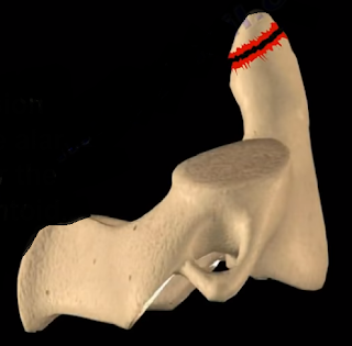
Type I fractures are a stable avulsion fracture of the alar ligament near the tip of the odontoid. A soft collar can be used to treat Type I fractures. Be aware of significant ligamentous injuries. Type II fractures are at the base of the odontoid process. Type II are the most common and are troublesome. The nonunion rate is about 20-80% due to interruption of the blood supply. The risk factors of nonunion include if the patient is over the age of 60 years old, if the patient has more than 6mm of displacement, smoking and diabetes, and you are unable to achieve reduction. In posterior displacement, extension injury (rare type) the anterior displacement is more common (flexion injury). Delay in treatment also increases the rate of nonunion. For treatment of young patients with no nonunion risks use a halo. The patient is younger than 60 years. The fracture is minimally displaced. Initial dens displacement is less than 6 mm, and the reduction is within one week of the injury. Healing will occur in the majority of cases.
If the patient has a nonunion risk, or when reduction of the fracture cannot be achieved or maintained, then we need to think about surgery and the fracture pattern. When the fracture pattern allows, you can put an anterior screw into the odontoid (to preserve the motion of C1/C2). Odontoid screw is used in younger patients instead of fusion to avoid loss of 50% of the neck rotation). Do not use the anterior screw fixation in patients with osteoporosis, in older patients, or in patients with a short neck. Another scenario is, if the patient has nonunion risks but the fracture pattern does not allow you to place an anterior odontoid screw, then you are going to fuse C1 to C2 (this will lose 50% of neck rotation). In general, C1/C2 fusion is used in cases of nonunion or it is used in cases of displaced fracture in the older patient and it can also be used if there is a failure of treatment with a halo. C1/C2 fusion can also be used if the fracture is comminuted and unstable. Posterior C1/C2 fusion can be done with different screw or wire constructs. A vascular watershed area exists between the apex of the odontoid, which is supplied by branches of the internal carotid artery and the base of the odontoid, which is supplied by branches of the vertebral artery. Type II fracture of the odontoid may get nonunion due to cortical bone and poor blood supply.
Type III fractures extend through the body of C2. This area is rich in blood supply and the fracture heals in the majority of cases. Treatment for Type III odontoid fractures includes external cervical orthosis (especially in the elderly patient) and a halo (if the fracture is displaced) (do not use in elderly patients). Odontoid fractures in the elderly can occur due to a simple fall and usually the diagnosis is missed. It is associated with increased complications and mortality. Do not use a halo in elderly patients. Use an external cervical orthosis of some sort. Fibrous union might be adequate if the fracture is not badly displaced, otherwise you will do fusion of C1/C2. For example, an 80 year old patient with osteoporosis, who is a smoker and has a displaced odontoid fracture that cannot be reduced, then this fracture will lead to nonunion and more complications. You need to do posterior C1/C2 arthrodesis. In general, if the elderly patient with an odontoid fracture is not a good surgical candidate, then you will give the patient a cervical orthosis. You can do the C1/C2 fusion by using transarticular screws, which you are not going to do if you have an aberrant vertebral artery. Another technique can be done for the fusion where fusion between C1/C2 is done with the screw placed into the C1 lateral mass and the C2 pedicle, plus a bone graft. There is increased survival for the elderly patient that undergoes surgery for Type II odontoid fracture. This may be a selection bias, because they have healthier patients who are physiologically active and young who are fit for surgery. The synchondrosis between the odontoid and the C2 body fuses by the age of 6 years.
Odontoid fracture in young children usually occurs by the age of 4 years. Physicians may confuse the synchondrosis with a fracture. The treatment of odontoid fracture in children is done with a Minerva brace or halo vest, if the fracture is displaced. You will use more pins and less torque. Finger tighten the pins. The Os Odontoideum looks like a fracture. It is oval shaped, it has sclerotic edges, and Os is smaller than the normal dens. The Os Odontoideum is a congenital process. The mechanism that causes the Os Odontoideum is unknown, but it is probably developmental or it can result from an old trauma.
Monday, February 18, 2019

Acute Low Back Pain Lumbar Disc Herniation


Acute Low Back Pain Lumbar Disc Herniation

Low back pain is a common condition. 90% of patients with low back pain will improve without surgery. Usually they get better with spontaneous resolution of the symptoms within 12 weeks. We usually advise the patient for early return to activity and function as the symptoms and the pain permits. The risk factors for development of low back pain are numerous, some include: vibration exposure, poor physical fitness, smoking and obesity, anxiety and depression, job dissatisfaction, or repetitive bending or “stooping” on the job. In summary, if the patient has no red flags and has a normal neurological exam, there is no reason to get early radiological studies. Getting early x-rays and early MRIs leads to a better patient satisfaction but does not give a better patient outcome. If there is no specific pain pattern, then there is no need for further workup. MRIs are good studies, but they give false positives. There is degeneration or a bulge of a disc in 35% of all asymptomatic subjects between 25-39 years of age. In patients 60 years old or older, the majority of the patients will have changes in the MRI. MRI abnormalities are common and must be correlated with the age and the clinical signs and symptoms of the patient. An MRI is good for diagnosing the lumbar disc herniation, which is sometimes called a ruptured disc, a slipped disc, or a herniated disc. The most common location of a disc herniation is a posterolateral herniation involving one nerve root. A foramninal L4-L5 herniation occurs in about 8%-10% of the cases. It involves the exiting nerve. A central herniation involves multiple nerve roots. It predominantly causes low back pain more than leg pain. It may cause bladder and bowel symptoms. This type of disc herniation causes Cauda Equina Syndrome which needs urgent diagnosis and surgical treatment. Clinical evaluation for a herniated disc examines sensory and motor reflexes. The Straight Leg Raising Test is the most important finding. It can be done in either the sitting or supine position. The test is positive as indicated by pain in the leg when the patient’s leg is raised to flex the hip with the knee extended. A positive straight leg test means a tension sign, something is putting tension or stress on the sciatic nerve. When the test is positive, it indicates possible disc herniation.
Treatment is typically non-operative. First, reassure the patient. Let the patient take some rest (no more than a few days), give the patient anti-inflammatory medication, and instruct them to attend physical therapy. Indications for surgery include progressive neurological deficits, Cauda Equina Syndrome, the patient is not getting better with time and treatment or if the symptoms are not getting better with conservative treatment, or the patient has a positive tension sign with persistent sever pain. Patients with sciatica and positive tension signs or patients with positive neurological findings on clinical exam with positive MRI findings make ideal surgical candidates. Surgery results in relief of leg pain in the majority of patients. Back pain may persist in some patients. Surgery results in neurological improvement, 50 % motor and sensory and 25% reflexes. In patients with discogenic back pain, they may need fusion which is a major procedure.The worst pressure on the disc occurs with prolonged sitting and bending over. This is the position that produces the highest pressure on the disc. If a patient has back pain but no radiation, by the patient’s history or physical examination and there are no red flags, then there is no reason to get x-rays or MRI early in the treatment of the patient. Red flags include a history of trauma, a tumor, infection, or Cauda Equina Syndrome symptoms. To rule out a history of trauma you should rule out fractures with x-rays, MRI, or CT scans. Tumors are a risk if the patient is older than 50 years old, if the patient had weight loss, or if the patient has pain at rest or at night. An infection may be present if the patient has fever and chills, if the patient has a history of diabetes, or if the patient has a history of IV drug abuse. Cauda Equina Symptoms may be present if the patient has back pain more than leg pain or if the patient also has bladder and bowel symptoms. Cauda Equina Syndrome needs to be diagnosed and surgically treated early. An MRI needs to be ordered urgently in the course of treatment. The MRI should be ordered STAT. There may need to be a wet read; a wet read is an early preliminary read of the radiographs. A wet read needs to be communicated with the physician and can be done while the patient is still on the table of the MRI.

Monday, February 4, 2019

Cervical Radiculopathy


Cervical Radiculopathy

Cervical radiculopathy is caused by cervical nerve root compression. The patient will have pain and/or progressive neurological deficit that results from conditions such as disc herniation that irritates a nerve in the cervical spine. Cervical radiculopathy is an irritation of the cervical nerve root. Cervical spine and shoulder problems overlap. The condition is of cervical spine etiology if the patient’s symptoms are relieved by shoulder abduction, by placing the hand over the head. The relief of the symptoms occurs due to decreased tension on the nerve roots. In cervical disc problems, be aware of false positive MRIs especially if the patient is above the age of 40 years old. Nerve conduction studies are not useful; they have a high false negative rate. EMG and nerve studies may differentiate radiculopathy from peripheral nerve entrapment. Cervical disc problems usually affect the lower numbered nerve root.
When you see the middle finger numbness, then this is C7. When compression of the C7 nerve root, there will be middle finger numbness, triceps weakness, and the triceps reflex will be affected. The cervical nerve roots are horizontal in orientation. It does not matter if cervical disc herniation is central or foraminal, it will compress the same nerve root. C7 nerve root runs above the pedicle of the C7 vertebra. C5-C6 is the most commonly affected disc and that will compress the C6 nerve root. The patient will come to the doctor with unilateral arm pain that is relieved by arm elevation. The numbness and paresthesia will occur in specific dermatomes. The patient may also have upper trapezius pain or interscapular pain. The patient may complain of occipital headache. When you examine the patient, do provocative tests such as the spurling’s test and the shoulder abduction test. The Spurling’s test is done by extending and rotating the neck towards the involved side. It reproduces the symptoms by narrowing the neuroforamen. The Spurling’s test differentiates cervical radiculopathy from peripheral nerve entrapment. Lifting the arm above the head relieves the symptoms if the cervical nerve roots are irritated. The Shoulder Abduction test differentiates cervical pathology from other causes of painful shoulder etiology. Make sure that you do not have a double crush syndrome, one in the neck and one in the peripheral nerve. Make sure that you differentiate radiculopathy from myelopathy. Make sure that you exclude a coexisting myelopathy. Examine the patient for upper motor neuron signs or cervical
myelopathy. Test the patient for gait instability. Test the patient for Hoffman’s sign. Test the patient for Babinski reflex. Test the patient for ankle Clonus. Check to see if the patient has hyperflexia in the upper and lower extremities (triceps/quadriceps). Even if there is a bad cervical spine disc problem on the MRI, treat it conservatively for about 3 months. Give the patient therapy and nonsteroidal anti-inflammatory medication (NSAIDS). 75% of the patients will improve with nonoperative treatment. Cervical radiculopathy is generally treated nonoperatively, in contrast to cervical myelopathy. Do surgery when there is persistent, severe pain for 6-12 weeks and/or progressive neurological deficit such as weakness or numbness. The procedure to treat cervical radiculopathy surgically is usually done anteriorly with direct removal of the lesion that causes the radiculopathy such as a herniated disc or spurs. When you place the anterior bone graft or the allograft in the disc space, you open the nueroforamen, and that will indirectly relieve the nerve. Then you will add the anterior plate. Some surgeons prefer to do a posterior approach.

Monday, January 14, 2019

Lumbar Spinal Stenosis


Lumbar Spinal Stenosis

Lumbar spinal stenosis is a narrowing of the spinal canal and narrowing of the intervertebral foramen (nerve root canal).
There are two types of lumbar spinal stenosis- central and lateral. Hypertrophy of the facet joints, hypertrophy of the ligamentum flavum, disc degeneration, or arthritis are all examples of conditions which constrict the nerve root canals causing compression of the spinal nerves and sciatica. Patients will have back pain that is better with flexion, or leaning forward like over a grocery cart. The pain will be worse with extension of the back. Leaning forward increases the foramen size by about 12%. Leaning backwards reduces the foramen size by about 20%. Neurological exam is normal in about 50% of the patients.
Central canal stenosis is responsible for giving neurogenic claudication. Patients may have leg pain, back pain, buttock pain, weakness, cramps of the calf, and a heavy sensation. Patients will exhibit grocery cart sign (flexion of the back). The patient history is key for making the diagnosis of spinal stenosis. Lateral recess stenosis will give radicular symptoms. It can occur in the nerve root canal. Neural foraminal stenosis occurs in the intervertebral foramen. Physicians should look for other conditions such as hip problems, metastatic tumors, or vascular conditions. You should always examine the pulses. Neurogenic claudication and vascular claudication may coexist. Walking is bad for both neurogenic and vascular claudication. Sitting will relieve the symptoms in both neurogenic and vascular claudication. Stopping and standing is good for the vascular claudication but still causes symptoms for lumbar spinal stenosis. Using a stationary bicycle will relieve symptoms of lumbar spinal stenosis, however it will aggravate the symptoms in vascular claudication. In vascular claudication, pain starts within the calf and leg. In neurogenic claudication, pain starts proximally and then spreads distally. It seems like postural changes of the spine will make the neurogenic claudication worse, however this will not affect the vascular claudication. Vascular claudication will be affected by muscle movement or muscle function, such as walking of riding a bicycle. In neurogenic claudication, leaning over while riding the bicycle will relieve the symptoms in the same way as the shopping cart sign. Spinal stenosis can be treated operatively. In central canal stenosis, you should do a decompression by laminectomy. In lateral recess stenosis, you should do a medial facetectomy. You should add fusion for instability or if more than 50% of the bilateral facets are removed. You should look at the x-rays or the MRI. If there is a slip of the vertebrae, do a fusion in addition to the laminectomy. The risk of pseudoarthrosis is increased 500% by smoking.
Depression and other comorbidities can affect the outcome. In two years, patients who are treated with surgery are better in pain and function than the patient who is treated conservatively. The most common reason for failed surgery is recurrence of the disease (residual foraminal stenosis). Walking is bad without the aid of a shopping cart. Leaning over the shopping cart will relieve the symptoms. If you have a patient with lower back pain and gait disturbance (hyperflexia), then you have an upper motor neuron lesion. Think about the cervical spine. You need to get an MRI of the cervical spine after you examine the patient. Think of cervical spine myelopathy because lumbar stenosis does not give these findings. Patient with spinal stenosis, spondylolisthesis, or facet disease will have pain with extension of the lumbar spine. Pain with lumbar spine flexion will suggest a disc related disorder.

Monday, January 7, 2019

Ankylosing Spondylitis- An Overview


Ankylosing Spondylitis- An Overview

Ankylosing Spondylitis is an inflammatory condition that affects young adults, occurs more in males, and affects the spine, sacroiliac joints, and large joints (ex. Hip). Ankylosing means “rigid” or fusion.
Spondy means “spine”. Spondylitis is inflammation of the spine. The patient may have inflammation followed by fusion of the spine and the sacroiliac joints. Other large joints (ex. Hip) may be affected, so the patient may complain of morning stiffness, low back pain, and maybe hip pain. The pain associated with Ankylosing Spondylitis gets better with exercises and not with rest. There is a difference between Reheumatoid arthritis and Ankylosing Spondylitis. Rheumatoid arthritis affects the synovial lining of joints and affects predominantly the cervical spine. Ankylosing Spondylitis affects ligaments, tendons, discs, and some joints, but it will affect the entire axial spine. Ankylosing Spondylitis is part of the seronegative spondyloarthropathy. This means that the rheumatoid factor is negative. Although the rheumatoid factor is negative, the HLA-B27 is positive. Ankylosing Spondylitis is a systemic problem that involves the immune system. It is almost like rheumatoid arthritis, but with a negative rheumatoid factor. Risk Factors of Ankylosing Spondylitis would include a young male with a positive family history + HLA-B27 gene positive. The HLA-B27 is part of the immune system. It is an antigen that will be on the surface of the cell. HLA-B27 probably has the same amino acid sequence as the protein produced by bacteria (klebsiella pneumonia), by food, or by other things. When the immune system identifies this protein and it goes through the blood stream, then these T-cells can recognize that antigen that protein is on the surface of cells (HLA-B27). Then the T-cells recruit other cells to attack it. Everything that contains HLA-B27 (tendons, ligaments, joints, etc) will be attacked because they think it is a bad protein.
The protein produced by the bacteria for example or by the HLA-B27 have the same sequence and the immune system cannot tell the difference between both of them so it is an autoimmune disease. These patient will have fusion of the spine. The spine will not have any free movement. The patient will complain of gradual stiffening of the spine and limited chest wall expansion. Less than 2 cm of chest wall expansion is more diagnostic than the HLA-B27 blood test. Ankylosing Spondylitis is a difficult condition to diagnose and there will be a “Bamboo Spine” seen on the x-ray. There will be sacroiliac joint involvement which is a characteristic for Ankylosing Spondylitis. There will also be fusion of the SI joint. There may be systemic autoimmune disease that will cause fever and malaise. There will be uveitis (redness and inflammation of the eye). There will be aorta inflammation that may lead to aortic aneurysm if the aorta is dilated or aortic regurgitation. The patient may have depression. When you do the blood test, it will be HLA-B27 positive. The sedimentation rate and CRP could be high. Treatment of Ankylosing Spondylitis uses things that decrease inflammation such as anti-inflammatory medications, physical therapy to improve flexibility and strength of the spine and joints, or TNF alpha blocking agents.

Monday, December 3, 2018

DISH Diffuse Idiopathic Skeletal Hyperostosis


DISH Diffuse Idiopathic Skeletal Hyperostosis

The DISH has flowing ossification along anterolateral aspect of at least four continuous vertebrae. When you look at the x-ray, you find ossification along the anterior aspect of the body but separate from the vertebrae and the disc height is preserved. It occurs in older patients (50 years and above). It affects all of the spine (more in the thoracic spine), especially on the right side, which is typical of DISH. The syndesmophytes are equal on the right and left sides in the lumbar and cervical vertebrae.

There is no involvement of the discs and there is no facet fusion or sacroiliac joint involvement. The patient may have other comorbidities such as gout or diabetes, and you need to get the hemoglobin A1c (HbA1c test) in these patients. Some patients may have high cholesterol levels. The patient will complain of back pain and spinal stiffness. DISH will have large syndesmophytes, and if the condition occurs in the neck, it will cause dysphagia, hoarseness of the voice, and sleep apnea. Diagnosis can be established by x-ray of the spine. On lateral x-ray of the cervical spine, you will find anterior bony fragments and the discs are preserved. The fractures in the spine are usually due to a hyperextension injury and can be occult, resulting from minor trauma and may have major instability. There is an increased mortality in c-spine trauma in DISH, high mortality especially in non-operative treatment. If the patient has a history of sudden neck or back pain, then the patient will be assumed to have an occult fracture, so try to get a CT scan or an MRI even if the pain is minimal and even if the x-rays appear normal. Heterotopic ossification after total hip arthroplasty is more in patients with DISH.

What is the difference between DISH and Ankylosing Spondylitis?


DISH


-Flowing large syndesmophytes

-No bamboo spine
-Sacroiliac (SI) join will not be involved
-Occurs in older patients
-some patients may have diabetes, check hemoglobin A1c

Ankylosing Spondylitis

-Diffuse ossification of the disc space without large osteophytes
-Bamboo spine
-The patient is usually young
-Sacroiliac (SI) joint is involved
-HLA-B27 is positive in about 90% of the time
-Limited chest expansion


Tuesday, August 21, 2018

Toe Deformities


Deformities of the toes are not uncommon and can occur from muscle imbalance, or other causes such as rheumatoid arthritis, diabetes, compartment syndrome, synovitis, or neurological disorders. Hammer toe occurs as flexion of the proximal interphalangeal (PIP) joint. Hammer toe is similar to the Boutonniere deformity of the finger. Claw Toe is a hyperextension deformity of the MTP joint and flexion of the PIP and DIP, resembling a pirate hook. Claw toe is similar to an intrinsic minus deformity of the hand, or “claw hand”. Mallet toe is similar in appearance to mallet finger, and is a flexion deformity of the DIP joint.

Tuesday, July 24, 2018

Lumbosacral Plexus


The sciatic nerve is the key nerve of the lumbosacral plexus. It arises from the spinal nerves of L4 through S3. The sciatic nerve is the cornerstone of the lumbosacral plexus. Knowledge of the lumbosacral plexus starts with the sciatic nerve because it is the most important nerve. The lumbosacral plexus is comprised of the sciatic nerve and S4. The branches of the lumbosacral plexus are the superior gluteal nerve (L4-S1), the inferior gluteal nerve (L5-S2), the posterior cutaneous nerve of the thigh (S1-S3), and the pudendal nerve (S2-S4) (SIPP).