Monday, January 28, 2019

Elbow Dislocation in Adults


Elbow Dislocation in Adults

With elbow dislocations, recognize the terrible triad: elbow dislocation, radial head fracture, and coronoid fracture. The terrible triad is not a simple elbow dislocation; it is a complex elbow dislocation. In addition to these three injuries of the elbow, there is always a tear of the lateral ulnar collateral ligament. The treatment usually is reduction and splinting of the elbow.
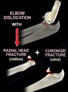
This cannot be the definitive treatment; it is the initial treatment. If no surgery is done, you will have recurrent dislocation of the elbow. You need to do surgery for reduction and fixation of the fractures and also to restore the elbow stability. This injury is unstable. Simple reduction and splinting is not going to work for this injury. You have to recognize the terrible triad which means surgery. There are multiple types of elbow dislocation based on the position of the olecranon relative to the humerus. The most common type of elbow dislocation is the posterolateral type. There are two basic types of elbow dislocations: simple and complex. Simple elbow dislocations have no fracture seen, and are usually a ligamentous injury. Complex elbow dislocations have associated fractures in addition to the ligamentous injury. With any elbow dislocation, you need to check the shoulder and the wrist for injuries and fractures because it can occur in up to 15%. When you have a simple dislocation of the elbow, you need to reduce it and then check the range of stability of the elbow. If you find that the elbow is stable with range of motion, then you will do a short period of immobilization with a posterior splint for approximately one week with the elbow in about 90 degrees of flexion. Then start active range of motion of the elbow. Recurrence of the dislocation is rare (less than 1%). If you keep the elbow immobilized more than 3 weeks, there will be severe stiffness of the elbow. Surgery should be done if the dislocation is irreducible, if there is associated fracture, or if you are unable to maintain stability of the elbow. After immobilization and early range of motion of the elbow, you will see the patient and do follow up x-rays to check joint congruity and to make sure that the elbow reduction is maintained. To treat the terrible triad, you should initially do a closed reduction. Open reduction and internal fixation of the coronoid (if possible), of the radial head or excise the radial head with radial head arthroplasty if the radial head is unreconstructable. In addition, you will do lateral ulnar collateral ligament (LUCL) repair. Never excise the radial head alone in this situation. For an elbow dislocation with olecranon fracture, do open reduction and plate fixation. K-wires and tension band is not strong enough to hold the fracture and stabilize the elbow at the same time. For an elbow dislocation with a radial head fracture, do fixation or replacement of the radial head (never do excision of the radial head alone in this situation). The LUCL is the most important lesion in recurrence or persistence of instability of the elbow following simple elbow dislocation.
The injury progresses from lateral to medial. The lateral collateral ligament fails first, and it avulses proximally at the lateral epicondyle. The medial collateral ligament (MCL) fails last. In varus posteromedial rotary instability, there is an elbow injury plus LUCL tear, plus coronoid fracture which involves the medial facet of the coronoid. Treatment for chronic dislocation is open reduction capsular releases with hinge external fixation and early range of motion. Loss of terminal extension is a complication of elbow dislocation. Usually for decreased range of motion of the elbow, you will do static progressive splinting between 6-10 weeks. No manipulation of the elbow is done, which is different from the knee after total knee replacement, where you can do manipulation up to three months. With heterotopic ossification, do excision. Remove the myositis and excise the posterior part of the MCL to allow more flexion. To be functional, the range of motion of the elbow should be between 30-130 degrees. Some physicians suggest that if the flexion is less than 100, you will do release of the posterior bundle of the MCL in addition to release of the ulnar nerve. If you want more flexion of the elbow, excise the posterior part of the MCL.

Monday, January 21, 2019

Patellar Fractures


Patellar Fractures

Patellar fractures can involve different topic, and I am going to try and highlight the important points related to patellar fractures. The medial patellofemoral ligament is the primary stabilizer of the patella, so when the patella dislocates, you will have an injury to that ligament and also an injury to the medial patellar facet articulation cartilage or an osteochondral fragment. In addition to the medial patellar facet injury, you will get a lateral femoral condyle injury. Bipartite patella occurs in about 8% of the population. It could be bilateral in about 50%, and it usually occurs in the superolateral aspect of the patella. You should observe it and not fix it. It is not an acute fracture that may need excision or lateral retinacular release. It can occur in children between 8-10 years old. It is a rare condition. The patient will be unable to do straight leg raising, so you suspect that the extensor mechanism is injured. The patient may have a high riding patella on x-ray with a palpable gap when you examine the patient. The x-ray may show small flecks of bone as the patellar tendon avulses with a portion of the distal pole of the patella. Sometimes the bony injury is so small that the condition can be missed. You should have a high index of suspicion. You may need to get an MRI to confirm diagnosis. The treatment is usually ORIF if the fracture is displaced.
The patella is a large sesamoid bone. The quadriceps muscle is inserted at the proximal pole and the distal pole gives attachment to the patellar tendon. The patella is triangular in shape. The proximal 3/4 of the patella is covered with cartilage, however the distal 25% of the patella is not covered with cartilage. The patella increases the power of the extensor mechanism by about 50% because it displaces the extensor mechanism anteriorly, and that will increase the moment arm.
Transverse fractures of the patella can be non-displaced or displaced. The patella can be pulled apart by the attached quadriceps tendon. The patient will be unable to do active extension of the knee. Upper or lower pole fractures of the patella are fractures at the site of attachment of the patellar tendon. Comminuted fractures of the patella can be non-displaced or displaced. Comminuted fractures have multiple pieces, are very unstable, and are difficult to fix. Vertical fractures of the patella are the most common, and they are stable and nondisplaced. Osteochondral fractures are small fractures of the patella usually associated with acute dislocation of the patella.
In examination, you may feel a palpable gap. The area of the knee is usually swollen. The patient will be unable to do straight leg raise. The lateral view of the knee is the best view to see the fracture. 2-3 mm of displacement will probably mean that the patient will need surgery.
If you think that the patient’s extensor mechanism is intact, and the patient is able to do straight leg raise, and the fracture is nondisplaced or minimally displaced, it is usually a transverse fracture in this situation, then immobilize the knee straight in a hinged knee brace for 4-6 weeks with weight bearing as tolerated. Sometimes the patient cannot move the knee because of the pan and injection of lidocaine inside the knee can help to assess the integrity of the extensor mechanism. If the patient has a total knee with 2mm displacement of the patella, and the extensor mechanism is intact, then the patient will be treated conservatively in a brace or in a knee immobilizer (no surgery).
Indication for surgery is a displaced patellar fracture and the inability to do straight leg raising.
First, preserve the patella (if possible). The tension band fixation technique is the gold standard for the treatment of displaced patellar fractures (the fracture is usually a transverse fracture), and the tension band technique is the one that gives us the most complications. The first step in the tension band technique is to reduce the fracture with reduction clamps. Next, at least two K-wires are placed across the fracture. An anterior tension band is applied, organized in a Figure-8 pattern. You need to put the Figure-8 tension band wire close to the patella superiorly and not far away from the patella because that may cause construct instability and fracture displacement. A second wire may be placed circumferentially around the patella. Bending the K-wires from both ends may decrease migration of the wires and decrease the complications. The wire that is bent at both ends may be difficult to remove. Tension band fixation technique may be done with K-wires or also with cannulated screws (through the cannulated screws, you place the wires). It does not matter if you have an open or closed fracture, you treat it the same way. When you place K-wires, it means symptomatic hardware and thus a secondary reoperation. It was found that the longitudinal screws and the tension band wires are a more superior fixation. The tension band construct when performed correctly will provide absolute stability and will convert the tension forces from the muscle pull into compression forces at the articular surface. You want to have anatomic reduction and stable fixation; don’t judge the reduction by what you see at the surface of the fracture. Try to see and feel the joint if you can. Check the x-rays carefully. The surface of the patella may be well reduced, however, the joint may be distracted or displaced. If you tighten the cerclage wire aggressively, you may have a good looking surface, but you may have a distracted join. After you fix the patella, you will do a range of motion of the knee before closure and give the patient a hinged knee brace, locked into extension with weight-bearing as tolerated. Weight-bearing is controversial. Some people start weight-bearing early, and some people start weight-bearing after 4-6 weeks. A can may be helpful to the patient. You will begin active flexion at 2-3 weeks (patient will lie prone, flexing and extending the knee). When the patient is prone, it avoids active knee extension and avoids excessive stress on the fracture site. At 6 weeks, you can unlock the brace and start moving the knee, gradually increasing the flexion.
If the patellar fracture is comminuted, you can use the peripatellar circumferential wire loop fixation, which is commonly used as an addition to other methods of fixation. You can also use a plate fixation utilizing a low profile implant and providing stable fixation. This technique is becoming more popular.
You can also excise the patella partially or completely. In a partial patellectomy, the distal pole is extra-articular, and if it is severely comminuted and less than 40% of the patella, then you can excise it (in general, you would like to preserve the patella). If you can’t preserve the patella and ORIF is not possible, then do partial patellectomy and preserve the largest piece. Partial patellectomy may be necessary, but open reduction and internal fixation (if possible) is associated with a better outcome. You will do the partial patellectomy in several comminuted inferior pole fractures. You will do medial and lateral retinacular repair, and a poor outcome may occur with removal of more than 40% of the patella. Total patellectomy will be done when the fractured patella cannot be fixed. Total patellectomy can cause extensor lag and loss of the extensor strength. The quadriceps torque is reduced by about 50%.
Symptomatic hardware and knee pain is the most common complication after patellar fracture fixation, especially if you use the tension band technique. It requires implant removal in about 50% of the time. This complication will include the hardware migration. Failure after patellar fracture fixation occurs in about 20% of the time due to increasing age, fixation with wires, technical errors and noncompliance.

Monday, January 14, 2019

Lumbar Spinal Stenosis


Lumbar Spinal Stenosis

Lumbar spinal stenosis is a narrowing of the spinal canal and narrowing of the intervertebral foramen (nerve root canal).
There are two types of lumbar spinal stenosis- central and lateral. Hypertrophy of the facet joints, hypertrophy of the ligamentum flavum, disc degeneration, or arthritis are all examples of conditions which constrict the nerve root canals causing compression of the spinal nerves and sciatica. Patients will have back pain that is better with flexion, or leaning forward like over a grocery cart. The pain will be worse with extension of the back. Leaning forward increases the foramen size by about 12%. Leaning backwards reduces the foramen size by about 20%. Neurological exam is normal in about 50% of the patients.
Central canal stenosis is responsible for giving neurogenic claudication. Patients may have leg pain, back pain, buttock pain, weakness, cramps of the calf, and a heavy sensation. Patients will exhibit grocery cart sign (flexion of the back). The patient history is key for making the diagnosis of spinal stenosis. Lateral recess stenosis will give radicular symptoms. It can occur in the nerve root canal. Neural foraminal stenosis occurs in the intervertebral foramen. Physicians should look for other conditions such as hip problems, metastatic tumors, or vascular conditions. You should always examine the pulses. Neurogenic claudication and vascular claudication may coexist. Walking is bad for both neurogenic and vascular claudication. Sitting will relieve the symptoms in both neurogenic and vascular claudication. Stopping and standing is good for the vascular claudication but still causes symptoms for lumbar spinal stenosis. Using a stationary bicycle will relieve symptoms of lumbar spinal stenosis, however it will aggravate the symptoms in vascular claudication. In vascular claudication, pain starts within the calf and leg. In neurogenic claudication, pain starts proximally and then spreads distally. It seems like postural changes of the spine will make the neurogenic claudication worse, however this will not affect the vascular claudication. Vascular claudication will be affected by muscle movement or muscle function, such as walking of riding a bicycle. In neurogenic claudication, leaning over while riding the bicycle will relieve the symptoms in the same way as the shopping cart sign. Spinal stenosis can be treated operatively. In central canal stenosis, you should do a decompression by laminectomy. In lateral recess stenosis, you should do a medial facetectomy. You should add fusion for instability or if more than 50% of the bilateral facets are removed. You should look at the x-rays or the MRI. If there is a slip of the vertebrae, do a fusion in addition to the laminectomy. The risk of pseudoarthrosis is increased 500% by smoking.
Depression and other comorbidities can affect the outcome. In two years, patients who are treated with surgery are better in pain and function than the patient who is treated conservatively. The most common reason for failed surgery is recurrence of the disease (residual foraminal stenosis). Walking is bad without the aid of a shopping cart. Leaning over the shopping cart will relieve the symptoms. If you have a patient with lower back pain and gait disturbance (hyperflexia), then you have an upper motor neuron lesion. Think about the cervical spine. You need to get an MRI of the cervical spine after you examine the patient. Think of cervical spine myelopathy because lumbar stenosis does not give these findings. Patient with spinal stenosis, spondylolisthesis, or facet disease will have pain with extension of the lumbar spine. Pain with lumbar spine flexion will suggest a disc related disorder.

Monday, January 7, 2019

Ankylosing Spondylitis- An Overview


Ankylosing Spondylitis- An Overview

Ankylosing Spondylitis is an inflammatory condition that affects young adults, occurs more in males, and affects the spine, sacroiliac joints, and large joints (ex. Hip). Ankylosing means “rigid” or fusion.
Spondy means “spine”. Spondylitis is inflammation of the spine. The patient may have inflammation followed by fusion of the spine and the sacroiliac joints. Other large joints (ex. Hip) may be affected, so the patient may complain of morning stiffness, low back pain, and maybe hip pain. The pain associated with Ankylosing Spondylitis gets better with exercises and not with rest. There is a difference between Reheumatoid arthritis and Ankylosing Spondylitis. Rheumatoid arthritis affects the synovial lining of joints and affects predominantly the cervical spine. Ankylosing Spondylitis affects ligaments, tendons, discs, and some joints, but it will affect the entire axial spine. Ankylosing Spondylitis is part of the seronegative spondyloarthropathy. This means that the rheumatoid factor is negative. Although the rheumatoid factor is negative, the HLA-B27 is positive. Ankylosing Spondylitis is a systemic problem that involves the immune system. It is almost like rheumatoid arthritis, but with a negative rheumatoid factor. Risk Factors of Ankylosing Spondylitis would include a young male with a positive family history + HLA-B27 gene positive. The HLA-B27 is part of the immune system. It is an antigen that will be on the surface of the cell. HLA-B27 probably has the same amino acid sequence as the protein produced by bacteria (klebsiella pneumonia), by food, or by other things. When the immune system identifies this protein and it goes through the blood stream, then these T-cells can recognize that antigen that protein is on the surface of cells (HLA-B27). Then the T-cells recruit other cells to attack it. Everything that contains HLA-B27 (tendons, ligaments, joints, etc) will be attacked because they think it is a bad protein.
The protein produced by the bacteria for example or by the HLA-B27 have the same sequence and the immune system cannot tell the difference between both of them so it is an autoimmune disease. These patient will have fusion of the spine. The spine will not have any free movement. The patient will complain of gradual stiffening of the spine and limited chest wall expansion. Less than 2 cm of chest wall expansion is more diagnostic than the HLA-B27 blood test. Ankylosing Spondylitis is a difficult condition to diagnose and there will be a “Bamboo Spine” seen on the x-ray. There will be sacroiliac joint involvement which is a characteristic for Ankylosing Spondylitis. There will also be fusion of the SI joint. There may be systemic autoimmune disease that will cause fever and malaise. There will be uveitis (redness and inflammation of the eye). There will be aorta inflammation that may lead to aortic aneurysm if the aorta is dilated or aortic regurgitation. The patient may have depression. When you do the blood test, it will be HLA-B27 positive. The sedimentation rate and CRP could be high. Treatment of Ankylosing Spondylitis uses things that decrease inflammation such as anti-inflammatory medications, physical therapy to improve flexibility and strength of the spine and joints, or TNF alpha blocking agents.