Showing posts with label knee pain. Show all posts

Monday, January 21, 2019

Patellar Fractures


Patellar Fractures

Patellar fractures can involve different topic, and I am going to try and highlight the important points related to patellar fractures. The medial patellofemoral ligament is the primary stabilizer of the patella, so when the patella dislocates, you will have an injury to that ligament and also an injury to the medial patellar facet articulation cartilage or an osteochondral fragment. In addition to the medial patellar facet injury, you will get a lateral femoral condyle injury. Bipartite patella occurs in about 8% of the population. It could be bilateral in about 50%, and it usually occurs in the superolateral aspect of the patella. You should observe it and not fix it. It is not an acute fracture that may need excision or lateral retinacular release. It can occur in children between 8-10 years old. It is a rare condition. The patient will be unable to do straight leg raising, so you suspect that the extensor mechanism is injured. The patient may have a high riding patella on x-ray with a palpable gap when you examine the patient. The x-ray may show small flecks of bone as the patellar tendon avulses with a portion of the distal pole of the patella. Sometimes the bony injury is so small that the condition can be missed. You should have a high index of suspicion. You may need to get an MRI to confirm diagnosis. The treatment is usually ORIF if the fracture is displaced.
The patella is a large sesamoid bone. The quadriceps muscle is inserted at the proximal pole and the distal pole gives attachment to the patellar tendon. The patella is triangular in shape. The proximal 3/4 of the patella is covered with cartilage, however the distal 25% of the patella is not covered with cartilage. The patella increases the power of the extensor mechanism by about 50% because it displaces the extensor mechanism anteriorly, and that will increase the moment arm.
Transverse fractures of the patella can be non-displaced or displaced. The patella can be pulled apart by the attached quadriceps tendon. The patient will be unable to do active extension of the knee. Upper or lower pole fractures of the patella are fractures at the site of attachment of the patellar tendon. Comminuted fractures of the patella can be non-displaced or displaced. Comminuted fractures have multiple pieces, are very unstable, and are difficult to fix. Vertical fractures of the patella are the most common, and they are stable and nondisplaced. Osteochondral fractures are small fractures of the patella usually associated with acute dislocation of the patella.
In examination, you may feel a palpable gap. The area of the knee is usually swollen. The patient will be unable to do straight leg raise. The lateral view of the knee is the best view to see the fracture. 2-3 mm of displacement will probably mean that the patient will need surgery.
If you think that the patient’s extensor mechanism is intact, and the patient is able to do straight leg raise, and the fracture is nondisplaced or minimally displaced, it is usually a transverse fracture in this situation, then immobilize the knee straight in a hinged knee brace for 4-6 weeks with weight bearing as tolerated. Sometimes the patient cannot move the knee because of the pan and injection of lidocaine inside the knee can help to assess the integrity of the extensor mechanism. If the patient has a total knee with 2mm displacement of the patella, and the extensor mechanism is intact, then the patient will be treated conservatively in a brace or in a knee immobilizer (no surgery).
Indication for surgery is a displaced patellar fracture and the inability to do straight leg raising.
First, preserve the patella (if possible). The tension band fixation technique is the gold standard for the treatment of displaced patellar fractures (the fracture is usually a transverse fracture), and the tension band technique is the one that gives us the most complications. The first step in the tension band technique is to reduce the fracture with reduction clamps. Next, at least two K-wires are placed across the fracture. An anterior tension band is applied, organized in a Figure-8 pattern. You need to put the Figure-8 tension band wire close to the patella superiorly and not far away from the patella because that may cause construct instability and fracture displacement. A second wire may be placed circumferentially around the patella. Bending the K-wires from both ends may decrease migration of the wires and decrease the complications. The wire that is bent at both ends may be difficult to remove. Tension band fixation technique may be done with K-wires or also with cannulated screws (through the cannulated screws, you place the wires). It does not matter if you have an open or closed fracture, you treat it the same way. When you place K-wires, it means symptomatic hardware and thus a secondary reoperation. It was found that the longitudinal screws and the tension band wires are a more superior fixation. The tension band construct when performed correctly will provide absolute stability and will convert the tension forces from the muscle pull into compression forces at the articular surface. You want to have anatomic reduction and stable fixation; don’t judge the reduction by what you see at the surface of the fracture. Try to see and feel the joint if you can. Check the x-rays carefully. The surface of the patella may be well reduced, however, the joint may be distracted or displaced. If you tighten the cerclage wire aggressively, you may have a good looking surface, but you may have a distracted join. After you fix the patella, you will do a range of motion of the knee before closure and give the patient a hinged knee brace, locked into extension with weight-bearing as tolerated. Weight-bearing is controversial. Some people start weight-bearing early, and some people start weight-bearing after 4-6 weeks. A can may be helpful to the patient. You will begin active flexion at 2-3 weeks (patient will lie prone, flexing and extending the knee). When the patient is prone, it avoids active knee extension and avoids excessive stress on the fracture site. At 6 weeks, you can unlock the brace and start moving the knee, gradually increasing the flexion.
If the patellar fracture is comminuted, you can use the peripatellar circumferential wire loop fixation, which is commonly used as an addition to other methods of fixation. You can also use a plate fixation utilizing a low profile implant and providing stable fixation. This technique is becoming more popular.
You can also excise the patella partially or completely. In a partial patellectomy, the distal pole is extra-articular, and if it is severely comminuted and less than 40% of the patella, then you can excise it (in general, you would like to preserve the patella). If you can’t preserve the patella and ORIF is not possible, then do partial patellectomy and preserve the largest piece. Partial patellectomy may be necessary, but open reduction and internal fixation (if possible) is associated with a better outcome. You will do the partial patellectomy in several comminuted inferior pole fractures. You will do medial and lateral retinacular repair, and a poor outcome may occur with removal of more than 40% of the patella. Total patellectomy will be done when the fractured patella cannot be fixed. Total patellectomy can cause extensor lag and loss of the extensor strength. The quadriceps torque is reduced by about 50%.
Symptomatic hardware and knee pain is the most common complication after patellar fracture fixation, especially if you use the tension band technique. It requires implant removal in about 50% of the time. This complication will include the hardware migration. Failure after patellar fracture fixation occurs in about 20% of the time due to increasing age, fixation with wires, technical errors and noncompliance.

Wednesday, August 8, 2018

Discoid Meniscus


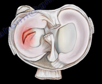
The meniscus is a cushion structure made of cartilage which fits within the knee joint between the tibia and the femur. The medial meniscus is C-shaped and the lateral meniscus in the more circular. The meniscus is made up of type I collagen that provides shock absorption and stability to the knee joint. The meniscus helps to protect the knee joint, allowing the bones to slide freely on each other. Discoid meniscus is a rare variation of the meniscus that usually affects the lateral meniscus of the knee in less than 5% of the population and could be bilateral in about 25% of the cases.
Discoid meniscus is a large meniscus with abnormal attachment causing increased mobility of the meniscus. It causes a pop, click, or snapping with locking and pain. There will be loss of full knee extension with tenderness on the lateral joint space. Symptoms occur more during extension of the knee. The discoid meniscus occurs due to the abnormal development and increase in size of the meniscus. An x-ray could show increased widening of the joint space. An MRI will show the “bow tie” sign in three or more sagittal continuous cuts. The coronal MRI will show a thick and flat meniscus extending beyond the halfway point of the condyle.


Watanabe Classification of Discoid Lateral Meniscus


  • Type I: Block-shaped stable
  • Type II: Block-shaped, stable, partial meniscus (has good peripheral attachment)
  • Type III: Unstable meniscus with stability arising only form the ligament of Wrisberg. (no posterior meniscal tibial attachment).

Treatment


An asymptomatic patient will be treated with observation. A symptomatic patient may receive a partial meniscectomy and saucerization with repair of type III (no posterior tibial meniscal attachment)

Tuesday, July 10, 2018

McMurray's Test- Meniscal Tear


Meniscal injuries are very common. The McMurray’s Test is a rotational maneuver of the knee that is frequently used to aid in the diagnosis of meniscal tears. With a meniscal tear, the patient usually complains of knee pain localized to the lateral or medial side of the knee joint. The patient will have locking, clicking, pain, or effusion.


During the physical examination, joint line tenderness is the most sensitive finding. Swelling of the knee and a possible extension lag (locked knee) is also a common finding. Pain at a higher level is usually associated with the medial collateral ligament. Pain at a lower level is usually associated with the pes anserine bursa.


What is the McMurrays test?             


The McMurray’s test is a knee examination test that provokes pain or a painful click as the knee is brought from flexion to extension with either internal or external rotation. The McMurray’s test uses the tibia to trap the meniscus between the femoral condyles of the femur and the tibia. When performing the test, the patient should be lying supine with the knee hyperflexed. The examiner then grasps the patient’s heel with one hand and places the other hand over the knee joint. To test the medial meniscus, the knee is fully flexed, and the examiner then passively externally rotates the tibia and places a valgus force. The knee is then extended in order to test the medial meniscus. To test the lateral meniscus, the examiner passively internally rotates the tibia and places a varus force. The knee is then extended in order to test the lateral meniscus. A positive test is indicated by pain, clicking or popping within the joint and may signal a tear of either the medial or lateral meniscus when the knee is brought from flexion to extension.


How reliable is the McMurray’s test?


There are mixed reviews for the validity of this test. An MRI is a very sensitive exam and makes the diagnosis easier, while excluding other associated injuries.


Tuesday, April 10, 2018

Pes Anserine Bursitis


Several bursa are seen around the knee area. These bursa include the suprapatellar, prepatellar, infrapatellar, and pes anserine. The pes answerine bursa is a small fluid filled sac located between the tibia and the three tendons of the Sartorius, Gracilis, and Semi-tendinosus.
These muscles are innervated by three separate nerves, the femoral, obturator, and the tibial branch of the sciatic nerve, respectively. Pes Anserine bursitis, or “breast stroke knee”, is an inflammatory condition of the medial knee at the pes anserine bursa that is common in swimmers.

What is the pes anserine?

The pes anserine is the common area of insertion for the three tendons along the proximal medial aspect of the tibia. This condition is also sometimes referred to as a “goosefoot” because the pes anserinus tendons resemble the shape of a goose foot. Pes Anserine bursitis is usually seen as causing pain, tenderness, and localized swelling after trauma or total knee replacement. The pain is seen below the joint line on the medial part of the proximal tibial with the bursa being deep to the tendons.


Treatment

Treatment consists of physical therapy, nonsteroidal anti-inflammatory medications, and injections. The physician will need to rule out meniscal tears, stress fractures, or osteonecrosis of the tibia, as these are all differential diagnosis.


Tuesday, March 6, 2018

Tibial Spine Fractures in Children


Tibial Spine fractures are similar to ACL injuries in adults. It occurs due to hyperextension of the knee and is commonly seen during falls from a bicycle. An injured child with a swollen knee and who has fallen from a bicycle should alert the clinician to the possibility of a tibial spine fracture. A meniscal injury may also occur, especially with the medial meniscus. The interposition of the meniscus or rotation of the fracture may prevent a closed reduction.
Tibial spine fractures are classified using the Meyers and McKeever Classification and are separated into three types. Type I classifications are nondisplaced, Type II fractures are identified as being minimally displaced with an intact posterior hinge, and Type III fractures are classified as being completely displaced.
The presentation and examination is similar to an ACL tear with immediate swelling as well as a positive Lachman’s Test or Anterior Drawer Test. An x-ray will show the fracture and a CT scan will help in planning for surgery. An MRI may be required to show a trapped or a meniscal injury.

Treatment


Treatment will consist of an aspiration of the large hematoma. Nonoperative treatment is used for Type I fractures and reducible Type II fractures; a closed reduction and immobilization in 0-20 of flexion. Surgery is performed in Type II fractures and unreducible Type II fractures. An ORIF or arthroscopic reduction and fixation will be performed. During surgery, the trapped meniscus will be moved out of the way and the surgeon will use sutures or screws for fixation. It is important for the surgeon to remember to avoid the physis.

Complication


ACL laxity is common but not clinically significant. Stiffness or arthrofibrosis occurs with surgical fixation. Growth arrest is rare.

Tuesday, January 2, 2018

Pivot Shift of the Knee—ACL tear


The anterior cruciate ligament is located at the front of the knee. Rupture of the anterior cruciate ligament (ACL) is a condition commonly seen in sports, usually due to a non-contact pivoting injury. The Pivot Shift test is a specific test for an ACL deficient knee (ACL injury). A pivot shift is pathognomonic for an ACL tear and is best demonstrated in a chronic setting. The Lachman’s test is the most sensitive examination test for an ACL injury.
The ACL keeps the tibia from sliding out in front of the femur and provides rotational stability to the knee. Rupture of the ACL causes anterolateral rotatory instability. The tibia moves anterolaterally in extension; however, when you flex the knee the IT band becomes a flexor of the knee and pulls back, reducing the tibia. The Pivot Shift Test goes from extension (tibia subluxed) to flexion, with the tibia reduced by the iliotibial band.
Both the Lachman’s test and the Pivot Shift test are associated with 20-30 degrees of knee flexion. The Lachman’s test starts at 20-30 degrees of flexion, but with the Pivot Shift test, you will feel the clunk at 20-30 degrees of flexion. Remember: 20-30 degrees of flexion is important for examination of the ACL. The femur is stabilized with one hand and the other hand pulls the tibia anteriorly and posteriorly against the femur. The tibia can be pulled forward more than normal (anterior translation). The examiner will have a sense of increased movement and lack of a solid end point.
When performing the Pivot Shift test, the patient should be totally relaxed and lying supine. The knee is in the subluxed position when in full extension. The pivot shift starts with extension of the knee and you can feel a “clunk” at 20-30 degrees of flexion. The physician will hold the knee in full extension, then add valgus force, and internal rotation of the tibia to increase the rotational instability of the knee. Then the physician will take the knee into flexion. A palpable clunk is very specific of an ACL tear. The iliotibial band will reduce the tibia and create the clunk on the outside of the knee. The physician should always compare the results with the other side.


The ACL prevents anterior translation of the tibia. It is a secondary restraint to tibial rotation and varus and valgus. The ACL consists of two bundles:

  • The Posterolateral Bundle
  • Anteromedial Bundle
The Posterolateral bundle prevents the pivot shift and contributes to rotational stability. This bundle also prevents internal rotation of the tibia with the knee in near extension (tight in extension, loose in flexion). If it is sectioned, it increases the anterior translation and tibial rotation at 30° of flexion. The Anteromedial bundle is tight in flexion and if sectioned, it increases the anterior translation at 90° of flexion.
The Lachman’s test is the most sensitive test, especially in acute settings. The examiner will find no end point with anterior translation of the knee and the physical examination can be difficult or limited due to pain. With the Pivot Shift test, the patient must be completely relaxed. The test is helpful in chronic situations, especially if the patient complains of the knee giving way.
During the Pivot Shift, the knee subluxes in extension and reduces at 20-30 degrees of flexion. The Pivot Shift correlates closely with patient satisfaction of their reconstructed knee. It is also a measure of functional instability following ACL reconstruction. Verticle femoral tunnel placement will cause rotational instability seen as a positive pivot shift, and the malposition of the bone tunnel will be seen in an AP view x-ray of the knee. The 9 or 10 o’clock position is better than the 12 o’clock. A vertical position is bad.

The patient with an ACL injury usually has a non-contact pivoting injury even with:

  • Awkward landing
  • Feeling a “Pop” sensation
  • Immediate swelling
  • Aspiration usually shows blood in the knee (75% chance of ACL tear with hemorrhage in the knee)
  • Positive Lachman’s Test (may be hard to examine due to pain)

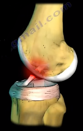
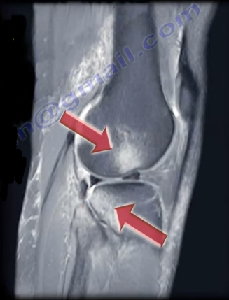
An MRI is going to be the best imaging technique. An MRI of the knee joint will show bone lesions or bruising in the typical location associated with tears of the ACL. These injuries are typically located at the middle of the femoral condyle and posterior part of the tibia laterally. It is also possible to find a triple injury within the MRI (O’Donoghue’s Unhappy Triad).


O’Donoghue’s Unhappy Triad consists of:

  1. Tear of the Lateral Meniscus
  2. Anterior Cruciate Ligament Injury (ACL tear)
  3. Medial Collateral Ligament Injury

In chronic ACL tears, the posterior horn of the medial meniscus is the most commonly injured structure. In acute ACL tears, send the patient for therapy for range of motion, brace the patient, and allow the MCL to heal and reconstruct the ACL later if needed. It is important to stress hamstring therapy in ACL tears. The patient will probably complain of instability immediately or later on.

Tuesday, October 10, 2017

Tibial Plateau Fracture Balloon Osteoplasty


A tibial plateau depression fracture occurs when axial forces from the femoral condyles against the articular surface of the tibia cause the injury. The compression fracture may also be associated with other fractures. Several techniques are used for the treatment of this fracture. Balloon osteoplasty is proposed as a useful tool for reduction and elevation of the tibial plateau depression fracture.

The technique of balloon osteoplasty can be done with or without the supplementation of screws and plate fixation. Trajectory lines are drawn on the medial side of the knee for access to the fracture area. A small incision is made at the point where the lines cross. A cannula and trocar tip stylet is used to access the fractured area under radiographic guidance. The cannula and trocar is advanced under the area of depression using a mallet or drill when needed. Once inserted, the trocar is then removed from the cannula.
 The precision drill is then inserted into the cannula and advanced until it passes under the depressed area of bone. Then, the precision drill is removed from the cannula and the inflatable bone tamp is inserted through the cannula to the area of the depressed fragments. The inflatable bone tamp is centered under the area of depression and then inflated to reduce the depression fracture. Once the depression has been adequately reduced, the inflatable bone tamp can be deflated and removed from the cannula.
The void that has been left by the inflatable bone tamp is now ready to be replaced with bone filler. The device loaded with injectable material is inserted and used until the void is completely filled. Finally, you will remove the injectable material device and the cannula. Plate fixation may be added before or after the injectable material is inserted.