Showing posts with label fracture. Show all posts

Monday, October 4, 2021

Fractures of the Calcaneus: Everything You Need to Know

Fractures of the Calcaneus: Everything You Need to Know

Written by Dominic Ruwe and Dr. Nabil Ebraheim

Fractures of the calcaneus can be open or closed.1 Open fractures are more serious than closed fractures.1 The primary fracture line is caused by an axial load injury.1 The primary fracture line goes from anterolateral to posteromedial.1 The primary fracture line divides the calcaneus into two main fragments: the superomedial fragment which is also called the constant or sustentacular (SAS) fragment and the superolateral or tuberosity fragment.1 The superomedial fragment includes the sustentaculum tali and is stabilized to the talus by ligaments. So, the talus is attached to the constant fragment.1 The sustentacular fragment is a useful reference point for fracture reduction.2 The flexor hallucis longus tendon lies underneath the sustentaculum. If screw placement to the sustentacular fragment is too long, the flexor hallucis longus tendon could be affected, causing fixed flexion of the big toe.3

                The Essex-Lopresti classification system is a useful way to differentiate between different joint fractures. There are two types of Essex-Lopresti fractures: a tongue-type fracture and a joint depression type fracture.1 In the tongue-type, the posterior facet is attached to the tuberosity. In the joint depression type, the posterior facet is not attached to the tuberosity.4 In the tongue-type, the primary fracture line exits anterolaterally and posteromedially.5 The secondary fracture line appears beneath the posterior facet and exits posteriorly through the tuberosity.5 The superolateral fragment and posterior facet are attached to the tuberosity. The tongue-type fracture can be treated with open reduction and internal fixation.6

                In the joint depression type, the primary fracture line splits the calcaneus obliquely through the posterior facet and exits anterolaterally and posteromedially.1 The secondary fracture line exits superiorly just behind the posterior facet.1 The posterior facet is a free fragment. The lateral portion of the posterior facet is usually involved and depressed.4

The Sander’s classification of calcaneal fractures is used to guide the treatment and predict the outcome of the treatment. This classification system is based on the number of posterior facet fracture fragments seen on a coronal CT scan.7 Type I is a nondisplaced fracture which requires nonoperative treatment.7 Type II is a two-part fracture of the posterior facet.7 Type III is a three-part fracture of the posterior facet.7 Type II and III calcaneal fractures benefit from surgery of reduction and fixation.1 Type III fractures normally result in more arthritis because it has more fracture fragments and may end by fusion.8 Type IV fractures are highly comminuted.9 They may require primary subtalar arthrodesis.1

Calcaneal avulsion fractures are typically serious. These types of fractures require urgent reduction and internal fixation to prevent skin complications.10 In joint depression fractures of the calcaneus, the swelling must go down before surgery. Avulsion fractures of the calcaneus are emergencies, so emergency surgery is performed before the swelling goes down. Open reduction and internal fixation of the calcaneus is generally delayed for 1-2 weeks to allow for improvement of the soft tissue swelling, except with avulsion fractures.1 Avulsion fractures can cause skin tenting and urgent reduction is recommended.10

There are many associated conditions with calcaneal fractures. Ten percent are associated with spinal fractures.11 Ten percent are associated with compartment syndrome of the foot.12 If this is neglected, it will lead to claw toes due to contracture of the intrinsic flexor muscles.12 Approximately ten percent are associated with bilateral fractures.13 Sixty percent are associated with calcaneocuboid joint fractures.14 Calcaneal fractures may also be associated with peroneal tendon subluxation. Peroneal tendon subluxation may be detected on axial CT scans or it may be seen as an avulsion fracture of the fibula on x-rays.15

                Complication rates for calcaneal fractures are high. Factors associated with poor outcomes are age greater than 50, smoking, early surgery, history of a fall, heavy manual labor, males, bilateral injury, workman’s compensation, and peripheral vascular disease.1,16,17 Men do worse with calcaneal fractures than women. Calcaneal fractures in men are normally associated with workman’s compensation, heavy labor, and a 0˚ Bohler angle.1 These fractures typically need subtalar fusion.18 Calcaneal fractures in females have a simple fracture pattern. Since calcaneal fractures in males are usually more severe, it follows that better outcomes are seen in females with calcaneal fractures.19

                The Bohler angle is measured on lateral x-rays.1 This angle is normally between 20˚-40˚.1 The Bohler angle is formed by a line drawn from the highest point of the anterior process of the calcaneus to the highest point of the posterior facet and a line drawn tangential to the superior edge of the tuberosity.1 A decrease in this angle indicates a collapse of the posterior facet.1 When viewing calcaneal fractures with the Harris view, the calcaneus appears to be shortened and widened with varus.1 When viewing calcaneal fractures through CT scans, the axial cut shows the calcaneocuboid joint and peroneal tendon subluxation.1,20 The sagittal view shows the subtalar joint and its depression.21 The coronal view shows the displacement of the posterior facet.22 Coronal CT scans can also show the number of the joint fracture fragments.1 The surgical outcome of calcaneal fractures correlate with the number of the joint fracture fragments and the quality of reduction.1 MR imaging shows stress fractures of the calcaneus and the integrity of the peroneal tendons.23,24

Stress fractures of the calcaneus may be misdiagnosed as plantar fasciitis.25 Stress fractures usually occur in female runners.26 It is characterized by swelling and tenderness with medial and lateral compression of the hindfoot during the squeeze test.27 If the X-ray is negative, an MRI should be obtained. The fracture will be seen in T1 MR imaging as a linear streak or a band of low signal intensity in the posterior calcaneal tuberosity.28 In T2 imaging, the signal will be increased.28

                There are several complications with calcaneal fractures. Wound-related complications are the most common complication.29 Wound-related complications occur more in smokers, diabetics, and patients with open fractures.1 Open fractures of the calcaneus is another common complication. Open fractures of the calcaneus can lead to amputation.30 There is also a high risk of infection with open fractures.30 Grade I and Grade II open fractures have wounds that open medially. Open reduction and internal fixation (ORIF) can be done to treat this complication.30 Open reduction and internal fixation should not be done in Grade III medial wounds and in most lateral wounds.30 Another complication is malunion of the calcaneus.31 This is characterized by widening of the heel, varus deformity, and loss of height.31 The talus is dorsiflexed, limiting dorsiflexion of the ankle.31 Peroneal tendon irritation and impingement from the lateral wall is another complication.32

                Surgery on the calcaneus decreases the risk of post-traumatic arthritis.33 Tongue-type and joint depression type fractures may benefit from open reduction and internal fixation.6 Subtalar distraction arthrodesis is a good operation to treat calcaneal fractures associated with loss of height and limited dorsiflexion of the ankle.31 This operation improves talar inclination and decreases anterior ankle impingement.31 Additionally, it takes care of arthritis in the subtalar joint.31 Another surgical approach is extensile lateral approach. The lateral calcaneal artery provides blood supply to the lateral flap associated with the calcaneal extensile approach.34 It is important to be aware that the Sural nerve is in the vicinity of the surgical area.35 Delayed wound healing is a common complication in the extensile lateral approach.35


References:

1. Trompeter A, Razik A, Harris M. Calcaneal fractures: Where are we now? Strategies in Trauma and Limb Reconstruction. 2017;13(1):1–11.

2. Berberian W, Sood A, Karanfilian B, Najarian R, Lin S, Liporace F. Displacement of the SUSTENTACULAR fragment in INTRA-ARTICULAR CALCANEAL FRACTURES. Journal of Bone and Joint Surgery. 2013;95(11):995–1000.

3. Carr JB. Complications of CALCANEUS fractures entrapment of the Flexor hallucis longus. Journal of Orthopaedic Trauma. 1990;4(2):166–8.

4. Rothberg DL, Yoo BJ. Posterior facet cartilage injury in OPERATIVELY Treated Intra-articular CALCANEUS FRACTURES. Foot & Ankle International. 2014;35(10):970–4.

5. White EA, Skalski MR, Matcuk GR, Heckmann N, Tomasian A, Gross JS, et al. Intra-articular tongue-type fractures of the calcaneus: Anatomy, injury patterns, and an approach to management. Emergency Radiology. 2018;26(1):67–74.

6. Chhabra N, Sherman SC, Szatkowski JP. Tongue-type calcaneus fractures: a threat to skin. The American Journal of Emergency Medicine. 2013;31(7).

7. Jiménez-Almonte JH, King JD, Luo TD, Aneja A, Moghadamian E. Classifications in Brief: Sanders classification OF INTRAARTICULAR fractures of the calcaneus. Clinical Orthopaedics & Related Research. 2018;477(2):467–71.

8. Rammelt S, Marx C. Managing severely malunited calcaneal fractures and fracture-dislocations. Foot and Ankle Clinics. 2020;25(2):239–56.

9. Piovesana LG, Lopes HC, Pacca DM, Ninomiya AF, Dinato MC, Pagnano RG. Assessment of reproducibility of sanders classification for calcaneal fractures. Acta Ortopédica Brasileira. 2016;24(2):90–3.

10. Berringer R. Avulsion fracture of the calcaneus. Canadian Medical Association Journal. 2018;190(45).

11. Rowe CR. Fractures of the os calcis. JAMA. 1963;184(12):920.

12. Myerson Mark, Manoli Arthur. Compartment syndromes of the foot after calcaneal fractures. Clinical Orthopaedics and Related Research. 1993;&NA;(290).

13. Popelka V. Súčasné trendy v liečbe intraartikulárnych zlomenín pätovej kosti [Current Concepts in the Treatment of Intra-Articular Calcaneal Fractures]. Acta Chir Orthop Traumatol Cech. 2019;86(1):58-64. Slovak. PMID: 30843515.14.

14. Kinner B, Schieder S, Müller F, Pannek A, Roll C. Calcaneocuboid joint involvement IN CALCANEAL FRACTURES. Journal of Trauma: Injury, Infection & Critical Care. 2010;68(5):1192–9.

15. Park C-H, Gwak H-C, Kim J-H, Lee C-R, Kim D-H, Park C-S. Peroneal tendon Subluxation and dislocation In CALCANEUS FRACTURES. The Journal of Foot and Ankle Surgery. 2021;60(2):233–6.

16. Su J, Cao X. Can operations achieve good outcomes in elderly patients with SANDERS II–III calcaneal fractures? Medicine. 2017;96(29).

17. Clare MP, Crawford WS. Managing complications of CALCANEUS FRACTURES. Foot and Ankle Clinics. 2017;22(1):105–16.

18. Csizy M, Buckley R, Tough S, Leighton R, Smith J, McCormack R, et al. Displaced Intra-articular CALCANEAL FRACTURES. Journal of Orthopaedic Trauma. 2003;17(2):106–12.

19. Barla J, Buckley R, McCormack R, Pate G, Leighton R, Petrie D, et al. Displaced intraarticular calcaneal fractures: Long-term outcome in women. Foot & Ankle International. 2004;25(12):853–6.

20. Toussaint RJ, Lin D, Ehrlichman LK, Ellington JK, Strasser N, Kwon JY. Peroneal tendon DISPLACEMENT Accompanying INTRA-ARTICULAR CALCANEAL FRACTURES. Journal of Bone and Joint Surgery. 2014;96(4):310–5.

21. Badillo K, Pacheco JA, Padua SO, Gomez AA, Colon E, Vidal JA. Multidetector CT evaluation Of CALCANEAL FRACTURES. RadioGraphics. 2011;31(1):81–92.

22. Buckley R. Displaced fracture of the calcaneus body [Internet]. AO Foundation Surgery Reference. [cited 2021Sep29]. Available from: https://surgeryreference.aofoundation.org/orthopedic-trauma/adult-trauma/calcaneous/displaced-body-fractures/definition

23. Kato M, Warashina H, Kataoka A, Ando T, Mitamura S. Calcaneal insufficiency fractures following ipsilateral total knee arthroplasty. Injury. 2021;52(7):1978–84.

24. Park HJ, Cha SD, Kim HS, Chung ST, Park NH, Yoo JH, et al. Reliability of MRI findings OF PERONEAL Tendinopathy in patients with LATERAL CHRONIC Ankle Instability. Clinics in Orthopedic Surgery. 2010Nov5;2(4):237.

25. Weber JM, Vidt LG, Gehl RS, Montgomery T. Calcaneal stress fractures. Clinics in Podiatric Medicine and Surgery. 2005;22(1):45–54.

26. Labronici P, Pires RE, Amorim L. Calcaneal stress fractures in civilian patients. Journal of the Foot & Ankle. 2021;15(1):54–9.

27. Kiel J, Kaiser K. Stress Reaction and Fractures. 2021 Aug 4. In: StatPearls [Internet]. Treasure Island (FL): StatPearls Publishing; 2021 Jan–. PMID: 29939612.

28. Lawrence DA, Rolen MF, Morshed KA, Moukaddam H. MRI of heel pain. American Journal of Roentgenology. 2013Apr18;200(4):845–55.

29. Ding L, He Z, Xiao H, Chai L, Xue F. Risk factors for postoperative wound complications of calcaneal fractures following plate fixation. Foot & Ankle International. 2013;34(9):1238–44.

30. Heier KA, Infante AF, Walling AK, Sanders RW. Open fractures of THE Calcaneus: Soft-tissue Injury DETERMINES OUTCOME. The Journal of Bone and Joint Surgery-American Volume. 2004;86(11):2569.

31. Guang-Rong Y, Xiao Y. Surgical management Of Calcaneal Malunion. Journal of Orthopaedics, Trauma and Rehabilitation. 2013;17(1):2–8.

32. Davis D, Seaman TJ, Newton EJ. Calcaneus Fractures. 2021 Aug 9. In: StatPearls [Internet]. Treasure Island (FL): StatPearls Publishing; 2021 Jan–. PMID: 28613611.

33. Vilá-Rico J, Ojeda-Thies C, Mellado-Romero MÁ, Sánchez-Morata EJ, Ramos-Pascua LR. Arthroscopic posterior subtalar arthrodesis for salvage of posttraumatic arthritis following calcaneal fractures. Injury. 2018;49.

34. Mehta CR, An VV, Phan K, Sivakumar B, Kanawati AJ, Suthersan M. Extensile lateral versus sinus Tarsi approach For displaced, intra-articular Calcaneal Fractures: A meta-analysis. Journal of Orthopaedic Surgery and Research. 2018;13(1).

35. Buckley R. Extended lateral approach to the calcaneus [Internet]. AO Foundation Surgery Reference. [cited 2021Sep29]. Available from: https://surgeryreference.aofoundation.org/orthopedic-trauma/adult-trauma/calcaneous/approach/extended-lateral-approach-to-the-calcaneus


Monday, August 12, 2019

Open Fractures of the Tibia


Open Fractures of the Tibia

25% of tibial shaft fractures can be open. Open fractures can lead to complications including wound problems, osteomyelitis, nonunions, and infected nonunions. The treatment of open fractures of the tibia can be challenging. A lot of the concepts are not black and white; they may be in the grey zone. We don’t know the best time for debridement. We don’t know what the optimal irrigation solution is and what the optimal pressure for the fluid is. We don’t know for sure the ideal duration of giving antibiotic prophylaxis, but we know that it is important to give the appropriate antibiotics early and do meticulous debridement. We know that the IM rod is better than the plate fixation or external fixator, and the result of the reamed IM rod or unreamed IM rod is the same. We need to close or cover the wound before 1 week and the vac can be used provisionally when we cannot close the wound, primarily at the optimal time. A grade I fracture is less than 1 cm. a grade II fracture is 1-10cm. A grade III fracture is more than 10 cm, and there is contamination. Grade III fractures are divided into three types. Grade IIIa fractures require adequate tissue for closure (or skin graft). Grade IIIb fractures require extensive periosteal stripping and the patient will need a flap (rotational or free flap). Grade IIIc fractures have a vascular injury that requires repair or amputation. The relative indication for amputation is warm ischemia for more than 6 hours, absent plantar sensation and severe ipsilateral foot trauma.
The most predictive factor for amputation is the severity of the soft tissue injury in the ipsilateral extremity. When comparing limb salvage versus amputation, the patient’s outcome is generally the same at 1-5 years. Lack of plantar sensation does not predict poor outcome after limb salvage. Segmental fractures are Grade III fractures, even if the open fracture is 1 cm. The ideal irrigation solution and the pressure used to controversial. Timing of the initial debridement is controversial. Irrigation and debridement within 6 hours was the gold standard in the past. Debridement is performed as a priority procedure no later than the morning after admission. There is no difference in infection rate for a patient who has the initial surgery before or after 6 hours, including patients with Type III open fractures. More than 40% of the patients usually wait longer than 6 hours for their initial surgery after arrival at the hospital. Delayed surgery for less severe fractures is acceptable as long as the debridement is done as a priority the following day. Unless there is a gross contamination, evidence is not clear as to when is the best time for the debridement. It seems like giving the patient antibiotics promptly is more important than the time of debridement. The preferred solution is normal saline and low pressure irrigation. Low pressure lavage may reduce reoperation rates due to infection, nonunions, and wound healing problems. Normally the tradition is to use 3, 6, and 9 liters of solution for Type I, Type II, and Type III open fractures (just recommendations). There is increased risk of wound healing with antibiotic solution. Meticulous irrigation and debridement of open fractures is important in decreasing the infection risk. Prophylaxis should be started as soon as possible. All patients with open fractures should receive first generation Cephalosporin’s that will cover gram-positive bacteria. You can give penicillin for farm injuries and clostridia prone wounds. You will give clindamycin if there is a penicillin allergy. In Type III open fractures, add aminoglycoside, such as gentamicin. It was found that local antibiotics delivery at the site of injury decreased the infection risk, such as cement beaded loaded with antibiotics. Antibiotic should be given within 3 hours of the time of injury (preferably given as soon as possible). There is reduction of 59% of acute infection in patients with open fractures treated with antibiotics. The infection rate is 1.6 times greater if antibiotics are given after 3 hours. Type I and Type II open fractures require antibiotic coverage for 24 hours after wound closure. For Type III open fractures, antibiotic administration should be given for a period of 72 hours after the injury and no more than 24 hours after wound closure. After the initial debridement, the patient will need staged debridement within 24-48 hours. There is a reduction infection rate, acute and chronic, for Type III open fractures with the use of systemic antibiotics and aminoglycoside cement beads compared with antibiotics alone. This combination of antibiotics lowers the infection rate for any open tibial fracture that is treated with an IM rod. Its affect is more noticed in Type III injuries. Plating of open fractures may cause chronic infection and infected nonunion. The healing time is doubled with plated open fractures. The IM rod resulted in a better alignment and lower reoperation rate than using external fixator. Also, no difference in the infection rate between the IM rod and the external fixator. You can use a reamed nail or an unreamed nail. They both have a comparable result and no difference in the outcome.
When reaming, you can use a bigger rod that provides better stability. Reamed nailing is superior in closed tibial fractures, but it is not superior in open tibial fractures. Reaming can cause increased pressure and disruption of the endosteal blood supply, can cause thermal necrosis and fat embolism with increased intramedullary risk of infection. The unreamed rod uses smaller nails and results in less stability but preserves the endosteal blood supply. Unreamed IM tibial rod appears to have a shorter time to union and fewer incidence of knee contractures when compared with circular wire external fixator. Nowadays, more and more orthopedic surgeons are using reamed nails for open fractures of the tibia. If you have a spiral fracture of the distal 1/3 of the tibia, you will need to get a CT scan of the ankle to identify a posterior malleolar fracture, which should be fixed before insertion of the IM rod. There is a lack of evidence to support the value of external fixator over the IM rod in open factures of the tibia. Due to patient discomfort, the high incidence of pin tract infection and loss of alignment, external fixator should not be used as a definitive fixation. Use external fixator temporarily (less than 4 weeks) and replace it with a rod in about 14 days. External fixator may be utilized for severely contaminated open fractures. Tibial fractures treated with a shorter duration of external fixator has reduction of the infection risk by 80%. When there is a shorter interval between removal of the external fixator and insertion of the IM rod of the tibia, there is a reduction of the risk of infection by 85%. In less severe soft tissue injuries, you do primary closure without tension. In cases of delayed closure, soft tissue coverage should be done within 7 days. Soft tissue coverage beyond 7 days will increase the infection. There is no difference in the incidence of infection in patients who had primary closure and delayed closure of the wound. It is recommended to do primary closure for Type I, Type II, and Type IIIa fractures with tension free closure and after timely antibiotic prophylaxis and adequate debridement. Intraoperative culture after debridement has no value. It does not predict future infection. In the upper 1/3 of the tibia, you can treat it by a medial gastrocnemius flap. In the middle 1/3 of the tibia, you can treat it by a soleus rotational flap. The use of a free flap for soft tissue coverage was less likely to have wound complications than the use of a rotational flap. The zone of injury may be larger than expected, and it may include the rotated muscle flap. The negative pressure wound therapy (the vac) is used frequently. The vac provides provisional coverage for wounds where the physician cannot do primary closure. There is decreased infection rate when using the vac. The vac is used for coverage after the initial debridement of the open fracture until the definitive coverage is done. It is a good temporizing dressing and can also be used in fasciotomy wounds. The vac promotes local wound healing. Bone morphogenetic protein (BMP2) decreases the need for secondary surgery and is used in acute open tibial fractures treated with IM rod.

Monday, April 22, 2019

Geleazzi Fracture


Galeazzi Fracture

Galeazzi fracture is a fracture of the distal 1/3 of the radius with disruption of the distal radioulnar joint (DRUJ). The fracture is always located above the proximal border of the pronator quadratus. The pronator quadratus rotates the distal fragment towards the ulna and pulls it proximally. We usually fix the fractured radius, and we then evaluate DRUF for instability after we fix the distal radius. If you have instability, make sure that the joint is reduced, then you will do percutaneous fixation of that joint. If you don’t have instability, you will do nothing or maybe give a long arm splint in supination if you think the patient needs the splint.
Basically, you will need intraoperative evaluation of the DRUJ. Not all distal radial fractures will be associated with distal radioulnar joint instability. They found that if the radius fracture is less than 7.5 cm from the join, then the distal radioulnar joint can be unstable. If the fracture radius is more than 7.5cm from the joint, then the distal radioulnar joint will be rarely unstable. So the closer the fracture of the radius is to the joint, the more likely that the distal radioulnar joint is involved, and we need to work diligently to find the problem and address it. The problem can be instability of the DRUJ. You may find an ulnar styloid fracture or you find that the radius is short (about 5 mm or more). In the AP view of the wrist, you may find widening of the joint or in the lateral view, you find that the ulna goes dorsally or volarly. The distal radioulnar joint has ligaments, volar and dorsal, that stabilize the joint, and that joint is usually stable in supination. Sometimes in old, complicated or difficult cases, you can’t really evaluate the distal radioulnar joint without getting a CT scan of both wrists (make sure that you position the wrist in the same position. Anatomic reduction and fixation of the radius with a volar plate. Then you assess the stability of the distal radioulnar joint. If the distal radial ulnar joint remains unstable, supination of the wrist may reduce that joint. If not, either a closed reduction or open reduction with pinning of the joint is done. If after anatomic restoration and plate fixation of the radius, the distal radioulnar joint remains irreducible, then the structure that is most likely obstructing the reduction is the extensor carpi ulnaris. It is imperative to recognize the problem of Galeazzi fracture, which is the distal radioulnar joint injury. The treatment of the problem acutely is better than late reconstruction. When you fix the radius, make sure that the radial bow is restored. The reduction of the joint is done by supination of the forearm, and you do immobilization in supination if the distal radioulnar joint is stable following open reduction of the distal radius. So there is an obvious injury that you will see, and you will test that injury and see if the joint is stable in supination. If it is, keep the forearm in supination. You will do pin fixation if the joint is reducible, but is unstable. The pin fixation will be done by cross pinning from the ulna to the radius and leave the pins for about 4 weeks. You can do open reduction of the joint if the joint is not reduced and something is blocking the reduction, such as the extensor carpi ulnaris tendon. If there is a large ulnar styloid process fracture, you probably need to open that fracture after you fix the radius, and then do open reduction and internal fixation of the large ulnar styloid fragment and immobilize the forearm in supination. It might be difficult to evaluate the stability of the distal radial ulnar joint. In general, the DRUJ is stable in most cases after anatomic reconstruction of the radius.

Monday, February 11, 2019

Femoral Neck Fracture Nonunion


Femoral Neck Fracture Nonunion

Femoral neck fracture nonunion has multiple facets and is important to understand all aspects of this important problem.

Example:

40 year old patient had a displaced femoral neck fracture, fixed with multiple cancellous screws about 9 months ago. The patient still has persistent groin pain. The patient cannot bear full weight on the hip. The patient has a painful limb, antalgic gait, and difficulty in walking. X-rays are not clear and show a possible nonunion. CT scan shows the nonunion with some Varus angulation. The treatment for this would be removal of the hardware and valgus osteotomy. The scenario can be more complicated by adding a healed femoral shaft fracture to the nonunion of the femoral neck. In this case, you will do removal of the hardware from the femur and removal of the screws from the femoral neck nonunion. You will do valgus osteotomy and fixation with a plate, preferably a blade plate, to treat the nonunion of the femoral neck.

Intracapsular fractures of the proximal part of the femur are not common in adults younger than 50 years old, but they are associated with a high incidence of avascular necrosis and nonunion. About 10-30 % of femoral neck fractures go to nonunion after ORIF. It is usually the vertical fracture pattern, such as Type III in Pauwels Classification. These fractures are more prone to nonunion due to shear stress, rather than compression forces across the fracture site. In Garden Classification fracture Type IV, where the fracture is completely displaced, the greater the displacement, the higher the incidence of nonunion and reoperation rate after fixation of the femoral neck. The inverted triangle pattern of fixation of femoral neck fractures is the one that is commonly used with the inferior screw posterior to the midline and adjacent to the calcar. Achieving and maintaining anatomic reduction is important for femoral neck fracture fixation and healing. The femoral neck fractures are intracapsular. There will be no abundant callus formation during the healing (healing is intraosseous only). Sometimes it is difficult to know if the fracture healed or not. There is no correlation between age, gender, and rate of nonunion. Varus malreduction correlates with failure of fixation after reduction and cannulated screw fixation. Posterior comminution of the fracture does not allow stable fixation and can lead to nonunion. The comminution of the femoral neck is usually posteriorly and inferiorly. Some recommend adding a fourth screw in this situation. High energy fractures have a worse prognosis for healing, especially in patients with metabolic bone disease and nutritional deficiency. When you see a femoral neck nonunion after fixation, you need to get blood work and rule out infection (get sedimentation rate and CRP).
For the high angle femoral neck fracture, follow the patient up closely with clinical exam and x-rays. There might be a Varus collapse on the x-rays. You may see a femoral neck nonunion or a failed internal fixation. The patient walks with a limp, the limb is shortened, and the patient may have rotational deformity of the extremity. In the young patient with a femoral neck nonunion, arthroplasty is not a desirable option. In a young patient with femoral neck fracture nonunion, valgus intertrochanteric osteotomy with plate fixation produces a good result in the majority of cases. Valgus intertrochanteric osteotomy with plate fixation produces approximately 80% union rate and the procedure makes a vertical fracture more horizontal, converting the shear forces into compressive forces. It is done in a healthy, young patient with no joint arthritis and when the femoral head is intact. This procedure also corrects the Varus malalignment. Basically, the procedure changes the vertical fracture orientation to a horizontal fracture to achieve compression. Other procedures done in the young patient include revision ORIF with or without bone graft, but this is rarely done. Other procedures done in the young patient also include free vascularized fibular graft which is done in some patients especially in the younger patient with a nonviable femoral head. Hemiarthroplasy is done in patients with low physical demands. The articular cartilage of the patient is preserved with no evidence of infection. Total hip arthroplasty is done in patients that are older, in patients that have hip arthritis, if the femoral head is not viable, or if the hardware is cut out. It can also be done in younger patients that are active, when the femoral head is not viable and the patient does not want a free vascularized fibular graft or if the patient had collapse of the femoral head with nonunion. The problem with total hip replacement in this situation is more dislocations of the hip postoperatively.

Monday, January 28, 2019

Elbow Dislocation in Adults


Elbow Dislocation in Adults

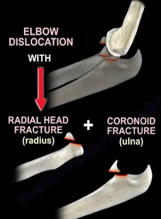
With elbow dislocations, recognize the terrible triad: elbow dislocation, radial head fracture, and coronoid fracture. The terrible triad is not a simple elbow dislocation; it is a complex elbow dislocation. In addition to these three injuries of the elbow, there is always a tear of the lateral ulnar collateral ligament. The treatment usually is reduction and splinting of the elbow.
This cannot be the definitive treatment; it is the initial treatment. If no surgery is done, you will have recurrent dislocation of the elbow. You need to do surgery for reduction and fixation of the fractures and also to restore the elbow stability. This injury is unstable. Simple reduction and splinting is not going to work for this injury. You have to recognize the terrible triad which means surgery. There are multiple types of elbow dislocation based on the position of the olecranon relative to the humerus. The most common type of elbow dislocation is the posterolateral type. There are two basic types of elbow dislocations: simple and complex. Simple elbow dislocations have no fracture seen, and are usually a ligamentous injury. Complex elbow dislocations have associated fractures in addition to the ligamentous injury. With any elbow dislocation, you need to check the shoulder and the wrist for injuries and fractures because it can occur in up to 15%. When you have a simple dislocation of the elbow, you need to reduce it and then check the range of stability of the elbow. If you find that the elbow is stable with range of motion, then you will do a short period of immobilization with a posterior splint for approximately one week with the elbow in about 90 degrees of flexion. Then start active range of motion of the elbow. Recurrence of the dislocation is rare (less than 1%). If you keep the elbow immobilized more than 3 weeks, there will be severe stiffness of the elbow. Surgery should be done if the dislocation is irreducible, if there is associated fracture, or if you are unable to maintain stability of the elbow. After immobilization and early range of motion of the elbow, you will see the patient and do follow up x-rays to check joint congruity and to make sure that the elbow reduction is maintained. To treat the terrible triad, you should initially do a closed reduction. Open reduction and internal fixation of the coronoid (if possible), of the radial head or excise the radial head with radial head arthroplasty if the radial head is unreconstructable. In addition, you will do lateral ulnar collateral ligament (LUCL) repair. Never excise the radial head alone in this situation. For an elbow dislocation with olecranon fracture, do open reduction and plate fixation. K-wires and tension band is not strong enough to hold the fracture and stabilize the elbow at the same time. For an elbow dislocation with a radial head fracture, do fixation or replacement of the radial head (never do excision of the radial head alone in this situation). The LUCL is the most important lesion in recurrence or persistence of instability of the elbow following simple elbow dislocation.
The injury progresses from lateral to medial. The lateral collateral ligament fails first, and it avulses proximally at the lateral epicondyle. The medial collateral ligament (MCL) fails last. In varus posteromedial rotary instability, there is an elbow injury plus LUCL tear, plus coronoid fracture which involves the medial facet of the coronoid. Treatment for chronic dislocation is open reduction capsular releases with hinge external fixation and early range of motion. Loss of terminal extension is a complication of elbow dislocation. Usually for decreased range of motion of the elbow, you will do static progressive splinting between 6-10 weeks. No manipulation of the elbow is done, which is different from the knee after total knee replacement, where you can do manipulation up to three months. With heterotopic ossification, do excision. Remove the myositis and excise the posterior part of the MCL to allow more flexion. To be functional, the range of motion of the elbow should be between 30-130 degrees. Some physicians suggest that if the flexion is less than 100, you will do release of the posterior bundle of the MCL in addition to release of the ulnar nerve. If you want more flexion of the elbow, excise the posterior part of the MCL.

Monday, January 21, 2019

Patellar Fractures


Patellar Fractures

Patellar fractures can involve different topic, and I am going to try and highlight the important points related to patellar fractures. The medial patellofemoral ligament is the primary stabilizer of the patella, so when the patella dislocates, you will have an injury to that ligament and also an injury to the medial patellar facet articulation cartilage or an osteochondral fragment. In addition to the medial patellar facet injury, you will get a lateral femoral condyle injury. Bipartite patella occurs in about 8% of the population. It could be bilateral in about 50%, and it usually occurs in the superolateral aspect of the patella. You should observe it and not fix it. It is not an acute fracture that may need excision or lateral retinacular release. It can occur in children between 8-10 years old. It is a rare condition. The patient will be unable to do straight leg raising, so you suspect that the extensor mechanism is injured. The patient may have a high riding patella on x-ray with a palpable gap when you examine the patient. The x-ray may show small flecks of bone as the patellar tendon avulses with a portion of the distal pole of the patella. Sometimes the bony injury is so small that the condition can be missed. You should have a high index of suspicion. You may need to get an MRI to confirm diagnosis. The treatment is usually ORIF if the fracture is displaced.
The patella is a large sesamoid bone. The quadriceps muscle is inserted at the proximal pole and the distal pole gives attachment to the patellar tendon. The patella is triangular in shape. The proximal 3/4 of the patella is covered with cartilage, however the distal 25% of the patella is not covered with cartilage. The patella increases the power of the extensor mechanism by about 50% because it displaces the extensor mechanism anteriorly, and that will increase the moment arm.
Transverse fractures of the patella can be non-displaced or displaced. The patella can be pulled apart by the attached quadriceps tendon. The patient will be unable to do active extension of the knee. Upper or lower pole fractures of the patella are fractures at the site of attachment of the patellar tendon. Comminuted fractures of the patella can be non-displaced or displaced. Comminuted fractures have multiple pieces, are very unstable, and are difficult to fix. Vertical fractures of the patella are the most common, and they are stable and nondisplaced. Osteochondral fractures are small fractures of the patella usually associated with acute dislocation of the patella.
In examination, you may feel a palpable gap. The area of the knee is usually swollen. The patient will be unable to do straight leg raise. The lateral view of the knee is the best view to see the fracture. 2-3 mm of displacement will probably mean that the patient will need surgery.
If you think that the patient’s extensor mechanism is intact, and the patient is able to do straight leg raise, and the fracture is nondisplaced or minimally displaced, it is usually a transverse fracture in this situation, then immobilize the knee straight in a hinged knee brace for 4-6 weeks with weight bearing as tolerated. Sometimes the patient cannot move the knee because of the pan and injection of lidocaine inside the knee can help to assess the integrity of the extensor mechanism. If the patient has a total knee with 2mm displacement of the patella, and the extensor mechanism is intact, then the patient will be treated conservatively in a brace or in a knee immobilizer (no surgery).
Indication for surgery is a displaced patellar fracture and the inability to do straight leg raising.
First, preserve the patella (if possible). The tension band fixation technique is the gold standard for the treatment of displaced patellar fractures (the fracture is usually a transverse fracture), and the tension band technique is the one that gives us the most complications. The first step in the tension band technique is to reduce the fracture with reduction clamps. Next, at least two K-wires are placed across the fracture. An anterior tension band is applied, organized in a Figure-8 pattern. You need to put the Figure-8 tension band wire close to the patella superiorly and not far away from the patella because that may cause construct instability and fracture displacement. A second wire may be placed circumferentially around the patella. Bending the K-wires from both ends may decrease migration of the wires and decrease the complications. The wire that is bent at both ends may be difficult to remove. Tension band fixation technique may be done with K-wires or also with cannulated screws (through the cannulated screws, you place the wires). It does not matter if you have an open or closed fracture, you treat it the same way. When you place K-wires, it means symptomatic hardware and thus a secondary reoperation. It was found that the longitudinal screws and the tension band wires are a more superior fixation. The tension band construct when performed correctly will provide absolute stability and will convert the tension forces from the muscle pull into compression forces at the articular surface. You want to have anatomic reduction and stable fixation; don’t judge the reduction by what you see at the surface of the fracture. Try to see and feel the joint if you can. Check the x-rays carefully. The surface of the patella may be well reduced, however, the joint may be distracted or displaced. If you tighten the cerclage wire aggressively, you may have a good looking surface, but you may have a distracted join. After you fix the patella, you will do a range of motion of the knee before closure and give the patient a hinged knee brace, locked into extension with weight-bearing as tolerated. Weight-bearing is controversial. Some people start weight-bearing early, and some people start weight-bearing after 4-6 weeks. A can may be helpful to the patient. You will begin active flexion at 2-3 weeks (patient will lie prone, flexing and extending the knee). When the patient is prone, it avoids active knee extension and avoids excessive stress on the fracture site. At 6 weeks, you can unlock the brace and start moving the knee, gradually increasing the flexion.
If the patellar fracture is comminuted, you can use the peripatellar circumferential wire loop fixation, which is commonly used as an addition to other methods of fixation. You can also use a plate fixation utilizing a low profile implant and providing stable fixation. This technique is becoming more popular.
You can also excise the patella partially or completely. In a partial patellectomy, the distal pole is extra-articular, and if it is severely comminuted and less than 40% of the patella, then you can excise it (in general, you would like to preserve the patella). If you can’t preserve the patella and ORIF is not possible, then do partial patellectomy and preserve the largest piece. Partial patellectomy may be necessary, but open reduction and internal fixation (if possible) is associated with a better outcome. You will do the partial patellectomy in several comminuted inferior pole fractures. You will do medial and lateral retinacular repair, and a poor outcome may occur with removal of more than 40% of the patella. Total patellectomy will be done when the fractured patella cannot be fixed. Total patellectomy can cause extensor lag and loss of the extensor strength. The quadriceps torque is reduced by about 50%.
Symptomatic hardware and knee pain is the most common complication after patellar fracture fixation, especially if you use the tension band technique. It requires implant removal in about 50% of the time. This complication will include the hardware migration. Failure after patellar fracture fixation occurs in about 20% of the time due to increasing age, fixation with wires, technical errors and noncompliance.

Monday, December 10, 2018

25 Vitamin D- Tests Orthopedic Surgeons Should Think About


25 Vitamin D, Tests Ortho Surgeons Think About-Everything You Need To Know

Vitamin D 25 is the most appropriate study to assess and monitor vitamin D status in the body. Vitamin D is important for proper maturation and development of bone. Vitamin D is also important in immunity and plays a role in other conditions. The main function of Vitamin D is absorption of the calcium and phosphate from the intestine. Vitamin D comes from diet, supplements, and exposure to the sun. Vitamin D is naturally found in fish. Exposure to the sun for 15 minutes will give a person about 10,000 units of Vitamin D. The average daily requirement of Vitamin D is approximately 400-800 International Units (IUs). Vitamin D gets activated metabolically in the liver and in the kidney. The activation occurs by hydroxylation. Hydroxylation to 25 Vitamin D3 occurs in the liver. The big organ takes the big number- 25, so 25(OH)-Vitamin D3. Another hydroxylation occurs in the kidneys. 2- Vitamin D3. This is the active form of Vitamin D and works mainly on the intestines and bones. The activation of Vitamin D to 1, 25 hydroxyvitamin D is controlled by the parathyroid hormone. Any deficiency or any problem in the process of activating Vitamin D3 to its active form will lead to deficiency of Vitamin D in the body. Vitamin D deficiency is very common and the majority of people are not aware of it. In fact, Vitamin D deficiency symptoms are subtle and nonspecific. 25-hydroxyvitamin D has a long half-life and a higher concentration. This is probably easier to measure and obtain 25 hydroxyvitamin D than the active form, which is 1,25-dihydroxyvitamin D. The half-life of 25-hydroxyvitamin D is 2-3 weeks. The half-life of 1,25-dihidroxyvitamin D is only 4-6 hours. The circulating levels of 25- hydroxyvitamin D is 1000x more than 1,25 dihydroxyvitamin D. therefore, 25-hydroxyvitamin D test is the best study to determine the Vitamin D deficiency in the body. A low level of 25- hydroxyvitamin D could mean that a person is not getting enough exposure to the sun, is not getting enough dietary Vitamin D, or there may be a problem with absorbing Vitamin D from the intestines. The patient may be taking Dilantin, which interferes with hydroxylation of Vitamin D in the liver. A low level of 1, 25-dihydroxyvitamin D usually indicates kidney disease. 40% of the United States population have Vitamin D deficiency.The small organ takes the small number-1. The result will be 1, 25 (OH)

Symptoms of Vitamin D deficiency may include:

·         Fatigue and tiredness
·         Not sleeping well
·         Muscle weakness
·         Bone pain
·         Osteoporosis/ Osteomalacia
·         Fractures


Elderly patients are vulnerable to Vitamin D deficiency because they usually live indoors or in nursing homes with no sun exposure or because these patients may not eat enough food containing Vitamin D or they may not receive enough supplements. Vitamin D deficiency may impair or affect wound healing. Vitamin D deficiency may cause bone loss and places the elderly patient at risk of fractures. Deficiency may cause slow healing of fractures or nonunion of the fractures. If you find a patient with fractures that are not healing well, or a patient with fractures due to low energy trauma where you find the bone mass is inadequate (osteoporosis), this is the time to get a 25- hydroxyvitamin D blood test. The Endocrine Society defines Vitamin D deficiency as 25 Vitamin D level below 20 ng/mL, and insufficiency as the level between 21-29 ng/mL. In general, a 25 Vitamin D level greater than 30 ng/mL is probably adequate, but these numbers are controversial.