Femoral Neck Fracture Nonunion
Femoral neck fracture nonunion has multiple facets and is
important to understand all aspects of this important problem.

Example:
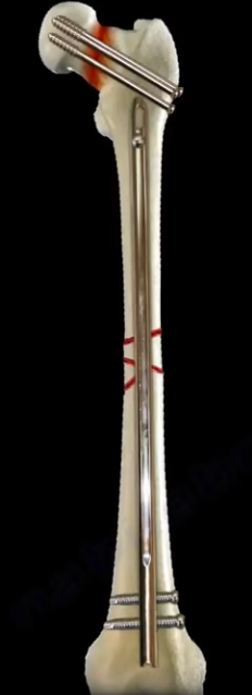
40 year old patient had a displaced femoral neck fracture, fixed with multiple cancellous screws about 9 months ago. The patient still has persistent groin pain. The patient cannot bear full weight on the hip. The patient has a painful limb, antalgic gait, and difficulty in walking. X-rays are not clear and show a possible nonunion. CT scan shows the nonunion with some Varus angulation. The treatment for this would be removal of the hardware and valgus osteotomy. The scenario can be more complicated by adding a healed femoral shaft fracture to the nonunion of the femoral neck. In this case, you will do removal of the hardware from the femur and removal of the screws from the femoral neck nonunion. You will do valgus osteotomy and fixation with a plate, preferably a blade plate, to treat the nonunion of the femoral neck.
Intracapsular fractures of the proximal part of the femur
are not common in adults younger than 50 years old, but they are associated
with a high incidence of avascular necrosis and nonunion. About 10-30 % of
femoral neck fractures go to nonunion after ORIF. It is usually the vertical
fracture pattern, such as Type III in Pauwels Classification. These fractures
are more prone to nonunion due to shear stress, rather than compression forces
across the fracture site. In Garden Classification fracture Type IV, where the
fracture is completely displaced, the greater the displacement, the higher the
incidence of nonunion and reoperation rate after fixation of the femoral neck. The
inverted triangle pattern of fixation of femoral neck fractures is the one that
is commonly used with the inferior screw posterior to the midline and adjacent
to the calcar. Achieving and maintaining anatomic reduction is important for
femoral neck fracture fixation and healing. The femoral neck fractures are
intracapsular. There will be no abundant callus formation during the healing
(healing is intraosseous only). Sometimes it is difficult to know if the fracture
healed or not. There is no correlation between age, gender, and rate of
nonunion. Varus malreduction correlates with failure of fixation after
reduction and cannulated screw fixation. Posterior comminution of the fracture
does not allow stable fixation and can lead to nonunion. The comminution of the
femoral neck is usually posteriorly and inferiorly. Some recommend adding a
fourth screw in this situation. High energy fractures have a worse prognosis
for healing, especially in patients with metabolic bone disease and nutritional
deficiency. When you see a femoral neck nonunion after fixation, you need to
get blood work and rule out infection (get sedimentation rate and CRP).