Showing posts with label ligaments. Show all posts

Monday, November 12, 2018

Ankle Ligaments Injury, Tests & Assessment


Ankle Ligaments Injury, Tests & Assessment

The ligaments of the ankle are complex. Injury to these ligaments are called ankle sprains. Sprain of the ankle is usually a low ankle sprain. Occasionally, it can be a high ankle sprain. Sprain of the ankle can be confused with other conditions that can happen around the ankle such as:
  • ·         Osteochondral lesion
  • ·         Peroneal tendon subluxation
  • ·         Fracture of the lateral talar process
  • ·         Fracture of the anterior process of the calcaneus
  • ·         High ankle sprain (syndesmotic injury)

Here are a few tests that are used to test for injury of these ligaments:

  • Anterior Drawer Test
  • Squeeze Test
  • External Rotation Stress Test
  • Talar Tilt Test (Inversion Test)


If the patient cannot bear weight on the ankle, the patient should get an x-ray. Injury of the deltoid ligament occurs at the medial side of the ankle, and it is usually associated with ankle fractures. Sometimes injury of the deltoid ligament is occult and the patient will need external rotation stress x-rays to demonstrate injury of the deltoid ligament. Injury to the lateral side ligaments is referred to as low ankle sprain. The anterior talofibular ligament is the weakest ligament on the lateral side. The anterior drawer test is done to test the competency of the anterior talofibular ligament. It is done in 20 degrees of plantar flexion and compare it to the other side. A shift of an absolute value of 9 mm on the lateral x-ray or 5 mm compared to the other side is positive. The calcaneofibular ligament is usually injured after the anterior talofibular ligament. The test used to diagnose injury of the calcaneofibular ligament is called the talar tilt test or the inversion test. Less than 5 degrees of tilt is usually normal. The final area of injury is called a high ankle sprain or injury to the syndesmosis. Contrary to a low ankle sprain, a high ankle sprain may require surgery. This is how injury to the syndesmosis occurs. Always check the fibula proximally to avoid missing a Maisonneuve fracture. The Maisonneuve fracture will have a proximal fibular fracture, a syndesmotic injury, and a deltoid ligament injury. This will require surgery. The tests used to diagnose high ankle sprains are the squeeze test and the external rotation stress test. The squeeze test is performed by squeezing the tibia and fibula at mid-calf. This will cause pain at the syndesmosis if a high ankle sprain is present. The external rotation test is the other test used to diagnose a high ankle sprain or an injury of the syndesmosis. The external rotation test is performed by first placing the ankle into a neutral position. Then, apply external rotation stress and finally, get a mortise view radiograph. There is a positive result for syndesmotic injury if the tibiofibular clear space is more than 5mm or if the medial clear space widening is more than 4mm.

Tuesday, February 20, 2018

Jefferson Fractures


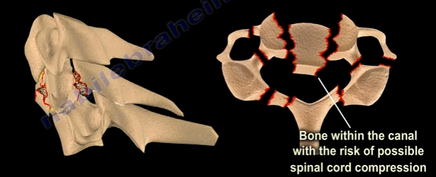
Fifty percent of patients with Jefferson fractures will have associated spine injuries. The canal is wide with a low risk of spinal cord injuries unless the transverse ligament is disrupted. It is difficult to view Jefferson Fractures on an x-ray (usually seen on the lateral side”. This fracture is considered a “Junctional Fracture” and could be missed. The classic Jefferson fracture is a burst fracture that results from an axial load. It could be a four part fracture with bilateral fractures of the anterior and posterior arch. There are variations which include two and three part fractures and incomplete formations of the posterior arch can be mistaken as a fracture.
When speaking of Jefferson fractures, it is important to be familiar with the structures that may be involved. These bony structures include: The Atlas (C1), Axis (C2), and the odontoid process. C1 and C2 are stabilized together by the transverse ligament and C1 and C2 provide a 50% of rotation of the neck. The C1 is a ring. At the upper cervical region, the spinal canal is 2.5 times larger than the cord size. The stability and treatment of Jefferson fractures depends on the integrity of the transverse ligament and the displacement of the fracture. You need to know about the important ligaments related to the Jefferson fracture. These ligaments include: the transverse ligament, the apical ligament, and the Alar ligament.

Diagnosing ligamentous injury


In order to determine a ligamentous injury, the physician will want to check the Atlanto-dens interval (A.D.I). Normally, this interval should be less than 3mm in adults and less than 5mm in children. If the ADI is between 3-5mm, this indicates an injury to the transverse ligament; the transverse ligament holds the odontoid and C1 together, alar and apical ligaments will be intact. If the A.D.I measures greater than 5mm, then there is an injury to the transverse, alar, and apical ligaments.


Fracture Types


A bony injury with the intact transverse ligament and a lateral mass displacement less than 7mm and the A.D.I is less than 3mm is considered a stable fracture. Nondisplaced fractures of this nature should be treated with a rigid orthosis. If the fracture is displaced, a halo will need to be used.
Another type of fracture can occur at C1 with a transverse ligament tear. The Atlanto-dens interval will be more than 3 mm in adults. The treatment will depend on the type of injury to the transverse ligament. With bony avulsions of the transverse ligament, the halo will need to be used cautiously. However, some surgeons prefer to do a fusion of C1 and C2. If there is an intrasubstance tear of the transverse ligament, the surgeon will perform a fusion at C1-C2. The surgeon will need to do early surgery as this is a significant injury with a risk of spinal cord compression.


In regards to “Open Mouth Views”, the normal overhang is visible during an “Open Mouth View”. If it is just a bony injury Jefferson fracture, the combined overhang will be less than 7mm and the transverse ligament is intact and it is a stable fracture. If a Jefferson fracture has a combined overhang of more than 7mm, then the transverse ligament is probably torn and there is an unstable fracture present.

Radiological Studies


A CT scan is probably the best study in diagnosing the characteristics of the bony injury. An MRI is the best study in diagnosing any associated transverse ligament injuries.

Friday, September 8, 2017

Orthopaedic Emergencies Part IV


In our final blog post regarding Orthopaedic Emergencies, we will review:

  1. Transverse Atlantal Ligament Rupture
  2. Bilateral Cervical Facet Dislocation
  3. Spinal Cord Compression
  4. Cauda Equina Syndrome


Transverse Atlantal Ligament Rupture


The normal Atlanto-Dental Interval is less than 3mm. An A.D.I measuring greater than 3mm will be translationally unstable in the sagittal plane due to transverse atlantal ligament rupture. This is usually apparent on x-rays or CT scan. If the condition is not diagnosed, it can result in spinal cord compression, respiratory arrest, and a catastrophic outcome. Treatment typically requires a posterior atlanto-axial arthrodesis.


Bilateral Cervical Facet Dislocation


Facet dislocations of the cervical spine:

  1. Unilateral Facet Dislocation
    1. Displacement is less than 50% of the vertebral body width
    2. May need surgery
  2. Bilateral Facet Dislocation
    1. Displacement greater than 50% of the vertebral body width
    2. Usually needs surgery
    3. Exclude disc herniation

Obtain a preoperative MRI to rule our disc herniation associated with facet dislocations.


Spinal Cord Compression


Spinal cord compression is more common with cervical spine injuries and thoracic spine injuries. Neurogenic shock resulting from spinal cord injury may complicate resuscitation of the patient and should be differentiated from hypovolemic shock. It is important to look for hypotension and bradycardia as well as thoracolumbar fractures which could be missed. Treatment consists of emergency management involving resuscitation and hemodynamic stabilization with concurrent neurologic examination. Protocol requires steroids given early. Definitive treatment consists of stabilization of unstable spinal injuries.


Cauda Equina Syndrome


Central disc herniation compressing the cauda equine. It results from injury to the lumbosacral nerve roots within the spinal canal. This syndrome presents with involvement of the bladder, bowel, and lower limbs and usually results from central disc herniation or fractures. Central disc herniation or bony fragments results in the compression of the nerve roots. Early diagnosis is imperative to find the cause of the compression on the nerve roots. Urgent decompression by the removal of the central disc herniation or stabilization of the fracture is necessary for treatment.