Showing posts with label disc herniation. Show all posts

Monday, February 18, 2019

Acute Low Back Pain Lumbar Disc Herniation


Acute Low Back Pain Lumbar Disc Herniation

Low back pain is a common condition. 90% of patients with low back pain will improve without surgery. Usually they get better with spontaneous resolution of the symptoms within 12 weeks. We usually advise the patient for early return to activity and function as the symptoms and the pain permits. The risk factors for development of low back pain are numerous, some include: vibration exposure, poor physical fitness, smoking and obesity, anxiety and depression, job dissatisfaction, or repetitive bending or “stooping” on the job. In summary, if the patient has no red flags and has a normal neurological exam, there is no reason to get early radiological studies. Getting early x-rays and early MRIs leads to a better patient satisfaction but does not give a better patient outcome. If there is no specific pain pattern, then there is no need for further workup. MRIs are good studies, but they give false positives. There is degeneration or a bulge of a disc in 35% of all asymptomatic subjects between 25-39 years of age. In patients 60 years old or older, the majority of the patients will have changes in the MRI. MRI abnormalities are common and must be correlated with the age and the clinical signs and symptoms of the patient. An MRI is good for diagnosing the lumbar disc herniation, which is sometimes called a ruptured disc, a slipped disc, or a herniated disc. The most common location of a disc herniation is a posterolateral herniation involving one nerve root. A foramninal L4-L5 herniation occurs in about 8%-10% of the cases. It involves the exiting nerve. A central herniation involves multiple nerve roots. It predominantly causes low back pain more than leg pain. It may cause bladder and bowel symptoms. This type of disc herniation causes Cauda Equina Syndrome which needs urgent diagnosis and surgical treatment. Clinical evaluation for a herniated disc examines sensory and motor reflexes. The Straight Leg Raising Test is the most important finding. It can be done in either the sitting or supine position. The test is positive as indicated by pain in the leg when the patient’s leg is raised to flex the hip with the knee extended. A positive straight leg test means a tension sign, something is putting tension or stress on the sciatic nerve. When the test is positive, it indicates possible disc herniation.
Treatment is typically non-operative. First, reassure the patient. Let the patient take some rest (no more than a few days), give the patient anti-inflammatory medication, and instruct them to attend physical therapy. Indications for surgery include progressive neurological deficits, Cauda Equina Syndrome, the patient is not getting better with time and treatment or if the symptoms are not getting better with conservative treatment, or the patient has a positive tension sign with persistent sever pain. Patients with sciatica and positive tension signs or patients with positive neurological findings on clinical exam with positive MRI findings make ideal surgical candidates. Surgery results in relief of leg pain in the majority of patients. Back pain may persist in some patients. Surgery results in neurological improvement, 50 % motor and sensory and 25% reflexes. In patients with discogenic back pain, they may need fusion which is a major procedure.The worst pressure on the disc occurs with prolonged sitting and bending over. This is the position that produces the highest pressure on the disc. If a patient has back pain but no radiation, by the patient’s history or physical examination and there are no red flags, then there is no reason to get x-rays or MRI early in the treatment of the patient. Red flags include a history of trauma, a tumor, infection, or Cauda Equina Syndrome symptoms. To rule out a history of trauma you should rule out fractures with x-rays, MRI, or CT scans. Tumors are a risk if the patient is older than 50 years old, if the patient had weight loss, or if the patient has pain at rest or at night. An infection may be present if the patient has fever and chills, if the patient has a history of diabetes, or if the patient has a history of IV drug abuse. Cauda Equina Symptoms may be present if the patient has back pain more than leg pain or if the patient also has bladder and bowel symptoms. Cauda Equina Syndrome needs to be diagnosed and surgically treated early. An MRI needs to be ordered urgently in the course of treatment. The MRI should be ordered STAT. There may need to be a wet read; a wet read is an early preliminary read of the radiographs. A wet read needs to be communicated with the physician and can be done while the patient is still on the table of the MRI.

Thursday, December 20, 2018

Acute Low Back Pain- Lumbar Disc Herniation


Acute Low Back Pain Lumbar Disc Herniation

Low back pain is a common condition. 90% of patients with low back pain will improve without surgery. Usually they get better with spontaneous resolution of the symptoms within 12 weeks. We usually advise the patient for early return to activity and function as the symptoms and the pain permits. The risk factors for development of low back pain are numerous, some include: vibration exposure, poor physical fitness, smoking and obesity, anxiety and depression, job dissatisfaction, or repetitive bending or “stooping” on the job. The worst pressure on the disc occurs with prolonged sitting and bending over.  In summary, if the patient has no red flags and has a normal neurological exam, there is no reason to get early radiological studies. Getting early x-rays and early MRIs leads to a better patient satisfaction but does not give a better patient outcome. If there is no specific pain pattern, then there is no need for further workup. MRIs are good studies, but they give false positives. There is degeneration or a bulge of a disc in 35% of all asymptomatic subjects between 25-39 years of age. In patients 60 years old or older, the majority of the patients will have changes in the MRI. MRI abnormalities are common and must be correlated with the age and the clinical signs and symptoms of the patient. An MRI is good for diagnosing the lumbar disc herniation, which is sometimes called a ruptured disc, a slipped disc, or a herniated disc. The most common location of a disc herniation is a posterolateral herniation involving one nerve root. A foramninal L4-L5 herniation occurs in about 8%-10% of the cases. It involves the exiting nerve. A central herniation involves multiple nerve roots. It predominantly causes low back pain more than leg pain. It may cause bladder and bowel symptoms. This type of disc herniation causes Cauda Equina Syndrome which needs urgent diagnosis and surgical treatment. Clinical evaluation for a herniated disc examines sensory and motor reflexes. The Straight Leg Raising Test is the most important finding. It can be done in either the sitting or supine position. The test is positive as indicated by pain in the leg when the patient’s leg is raised to flex the hip with the knee extended. A positive straight leg test means a tension sign, something is putting tension or stress on the sciatic nerve. When the test is positive, it indicates possible disc herniation. Treatment is typically non-operative. First, reassure the patient. Let the patient take some rest (no more than a few days), give the patient anti-inflammatory medication, and instruct them to attend physical therapy. Indications for surgery include progressive neurological deficits, Cauda Equina Syndrome, the patient is not getting better with time and treatment or if the symptoms are not getting better with conservative treatment, or the patient has a positive tension sign with persistent sever pain. Patients with sciatica and positive tension signs or patients with positive neurological findings on clinical exam with positive MRI findings make ideal surgical candidates. Surgery results in relief of leg pain in the majority of patients. Back pain may persist in some patients.
Surgery results in neurological improvement, 50 % motor and sensory and 25% reflexes. In patients with discogenic back pain, they may need fusion which is a major procedure.This is the position that produces the highest pressure on the disc. If a patient has back pain but no radiation, by the patient’s history or physical examination and there are no red flags, then there is no reason to get x-rays or MRI early in the treatment of the patient. Red flags include a history of trauma, a tumor, infection, or Cauda Equina Syndrome symptoms. To rule out a history of trauma you should rule out fractures with x-rays, MRI, or CT scans. Tumors are a risk if the patient is older than 50 years old, if the patient had weight loss, or if the patient has pain at rest or at night. An infection may be present if the patient has fever and chills, if the patient has a history of diabetes, or if the patient has a history of IV drug abuse. Cauda Equina Symptoms may be present if the patient has back pain more than leg pain or if the patient also has bladder and bowel symptoms. Cauda Equina Syndrome needs to be diagnosed and surgically treated early. An MRI needs to be ordered urgently in the course of treatment. The MRI should be ordered STAT. There may need to be a wet read; a wet read is an early preliminary read of the radiographs. A wet read needs to be communicated with the physician and can be done while the patient is still on the table of the MRI.

Tuesday, April 3, 2018

Cauda Equina—Central Disc Herniation


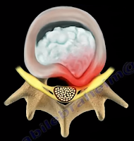
Disc herniations usually occurs posterolaterally, but it may also be central. The cauda equina is composed of several nerves within the lower end of the spinal canal. The top of the spinal cord is a tubular bundle of nervous tissue extending from the brain.

The following section of the spinal cord is called the Conus Medullaris and is the lower end of the spinal cord. The Cauda Equina is made up of multiple nerve roots beginning at the level of L1.


The most common disc herniation is the Posterolateral Disc Herniation. This type of herniation is a nerve root injury, which will cause changes to both the sensory and motor skills as well as the reflexes. A posterolateral disc herniation usually affects the foot and ankle, and may cause unilateral leg pain and weakness. Observe for a positive straight leg raising test. These herniations are usually initially treated with conservative methods.  


A central disc herniation will cause cauda equina syndrome, which is a compression over the lumbosacral nerve roots. This compression will cause more back pain than leg pain, and bladder and bowel symptoms will be evident. This herniation is considered a surgical emergency.

Tuesday, February 27, 2018

Sciatica, Disc Herniation, and Piriformis Syndrome


The sciatic nerve is a large nerve that comes from the lumbosacral plexus. The sciatic nerve has five nerve roots, L4, L5, S1, S2, and S3. The sciatic nerve runs from the lower spine, through the buttock to the lower leg and foot. The sciatic nerve initially emerges from the pelvis and exits the greater sciatic notch anteriorly and deep to the piriformis muscle, exiting below the piriformis muscle.
The sciatic nerve then enters the thigh between the ischial tuberosity and the greater trochanter of the femur. In about 10% of patients, the sciatic nerve is separated by all or part of the piriformis muscle. The sciatic nerve enters the thigh beneath the lower border of the gluteus maximus muscle. The nerve then runs down and branches out within the posterior aspect of the thigh, down to the leg and foot. The sciatic nerve gives multiple sensory and motor branches to specific areas and muscles in the leg and foot. The two main branches of the sciatic nerve are the posterior tibial nerve and the common peroneal nerve.
Irritation of the sciatic nerve may occur at multiple sites. The first site that we need to look at is the spine—which is where irritation may occur, usually from lumbar disc herniation. This is considered true sciatica or lumbar radiculopathy. Another site for irritation of the sciatic nerve is at the piriformis muscle. The sciatic nerve may become compressed by the piriformis muscle (piriformis syndrome). Piriformis Syndrome is a diagnosis of exclusion! If the patient has symptoms of sciatica, then there must be an MRI of the spine that is negative, proving that the symptoms are not associated with a possible disc problem. Once the MRI is negative, then you can say that the condition of sciatica may come from piriformis syndrome.
Disc Herniation