Showing posts with label heel pain. Show all posts

Monday, November 26, 2018

Rupture of the Plantar Fascia


Rupture of the Plantar Fascia

Plantar fascia rupture is not a very common injury, and it has the characteristic of acute pain in the arch of the foot. It occurs due to a tear in the plantar fascia, and that tear is painful. Rupture is often associated with long standing flat feet deformity or can occur from steroid injections. Another predisposing factor for plantar fascia rupture is plantar fasciitis.

Anatomy

The plantar fascia is formed by three bands: the medial, the central, and the lateral. The plantar aponeurosis is the central part of the plantar fascia. The plantar fascia is inserted into the medial tuberosity of the
calcaneus and extends distally, becoming broader and thinner. The plantar fascia acts as a bow string.

The rupture of the plantar fascia may be misdiagnosed as plantar fasciitis. When the plantar fascia tears, the patient will describe a tearing pain that usually occurs during athletic activity. The tear may be complete or incomplete. Complete tear of the plantar fascia occurs from sudden trauma or injury. The patient feels “popping” or “snapping” suddenly. Walking will be very difficult with tenderness, swelling and significant bruising on the sole of the foot (the condition is painful). Some patients may have a noticeable tightness of the calf muscle (equinus contracture) in association with rupture of the plantar fascia. Partial rupture is less common and occurs from overuse, as in running. MRI will identify the rupture, and it can also identify if the rupture is partial or incomplete. Rupture is often in the arch of the foot opposed to where the plantar fascia inserts into the heel (calcaneus). Ultrasound has the same accuracy as MRI for imaging the plantar fascia. Interpretation of the plantar fascia rupture may be difficult. You may need dynamic maneuvers with dorsal flexion of the forefoot to stretch the plantar fascia. Usually the proximal part of the plantar aponeurosis is clearly visualized on ultrasound. MRI is probably better in diagnosis plantar fascia rupture.

Treatment of Plantar Fascia Rupture

-Non-Weight Bearing for 2-3 Weeks
-Walking Boot
-Crutches
-Physical Therapy
-Surgical Treatment is the last resort.
-Could be used in some athletes who continue to have pain despite a well conducted conservative treatment
-Surgery is done to release the fascia and the excise the scar

Patients with rupture of the plantar fascia typically achieve a favorable outcome with return to full activity. 


Monday, November 12, 2018

Ankle Ligaments Injury, Tests & Assessment


Ankle Ligaments Injury, Tests & Assessment

The ligaments of the ankle are complex. Injury to these ligaments are called ankle sprains. Sprain of the ankle is usually a low ankle sprain. Occasionally, it can be a high ankle sprain. Sprain of the ankle can be confused with other conditions that can happen around the ankle such as:
  • ·         Osteochondral lesion
  • ·         Peroneal tendon subluxation
  • ·         Fracture of the lateral talar process
  • ·         Fracture of the anterior process of the calcaneus
  • ·         High ankle sprain (syndesmotic injury)

Here are a few tests that are used to test for injury of these ligaments:

  • Anterior Drawer Test
  • Squeeze Test
  • External Rotation Stress Test
  • Talar Tilt Test (Inversion Test)


If the patient cannot bear weight on the ankle, the patient should get an x-ray. Injury of the deltoid ligament occurs at the medial side of the ankle, and it is usually associated with ankle fractures. Sometimes injury of the deltoid ligament is occult and the patient will need external rotation stress x-rays to demonstrate injury of the deltoid ligament. Injury to the lateral side ligaments is referred to as low ankle sprain. The anterior talofibular ligament is the weakest ligament on the lateral side. The anterior drawer test is done to test the competency of the anterior talofibular ligament. It is done in 20 degrees of plantar flexion and compare it to the other side. A shift of an absolute value of 9 mm on the lateral x-ray or 5 mm compared to the other side is positive. The calcaneofibular ligament is usually injured after the anterior talofibular ligament. The test used to diagnose injury of the calcaneofibular ligament is called the talar tilt test or the inversion test. Less than 5 degrees of tilt is usually normal. The final area of injury is called a high ankle sprain or injury to the syndesmosis. Contrary to a low ankle sprain, a high ankle sprain may require surgery. This is how injury to the syndesmosis occurs. Always check the fibula proximally to avoid missing a Maisonneuve fracture. The Maisonneuve fracture will have a proximal fibular fracture, a syndesmotic injury, and a deltoid ligament injury. This will require surgery. The tests used to diagnose high ankle sprains are the squeeze test and the external rotation stress test. The squeeze test is performed by squeezing the tibia and fibula at mid-calf. This will cause pain at the syndesmosis if a high ankle sprain is present. The external rotation test is the other test used to diagnose a high ankle sprain or an injury of the syndesmosis. The external rotation test is performed by first placing the ankle into a neutral position. Then, apply external rotation stress and finally, get a mortise view radiograph. There is a positive result for syndesmotic injury if the tibiofibular clear space is more than 5mm or if the medial clear space widening is more than 4mm.

Tuesday, March 27, 2018

Heel Pain & The Baxter's Nerve


Pain located at the heel can have several different causes. It is important to make the correct diagnosis for the cause of the heel pain, so that appropriate treatment can be given to the patient. Common causes of heel pain include:


  1. Baxter’s Nerve Compression
  2. Plantar Fasciitis
  3. Fat Pad Atrophy
  4. Achilles Tendonitis
  5. Stress Fractures of the Calcaneus
  6. Lumbar Spine Radiculopathy
The sites and locations from different causes of the pain are all very close to each other, making it difficult to determine the source of the pain and to diagnose injuries.
In plantar fasciitis, irritation and swelling will occur at the thick tissue on the bottom of the foot. This fascia can become inflamed and painful, making walking more difficult. Plantar fasciitis is most severe in the morning when patients first stand on their feet. Stretching exercises or walking, often helps in relieving the painful tightening associated with plantar fasciitis. Pain symptoms will intensify with prolonged exercise or standing.  Plantar fasciitis is usually associated with a tight heel cord.


During fat pad atrophy, the fat that cushions the calcaneus is thinned. This condition is common in elderly people and can cause significant pain while walking. It also commonly occurs in patients with a history of steroid injections.
Achilles Tendonitis is a chronic injury to the tendon that joins the heel to the muscles of the lower leg, primarily occurring from overuse. The Achilles tendon gives us the ability to rise up on our toes, allowing for the act of walking. If a patient has Achilles tendonitis, walking can be painful and difficult.


Stress fractures of the calcaneus can occur due to overuse injuries. The patient will experience weight bearing pain. Stress fracture injuries of the heel are typical in patients who engage in running sports, due to the repetitive shock being placed on the heel. Side to side compression of the calcaneus is painful.
Pain on the lateral side of the foot may be a result of L5-S1 radiculopathy.
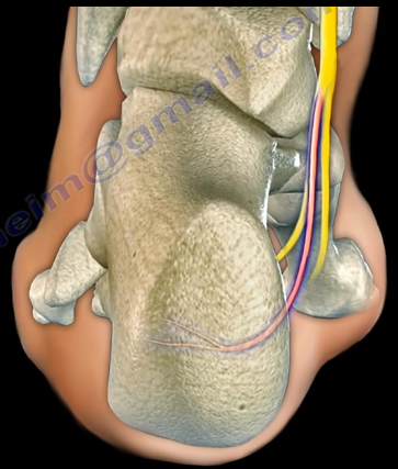
The Baxter’s nerve is the first branch of the lateral plantar nerve. It also contributes to 20% of all heel pain causes. The Baxter’s nerve provides motor innervation to the abductor digiti minimi muscle. When the nerve is affected by compression, the symptoms are similar to planter fasciitis. Entrapment of the Baxter’s nerve may cause up to 20% of heel pain, but is still overlooked. The first branch of the lateral planter nerve is the inferior calcaneal nerve (Baxter’s Nerve).


The nerve courses vertically between the abductor hallucis and the quadratus plantae muscles, then makes a 90° horizontal turn, coursing laterally beneath the calcaneus to innervate the abductor digiti minimi muscle. The Baxter’s nerve can become entrapped within the medial heel. The entrapment will result in heel pain, paresthesia, abnormal sensations on the plantar aspect (bottom) of the heel, and medial heel tenderness. There are two sites of entrapment that are typical with Baxter’s nerve impingements. The first site is located between the fascia of the abductor hallucis and quadratus plantae muscles. The second site is where the nerve passes along the anterior aspect of the medial calcaneal tuberosity.


Treatment


Management is usually conservative and consists of heel stretching exercises, rest, NSAIDs, and Orthotics. Surgery is done as a last resort with neurolysis of the nerve after release of the deep fascia of the abductor hallucis muscle. There is a good-excellent result in about 90% of patients after decompression of the nerve. Involvement of the Baxter’s nerve may affect running athletes causing pain on the medial plantar aspect of the foot.