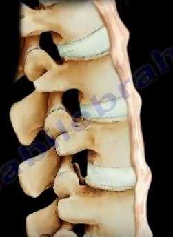
The DISH has flowing ossification along anterolateral aspect
of at least four continuous vertebrae. When you look at the x-ray, you find
ossification along the anterior aspect of the body but separate from the
vertebrae and the disc height is preserved. It occurs in older patients (50
years and above). It affects all of the spine (more in the thoracic spine),
especially on the right side, which is typical of DISH. The syndesmophytes are
equal on the right and left sides in the lumbar and cervical vertebrae.
There is
no involvement of the discs and there is no facet fusion or sacroiliac joint involvement.
The patient may have other comorbidities such as gout or diabetes, and you need
to get the hemoglobin A1c (HbA1c test) in these patients. Some patients may
have high cholesterol levels. The patient will complain of back pain and spinal
stiffness. DISH will have large syndesmophytes, and if the condition occurs in
the neck, it will cause dysphagia, hoarseness of the voice, and sleep apnea. Diagnosis
can be established by x-ray of the spine. On lateral x-ray of the cervical
spine, you will find anterior bony fragments and the discs are preserved. The fractures
in the spine are usually due to a hyperextension injury and can be occult,
resulting from minor trauma and may have major instability. There is an
increased mortality in c-spine trauma in DISH, high mortality especially in non-operative
treatment. If the patient has a history of sudden neck or back pain, then the
patient will be assumed to have an occult fracture, so try to get a CT scan or
an MRI even if the pain is minimal and even if the x-rays appear normal. Heterotopic
ossification after total hip arthroplasty is more in patients with DISH.
What is the difference between DISH and Ankylosing
Spondylitis?
DISH
-Flowing large syndesmophytes
-No bamboo spine
-Sacroiliac (SI) join will not be involved
-Occurs in older patients
-some patients may have diabetes, check hemoglobin A1c
Ankylosing Spondylitis
-Diffuse ossification of the disc space without large
osteophytes
Fibular fractures are usually associated with a complex
injury, however they can be an isolated fracture. Complex injuries where a
fibula fracture can occur include: fracture of the fibula and tibia, ankle
fracture, pilon fracture, and Maisonneuve fractures.
Maisonneuve fractures
involve a fracture of the proximal fibula associated with an occult injury of
the ankle. Isolated fibular fractures are rare and usually the result of direct
trauma. The fibula carries about 15% of the axial load and is the site of
muscle attachment for the peroneus muscles and the flexor hallucis longus
muscle. Check the patient who has a fibular fracture and no other fracture
involving the tibia to rule out a possible Maisonneuve fracture, especially if
there is no history of direct trauma to the leg. A high index of suspicion is
necessary to diagnose and treat this injury. For high fibular fractures, the
physician should look for signs of syndesmotic injury. Syndesmotic injury may
include an unexplained increase in the medial clear space or the tibiofibular
clear space is widened (should be less than 5mm). The x-ray will show the
fracture to be rotational or oblique. Maisonneuve fractures require surgery to
fix the syndesmosis.
Treatment will consist of reduction and fixation. It is
important to determine if the injury is a Maisonneuve fracture or an isolated
fibular fracture. An isolated fibular fracture will not need surgery.
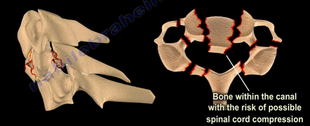
Fifty percent of patients with Jefferson fractures will have
associated spine injuries. The canal is wide with a low risk of spinal cord
injuries unless the transverse ligament is disrupted. It is difficult to view
Jefferson Fractures on an x-ray (usually seen on the lateral side”. This
fracture is considered a “Junctional Fracture” and could be missed. The classic
Jefferson fracture is a burst fracture that results from an axial load. It
could be a four part fracture with bilateral fractures of the anterior and
posterior arch. There are variations which include two and three part fractures
and incomplete formations of the posterior arch can be mistaken as a fracture.
When speaking of Jefferson fractures, it is important to be
familiar with the structures that may be involved. These bony structures
include: The Atlas (C1), Axis (C2), and the odontoid process. C1 and C2 are
stabilized together by the transverse ligament and C1 and C2 provide a 50% of
rotation of the neck. The C1 is a ring. At the upper cervical region, the
spinal canal is 2.5 times larger than the cord size. The stability and
treatment of Jefferson fractures depends on the integrity of the transverse ligament
and the displacement of the fracture. You need to know about the important
ligaments related to the Jefferson fracture. These ligaments include: the
transverse ligament, the apical ligament, and the Alar ligament.
Diagnosing ligamentous injury
In order to determine a ligamentous injury, the physician
will want to check the Atlanto-dens interval (A.D.I). Normally, this interval should
be less than 3mm in adults and less than 5mm in children. If the ADI is between
3-5mm, this indicates an injury to the transverse ligament; the transverse
ligament holds the odontoid and C1 together, alar and apical ligaments will be
intact. If the A.D.I measures greater than 5mm, then there is an injury to the
transverse, alar, and apical ligaments.
Fracture Types
A bony injury with the intact transverse ligament and a
lateral mass displacement less than 7mm and the A.D.I is less than 3mm is
considered a stable fracture. Nondisplaced fractures of this nature should be
treated with a rigid orthosis. If the fracture is displaced, a halo will need
to be used.
Another type of fracture can occur at C1 with a transverse
ligament tear. The Atlanto-dens interval will be more than 3 mm in adults. The
treatment will depend on the type of injury to the transverse ligament. With
bony avulsions of the transverse ligament, the halo will need to be used
cautiously. However, some surgeons prefer to do a fusion of C1 and C2. If there
is an intrasubstance tear of the transverse ligament, the surgeon will perform
a fusion at C1-C2. The surgeon will need to do early surgery as this is a
significant injury with a risk of spinal cord compression.
In regards to “Open Mouth Views”, the normal overhang is visible
during an “Open Mouth View”. If it is just a bony injury Jefferson fracture, the
combined overhang will be less than 7mm and the transverse ligament is intact
and it is a stable fracture. If a Jefferson fracture has a combined overhang of
more than 7mm, then the transverse ligament is probably torn and there is an
unstable fracture present.
Radiological Studies
A CT scan is probably the best study in diagnosing the
characteristics of the bony injury. An MRI is the best study in diagnosing any
associated transverse ligament injuries.
Pelvic fractures may cause significant bleeding. The
superior gluteal artery is responsible for the majority of bleeding in pelvic
fractures with an arterial injury. Most of the bleeding in pelvic fractures is
from the veins and the fracture itself. Hemorrhage is the most life-threatening
complication associated with pelvic injuries and will typically occur at the
Superior Gluteal Artery. Hemorrhage of the Superior Gluteal Artery can be
fatal. Approximately 10% of patients will have severe bleeding. Severe bleeding
usually occurs in fracture patterns that are highly unstable to both rotational
and vertical forces.
APC III (open book like type) is the complete disruption of
anterior SI, sacrotuberous, and sacrospinous ligaments; disrupted posterior SI
ligaments.
Vertical Shear Fractures are very bad fractures as they
cause the complete disruption of the anterior and posterior ligaments;
cephaloposterior displacement. Anteroposterior compression or vertical shear
injuries are consistently associated with a higher risk of mortality from bleeding.
The mortality rate is directly related to the amount of shock the patient is in
at the time of presentation.
When treating patients with pelvic fractures and massive
bleeding, it is important to remember that the patient will lose approximately
35% of their blood volume with acute hemorrhage before a sustained decrease in
systolic blood pressure occurs. Immediate application of a pneumatic anti-shock
garment is absolutely contraindicated in patients with a rupture of the
diaphragm. Ringer’s lactate is the preferred initial fluid replacement used to
resuscitate hypovolemic trauma patients in shock. A hypotensive blunt trauma
patient will be given an initial fluid push with 2,000mL of Ringer’s lactate. A
patient with bleeding and in shock will probably require O negative blood. If
the patient is given 4 units of blood but remains hemodynamically unstable,
then angiography and embolization is needed. Immediate application of an
external fixator is another method to control bleeding, especially if the
pelvis is unstable in external rotation. An abdominal and pelvic CT scan will
clearly define the bony injury as well as the extent and source of the
bleeding.
The best treatment for pelvic fractures with bleeding is a
blood transfusion with correction of hypothermia and coagulopathy.
The bulbocavernosus reflex indicates the absence or presence
of spinal shock. Spinal shock usually occurs between 24-72 hours after a spinal
injury. Spinal shock is manifested by the absence of the bulbocavernosus
reflex, hypotension, bradycardia, and complete loss of motor sensation and
reflexes. When the reflex is absent, this means that the patient is in spinal
shock because the anal sphincter will not contract when the reflex is absent.
When the reflex is present, this signals the end of spinal
shock; the anal sphincter will contract when the reflex is present. The reflex
is check by monitoring anal sphincter contraction in response to squeezing of
the penis of clitoris, or by pulling on an indwelling Foley catheter. It is a
polysynaptic response mediated by S2-S4.
What is Spinal Shock?
Spinal shock is the loss of sensation and motor power
following a spinal cord injury. Spinal shock is the loss of sensation and motor
power following a spinal cord injury. After an injury to the spine, if the
patient has no motor or sensory below the level of the lesion, the physician
must determine if the patient is in spinal shock by checking the
bulbocavernosus reflex.
If there is no anal contraction (absent bulbocavernosus
reflex), this indicates that the patient is still in shock and the prognosis
cannot be determined. If anal contraction is present (positive bulbocavernosis
reflex), this indicates the end of spinal shock. The patient’s condition and
prognosis at this point can be determined by examining sacral sparing (positive
sacral sparing indicates an incomplete lesion).
Loss of sensation and motor power below the level of injury
indicates complete spinal cord injury. Once the diagnosis of neurogenic shock
is established, the blood pressure should be managed with vasopressors to prevent
fluid overload. With the end of spinal shock, the prognosis can be determined. Examine
the patient thoroughly, including sacral sparing. The patient may have normal,
partial, or complete indications.
Lactic acid is a byproduct of anaerobic metabolism (Metabolism
without oxygen). Normally, the cells use anaerobic metabolism and it breaks
down the glucose to form energy. Normally, the cells use oxygen available to
breakdown the glucose and produce ATP. This occurs when you have oxygen. If the
body does not have the oxygen, it then converts the metabolism to an anaerobic
metabolism. The end product is lactic acid. If the body is acidic, and the pH
drops, this means that there is a tissue ischemia. If we take the glucose
(CCCCCC), and break it down, you will have two pyruvates—each attached to a
CCC. That’s not a lot of energy and the pyruvate is a mild acid.
There are different ways to get more energy, such as going
to the mitochondria to use the oxygen and produce more ATP. When we break down
the glucose to produce energy in the presence of oxygen, so the glucose will
split into two separate three carbon molecules called the pyruvate and produces
ATP. If you don’t have oxygen, then the pyruvate will be attached to hydrogen
atoms, which is H+ (proton that is an acid). If you are acidotic and a sick
patient, you will have a lot of floating hydrogen atoms and a lot of floating
protons.
The pyruvate will attach to the free hydrogen protons. The
H+ pyruvate is called lactic acid. It is a pyruvate that is holding onto
hydrogen. The lactic acid is the end product of anaerobic metabolism.
Now, with a base deficit, as the number of protons goes up,
which is hydrogen, then the pH will go down and the patient will become
acidotic. The body uses bicarbonate as a buffer if the pH goes down. There are
a lot of protons within the body and the bicarbonate will be exhausted in this
situation. The bicarbonate goes down because we are combining it with the
protons. When the bicarbonate goes down, this becomes metabolic acidosis.
When you combine the bicarbonate with the protons, this will
make carbonic acid (H2CO3), rather than HCO3-,
which is bicarbonate. The carbonic acid can give water (H2O), and CO2.
The lactic acid will give out the protons and be buffered by the bicarbonate,
leaving lactic acid. When this occurs, the bicarbonate will go down, and this
will be the base deficit.
When checking if a patient has been resuscitated, you can
check in several ways
The two ways this is asked on exams:
Base deficit from -2 to +2
Serum lactate level (normal is less than 2.5, some sources use normal less than 2)
In our final blog post regarding Orthopaedic Emergencies, we will
review:
Transverse Atlantal Ligament Rupture
Bilateral Cervical Facet Dislocation
Spinal Cord Compression
Cauda Equina Syndrome
Transverse Atlantal Ligament Rupture
The normal Atlanto-Dental Interval is less than 3mm. An
A.D.I measuring greater than 3mm will be translationally unstable in the sagittal
plane due to transverse atlantal ligament rupture. This is usually apparent on
x-rays or CT scan. If the condition is not diagnosed, it can result in spinal
cord compression, respiratory arrest, and a catastrophic outcome. Treatment
typically requires a posterior atlanto-axial arthrodesis.
Bilateral Cervical Facet Dislocation
Facet dislocations of the cervical spine:
Unilateral Facet Dislocation
Displacement is less than 50% of the vertebral
body width
May need surgery
Bilateral Facet Dislocation
Displacement greater than 50% of the vertebral
body width
Usually needs surgery
Exclude disc herniation
Obtain a preoperative MRI to rule our disc herniation
associated with facet dislocations.
Spinal Cord Compression
Spinal cord compression is more common with cervical spine
injuries and thoracic spine injuries. Neurogenic shock resulting from spinal cord
injury may complicate resuscitation of the patient and should be differentiated
from hypovolemic shock. It is important to look for hypotension and bradycardia
as well as thoracolumbar fractures which could be missed. Treatment consists of
emergency management involving resuscitation and hemodynamic stabilization with
concurrent neurologic examination. Protocol requires steroids given early.
Definitive treatment consists of stabilization of unstable spinal injuries.
Cauda Equina Syndrome
Central disc herniation compressing the cauda equine. It results
from injury to the lumbosacral nerve roots within the spinal canal. This
syndrome presents with involvement of the bladder, bowel, and lower limbs and
usually results from central disc herniation or fractures. Central disc
herniation or bony fragments results in the compression of the nerve roots.
Early diagnosis is imperative to find the cause of the compression on the nerve
roots. Urgent decompression by the removal of the central disc herniation or
stabilization of the fracture is necessary for treatment.
Open fractures are categorized with the Gustilo-Anderson Classification. A Grade I Injury indicated a clean wound, less than one centimeter long with minimal injuries to the soft tissue and minimal bone comminution. A Grade II injury consists of a moderately contaminated wound greater than one centimeter long with moderate tissue injury and moderate bone comminution. A highly contaminated wound, usually greater than ten centimeters, segmental fractures, farm yard injuries, high velocity gunshot wounds and fractures occurring in a highly contaminated environment regardless of the size of the wound.
Grade III injuries are classified further into A, B, and C. Grade III A is a severe soft tissue injury with a crushing comminuted fracture; soft tissue coverage of bone possible. Grade IIIB consists of a very severe loss of soft tissue cover with poor bone coverage and variable—may be moderate to severe bone comminution. Grade IIIB usually requires a soft tissue reconstructive surgery in the form of local or distant flaps. Grade IIIC fractures consists of a vascular injury requiring repair or amputation. There is a very severe loss of soft tissue cover with moderate to severe bone comminution. Injury of the femoral artery from the posteriorly displaced proximal fragment of a Grade III C open supracondylar fracture of the femur. Grade III C has a high rate of amputation, nonunion and infection.
Hip Infection (Septic Arthritis)
An infection in the hip is a serious disease especially in children. The intraarticular structures will be inflamed and the increased intracapsular pressure will decrease the blood supply to the femoral head. Infection is associated with a high risk of avascular necrosis. The position of the limb in the stage of effusion, flexion, abduction, and external rotation. Complications are severe and much more common in children. Complications include: pathological dislocation, avascular necrosis, osteomyelitis, and pelvic abscesses. Urgent aspiration followed by drainage of the hip joint combined with intravenous antibiotics are the typical treatment for hip infections.
Necrotizing Fasciitis
Necrotizing Fasciitis is an insidiously advancing soft tissue infection characterized by widespread tissue necrosis. The most common causative organism—group A beta—hemolytic streptococcus. There is a high mortality rate with sepsis and renal failure. Amputation and the mortality rate is increased due to a delay in diagnosis. Predisposing factors for necrotizing fasciitis include: trauma, surgery, as well we urogenital and anogenital infections. There are three types of necrotizing fasciitis: Type I—which is Polymicrobial, Type II—which is a Group A beta-hemolytic streptococcus, and Type III—which is gas gangrene-clostridial myonecrosis. Treatment consists of an immediate surgical debridement combined with intravenous antibiotics and hyperbaric oxygen if necessary.
Fracture with Soft Tissue Compromise
Soft tissue compromise associated with fracture blisters, ecchymosis, and severe bruising which indicate a greater degree of deep soft tissue damage. Blood filled fracture blisters are associated with high wound complications. Initial management involves application of a spanning external fixator with the fracture dislocation held in reduction with traction. The definitive management involves replacing the spanning external fixation with a hybrid fixator or plate once the soft tissue edema is resolved and the skin is wrinkled, usually in one to three weeks. Spanning external fixation can often be combined with percutaneous fixation of large articular fragments. A soft tissue compromise is more common with tibial plateau fractures and tibial pilon fractures with diaphyseal extension. A calcaneal avulsion fracture is considered an emergency. Urgent reduction and fixation is mandatory to avoid soft tissue complications. Type I—is a “sleeve” type tuberosity fracture. This pressure will create skin necrosis and significant soft tissue complication.