Wednesday, December 21, 2016


 Stress Fractures:

Common for Athletes and Those with
 
Active Lifestyles


Figure 1
Fractures are typically caused by a single traumatic event such as a car accident or fall. Here, bones are exposed to forces so extreme they are unable to support energy that is placed upon them. In contrast, there are fractures caused by a repetitive application of force. These fractures, called stress fractures, occur when muscles become so fatigued they lose their ability to absorb additional shock. These fractures are categorized as overuse injuries and are characterized by tiny cracks in the bone.

Bone is incredibly resilient. Each day, bone broken down by daily living is replaced by the body in a necessary balancing act. However, this balance is disturbed with excessive physical training.
Stress fractures often present in athletes such as tennis players, basketball players, gymnasts and track and field participants. Typical stress fracture causes include improper equipment, increased physical stress, increasing the amount or intensity of an activity too rapidly and the impact of an unfamiliar surface.
Figure 2

Stress fractures are usually categorized one of two ways: a fatigue fracture or an insufficiency fracture. Fatigue fractures are the result of bone being overused or being exposed to repetitive stress beyond its ability to repair itself. In contrast, insufficiency fractures are the result of bone being deficient in minerals or vitamins.
 
A typical cause of an insufficiency fracture is if a bone has been weakened by osteoporosis.
Figure 3

 While stress fractures can occur anywhere on the body, more than 50 percent occur in the lower leg. (Figure 1) (Figure 2). Stress fractures rarely occur in the upper extremities because they are not weight bearing bones, like the tibia and bones that run from the mid-foot to the toes. (Figure 3).

There are certain red flags that may signal stress fractures. These red flags include: pain that increases over time, pain that increases with activity and decreases with rest, pain that persists

Figure 5
Figure 4
even at rest, pain that occurs earlier in each successive workout and swelling. A runner with groin pain and a negative x-ray must have a back scan or an MRI to rule out hip stress fractures.

Early diagnosis of hip fractures is necessary to avoid displacement of the fracture.

The best treatment for stress fractures is rest. After about 6 to 8
weeks, patients should be able to return to their regular activities.
It’s important to do your best to safeguard against stress fractures.
This includes implementing a prevention plan that includes the following:
alternating activities that accomplish the same fitness goals (crosstraining); setting incremental goals when participating in a new sports activity; and maintaining a healthy diet(Figure 4) with
bone strengthening vitamins and minerals such as calcium and vitamin D (Figure 5).
In some situatoins, however, surgery is needed to treat stress fractues.(Figure 6)


Figure 6


Thursday, December 8, 2016

Raynaud’s Phenomenon


Raynaud’s phenomenon is a discoloration of the fingers due to vasospasms and decreased circulation of the fingers. This condition causes the fingers to feel numb and cool and become discolored in response to cold temperatures or stress. This typically occurs more in women.


Fingers may become white due to lack of blood flow. Then as the vessels dilate in order to keep the blood within the tissues, the fingers become blue. Finally, as the blood flow returns, the fingers become red. This condition is usually bilateral.
There are two types Raynaud’s disease and Raynaud’s syndrome. Raynaud’s disease occurs on its own without a cause. Raynaud’s syndrome is caused by a connective tissue disorder such as systemic lupus erythematosus, polymyositis and scleroderma. Raynaud’s phenomenon is the initial presentation for 70% of patients with scleroderma. If you have a patient with Raynaud’s phenomenon, study them for connective tissue disorders.

In chronic cases, the nails may become brittle with ridges, with ulcers of the fingers.

Tuesday, November 29, 2016

Vitamin C


Osteoporosis is a decrease in bone mass, which can lead to an increased risk of fracture.


Vitamin C actively protects against osteoporosis and has shown to prevent bone loss in animal models. Foods such as oranges, tangerines, pineapple, cantaloupe, broccoli and cauliflower contain high amounts of Vitamin C. The treatment of osteoporosis is going beyond giving calcium and vitamin D. High vitamin C intake is associated with lower 4-year bone loss in elderly men.


Vitamin C mineralizes bone and stimulates bone forming cells to grow, while preventing too much degradation of bone by inhibiting bone absorbing cells. Vitamin C is also used for prevention of the common cold in marathon runners. Vitamin C is vital to collagen synthesis and lessens oxidative stress. It helps cross-linking of the collagen. The collagen is the glue that is present in ligaments, tendons and bones.


Can vitamin C prevent complex regional pain syndrome in patients with wrist fractures? About 10% of wrist fracture patients will develop a chronic pain syndrome called reflex sympathetic dystrophy. Taking vitamin C reduces this risk substantially. Taking 500mg of vitamin C per day for 50 days following a wrist fracture is effective for preventing RSD. This double-blind study shows that vitamin C was associated with a lower risk of RSD after wrist fractures.

Tuesday, November 22, 2016

Phantom Pain Amputation


Phantom pain is a common symptom after amputation of a limb. Up to 80% of amputee patients will have phantom pain symptoms. The patient will feel the sensation of pain in the area where the limb was amputated.


Tingling, cramping, heat or cold sensation may be felt in addition to shooting pain and these symptoms usually decrease or disappear with time.


The patient may feel like the foot or toes are moving or itchy. If the symptoms continue for more than 6 months, the prognosis is usually not good. The causes of phantom pain are not very clear. The brain thinks that the limb is still present. The memory of pain is returned. The signal is interpreted as pain, regardless of the signal from the injured nerve. Peripheral and psychological factors may contribute to the condition of phantom pain.


Peripheral Theory

The pain may result from the nerve endings making a neuroma. The neuroma may generate an abnormal electric impulse that the brain will interpret as pain.

The term “phantom” does not mean that the symptoms are imaginary or “in the patient’s head”. Phantom pain is a real phenomenon. The symptoms of phantom pain may range from mild to severe and the pain may be constant.

Treatment

There are multiple treatment options that will depend on the level of the pain. Heat, massage, biofeedback and relaxation techniques, physical therapy, TENS unit, medications (including anticonvulsant or antidepressants) and surgery may be utilized. Surgery is usually used for neuromas. Neurostimulation technique is used as a spinal cord or deep brain stimulation.

Mirror Box Visual Feedback Therapy

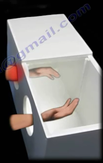
A mirror box is a box with two mirrors in the center (one facing each way) that is used to help patients feel like they still have a limb after having it amputated. The patient placed both the amputated limb and the other limb into the holes of the mirror box. The mirror is used to create a reflection of the non-amputated limb. Some people find that by doing exercises and moving the limb it can help to relieve the pain from phantom pain.


Thursday, November 10, 2016

Bone Remodeling in Children


Children have a unique ability for healing of their fractures and remodeling of their deformities. The physician should strive to achieve anatomic reduction of fractures in children.


The surgeon may not be able to achieve acceptable reduction every time, fractures do not always remodel. The deformity may lead to unacceptable results in cosmesis and function.

What are the rules for remodeling in children?

Age of the patient:

Younger children have better remodeling potential. The younger, the better. Children with two or more years of growth left have a chance for remodeling. The periosteum in children is thick and promotes faster healing of the fracture as well as increased potential for remodeling.

Distance:

The distance of the fracture from the end of the bone. Fracture in the metaphysis remodels better than in the middle of the bone. A fracture in the middle of the bone has less potential for remodeling.

Severity of Angulation

If there is minimal angulation, the bone could remodel completely. Where angulation is more severe, the bone will partially remodel. Angulation in the plane of joint movement is most likely to improve with growth and remodeling.

Remodeling will not improve displaced fractures involving the joint or the growth plate.

Wednesday, November 2, 2016

Dr. Nabil Ebraheim photos: 11/2/2016

Back Pain

There is certain etiology of low back pain in 85% of cases. Patients with a single occurrence of low back pain return to work within 6 weeks 90%of the time. Moreover, most patients get better with time. In fact, about 60% of patients get better in approximately 10 days.


Low back pain is the second most common cause of work absenteeism. If a person has a history of low back pain, it is likely they could develop occupational low back pain. Persistent back pain for more than 6 months constitutes only four percent of cases. Disability is usually closely related to compensation and litigation.

The least amount of pressure on the discs is measured with the patient lying in the supine position. The highest amount of disc pressure is measured while sitting with 20 degrees of forward leaning with a 20 kg load in the arms. It is better to keep the weight of the load close to the body. This will reduce the compressive forces placed on the lumbar spine. Yoga activities and exercises performed during sitting probably have less pressure being placed on the discs.

Physical factors which lead to low back pain include the following: lack of fitness; heavy lifting of objects; operating motor vehicles; prolonged sitting; operating motor vehicle accidents; prolonged sitting; operating vibrating tools; and cigarette smoking (nicotine causes disc degeneration).

There are many sports-related activities related to low back pain. When golfing, pain occurs as the result of twisting, bad forward bending, and most importantly overarching the spine during the swing. After the age of 40, we lose about 50% of our rotational spine movement. It is important to stretch and warm-up before starting the game. Vibration caused by horseback riding increases the load on the discs. The back muscles work constantly to keep posture straight. Caring for horses could also be detrimental to the back due to the bending and lifting associated with their care.

Virtually any structure in the spine can hurt including: the facet Joints; invertebral discs; spinal canal; sacroiliac joints; muscles; ligaments; nerves; hip joints/Piriformis muscles; and trochanteric bursitis. Red flags for cancer include: over 50 years of age; pain at rest and night; unexplained weight loss; history of cancer; and bone destruction involving the pedicle is pathognomonic. Red flags for infection include: diabetes; fever; drug abuse; urinary tract infection; and previous surgery

Treatment for acute low back pain, without sciatica (leg pain), involves a short period of bed rest, anti-inflammatory medications, and physical therapy for a short period of time. Patients will also be advised to work within the limits of pain.

Tuesday, November 1, 2016

X Linked Dominant Inheritance

Some orthopaedic conditions that Dr. Nabil Ebraheim has seen throughout his years as a surgeon are caused by genes.
X-linked dominance will not skip a generation.
One example of an x-linked dominant issue is Leri-Weill Dyschondrosteosis (LWD). LWD is a dominantly inherited skeletal dysplasia characterized by short stature, mesomelia, and Madelung wrist deformity. This is a rare genetic disorder caused by a mutation of the SHOX gene. The shortened stature is caused by the homeobox containing gene.
Hypophosphatemic Rickets is a vitamin D resistant rickets. X-linked hypophosphatemic rickets with inability of the renal tubules to absorb phosphates (phosphates levels are down linked to the PHEX gene - Phosphate Regulating Endopeptidase Homolog, X-linked). This is the most common cause of rickets within the United States of America. Dr. Nabil Ebraheim suggests that the doctor will look for a child short in stature, bowing of the lower limbs, and low serum phosphate levels. The Alkaline phosphatase (ALP) levels will also be high. Calcium levels in these patients will be normal, as well as the Parathyroid hormone (PTH). The treatment for this is usually high doses of phosphate replacement and high doses of vitamin D to facilitate the phosphate absorption.
In x-linked dominance, the affected individual has an affected parent due to the dominant gene. All affected males must have an affected mother. All affected fathers will have affected daughters and no affected sons. Dr. Nabil Ebraheim suggests looking to the males. Looking and the sons and fathers will be your clues.

Dr. Nabil Ebraheim's YouTube Channel
Dr. Ebraheim's Huffington Post

Monday, October 31, 2016

AO/ASIF Foundation


Prior to becoming the Trauma Fellowship Program Director and Orthopaedic Residency Program Director at the University of Toledo Medical Center, Dr. Nabil Ebraheim completed an AO Fellowship (technique of internal fixation) at Kantonsspital Chur in Switzerland. Possessing particular experience in handling spinal fusions and bone fractures, Dr. Nabil Ebraheim is considered a leading expert in the field of orthopedics.

Established in Switzerland in 1958, the AO technique of internal fixation, also known as the Association of the Study of Internal Fixation (ASIF) set out to change the way fracture treatments were performed. The association developed revolutionary instruments for use in treatment procedures along with advanced implants to help with recovery.

The AO/ASIF Foundation included 90 of the world’s leading trauma surgeons, and it created more teaching and training opportunities for these experts to promote their research. The clinical documentation and trauma treatment taught by the organization is responsible for many modern advancements in the field of orthopedics.

Tuesday, September 27, 2016

The Causes and Symptoms of Carpal Tunnel Syndrome


Professor and chairman of the Department of Orthopaedic Surgery at the University of Toledo Medical Center, Dr. Nabil Ebraheim provides care to patients with complex fractures involving the ankles and joints. Additionally, Dr. Nabil Ebraheim maintains a YouTube channel that provides viewers an in-depth look at various issues in his area of medical focus, such as carpal tunnel syndrome. 

Situated on the palmar side of the wrist, the carpal tunnel is a narrow passageway of ligaments and bones. The median nerve passes through this area, as do nine flexor tendons that help control finger and thumb movements. Carpal tunnel syndrome occurs when the transverse carpal ligament places pressure on structures such as the median nerve. Symptoms include numbness, tingling, weakness, and pain in certain regions of the hand.

In certain cases, the carpal tunnel needs to be released, or widened, to provide more space for the ligaments and nerve to pass. A key surgical concern is avoiding cutting the recurrent motor branch of the median nerve, which allows for opposition of the thumb.

Monday, May 2, 2016

University of Toledo - Dr Nabil Ebraheim

Dr Nabil Ebraheim

Dr. Nabil Ebraheim's doctor page at the University of Toledo's Medical Center can be found here.