A patient with a complete anterior interosseous nerve injury
or a high medial nerve injury should be asked to make a fist. The first and
second digits will have difficulty in flexing, while the other digits will
flex. The third digit will be weak, while the fourth and fifth digits are
normal. This position of the hand is similar to the position taken during a
hand blessing. The Benedictine sign is different from an “ulnar claw hand”.
Ulnar claw hand refers to damage to the ULNAR nerve and is seen when attempting
to extend all the digits (leaving the 4th and 5th digits
flexed). The O.K. sign is used to check for paralysis of the anterior
interosseous nerve due to entrapment or compression injury. A patient with
paralysis of the anterior interosseous nerve will be unable to make the O.K.
sign. This is due to weakness of the flexor pollicis longus and flexor
digitorum profundus muscles. A typical pinch attitude is associated with
anterior interosseous nerve injury.
The anterior interosseous nerve arises from the median nerve
about 4-6cm distal to the elbow, which is about 1/3 of the way down the
forearm. It exits from the anterolateral aspect of the median nerve and it runs
between the radius and the ulna on the interosseous membrane between and below
the muscles of the flexor digitorum profundus and the flexor pollicis longus.
The anterior interosseous nerve supplies the flexor digitorum profundus muscle
for the index and long fingers. It also supplies the flexor pollicis longus and
the pronator quadratus muscles. The flexor digitorum profundus muscle for the
index and long fingers is supplied by the anterior interosseous nerve. The
medial part of the FDP is supplied by the ulnar nerve (FDP has dual
innervation). The anterior interosseous nerve passes dorsal to the pronator
quadratus with the anterior interosseous artery and provides innervation to the
volar wrist capsule. The terminal branch of the anterior interosseous nerve
innervates the carpal joint capsule.
In patients with Martin-Gruber Connection, the median nerve,
or anterior interosseous nerve to the ulnar nerve in the forearm may present
with intrinsic muscle weakness. It may be differentiated also from
Parsonage-Turner Synrome (acute brachial plexus neuritis) and patient may have
pain in the affected extremity. In anterior interosseous nerve entrapment, the
median nerve conduction study result will be normal, however the needle EMG of
the anterior interosseous innervated muscles will be abnormal.
A SLAP tear is a tear that occurs where the biceps tendon
inserts into the superior labrum. A SLAP tear is different from a Bankart
lesion. SLAP tears are not common and can be hard to diagnose. Symptoms of a
SLAP tear include: pain deep within the shoulder or in the back of the
shoulder, as well as catching, popping, or clicking sensations. The patient may
also experience pain when throwing a ball with a decrease in velocity and the
feeling of having a dead arm after pitching. Patients will also experience pain
with overhead activity which mimics impingement syndrome. This typically
affects throwing athletes. When the biceps tendon is involved, pain may also be
located at the front of the shoulder. A SLAP tear can be an isolated lesion or
it can be associated with internal impingement, articular sided cuff tear, or
instability.
A SLAP tear is diagnosed with a clinical examination and
testing. The O’Brien’s test is the most commonly used test. Multiple tests are
usually used including the anterior slide test and the clunk test. An MRI with
contrast is the best imaging technique. When performing the O’Brien’s test, the
patient is standing or sitting with the arm at 90° of flexion, 10° of
adduction, and full internal rotation with the forearm pronated. The examiner applies pressure to the forearm
and instructs the patient to resist the applied downward force. Pain at the
shoulder joint suggests a SLAP lesion. Decrease in pain of the shoulder joint
on supination of the arm is suggestive of a SLAP tear.
Treatment consists of physical therapy, anti-inflammatory
medications, injections, and surgery (when conservative treatment fails). If
surgery is necessary, a labral debridement will be performed for minor tearing
and fraying. Biceps Tenodesis is becoming popular, as it is a procedure that
cuts the biceps tendon where it attaches to the labrum and reinserts it in
another area, usually in front of the shoulder. A biceps tenotomy is a
procedure that cuts the biceps tendon from the glenoid, releasing the long head
of the biceps tendon from its attachment allowing it to fall into the upper arm
out of the shoulder joint. A biceps tenotomy is probably best suitable for some
elderly patients. A SLAP repair is a procedure which uses sutures to anchor the
torn labrum to the glenoid. This repair is usually done for athletes and
patients under the age of 40 years.
To view my Youtube video, 'Massive Rotator Cuff Tear- Classic', click here.
Massive rotator cuff tears are a disabling problem. These
tears can cause pain, weakness, and sometimes swelling of the shoulder. The
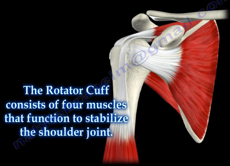
rotator cuff consists of four muscles that function to stabilize the shoulder
joint: supraspinatus rotator cuff tendon, subscapularis tendon, infraspinatus
rotator cuff tendon, and the teres minor rotator cuff tendon. The supraspinatus
tendon is the most common of the rotator cuff tendons to become ruptured. Massive
tears of the rotator cuff that are greater than 5cm usually involving both the
supraspinatus and infraspinatus tendons.
Massive tears of the rotator cuff are defined as tears
greater than 5cm, usually involving both the supraspinatus and infraspinatus
tendons. Retraction of the rotator cuff tendons along with muscle atrophy and
fatty infiltration can occur. This makes surgical reconstruction difficult with
the surgical outcome being unpredictable and less than satisfactory.
Treatment varies from physiotherapy to replacement of the
humeral head. Arthroscopic or open repair is usually the selected treatment.
Reconstruction can be done in selected cases. A rotator cuff arthropathy is
performed on massive cuff tears that are associated with superior migration of
the humeral head as well as instability and arthritis of the shoulder. The
patient will have pseudoparalysis and an x-ray will show shift of the humerus
proximally. An MRI will show massive cuff tear with retraction at the level of
the glenoid with atrophy of the muscle and fatty infiltration. A reverse shoulder
is the treatment of choice for the elderly with rotator cuff arthropathy as it
improves the pain and function. Hemiarthroplasty is the treatment for younger
patients. A standard head or a big humeral head can be selected.
A patient with a massive tear of the cuff usually develops
weakness of the shoulder and becomes unable to actively lift the arm without
assistance. Fluid collection within the shoulder may occur with a massive tear
of the rotator cuff.
There are two types of hip joint
dislocations: posterior and anterior. The position of the leg is important in
determining the type of hip dislocation. When the hip is dislocated, the leg is
usually shortened and it assumes a different position than the normal leg (the
other leg). If the dislocation is posterior, the leg will be in adduction and
internal rotation. If the dislocation is anterior, the leg will be in abduction
and external rotation. Notice that the affected extremity is shortened and
externally rotated. Leg shortening can also be seen in hip fractures and the
leg will be shortened and externally rotated.
Dislocation of the hip following
total hip surgery may require revision surgery, but it is rare. The majority of
hip dislocations after total hip dislocations are posterior, and they are
usually treated without surgery. Most occur within the first month of THA; 1-4%
in primary, 16% in revision. There is more incidence of dislocation in revision
hip replacement.
Causes & Risk Factors:
Posterior Approach (try to repair the capsule
adequately)
Malposition of the component
Ideally, the normal cup component will be in 20°
of anteversion and 40° of abduction
When the hip dislocates posterior, always check
for retroversion of the cup.
Prior hip fracture surgery, especially in the
elderly
Weakness of the abductor muscle—must achieve
soft tissue tension and function
Alcohol abuse
Improper neck length—looseness of the hip
The patient should be careful to
avoid all activities that cause dislocation after total hip surgery. The
patient should use a pillow between the legs while sleeping on their back and
they should be careful to not cross their legs in their sleep. Patients cannot
sleep on their sides as well. The patient should not bend the body at the waist
farther than 90°. When sitting, the patient must avoid chairs that make it
difficult to stand up, and sit at more than a 90° angle. The patient must not
sit with their legs crossed in the chair. The patient must be made aware that
if the leg is changed from its usual position, or becomes shortened, then the
hip is probably dislocated and their doctor should be consulted.
X-rays of the dislocated total hip
should include AP and lateral views. Look for eccentric wear and look for the
position of the prosthesis. CT scans may be needed before or after reduction of
the dislocation to check the version of the components. Treatment is variable
and depends on the situation. The treatment should be tailored for each case.
The majority of these cases with early dislocations can be treated successfully
with closed reduction and immobilization.
The treatment should start with
closed reduction of the total hip and immobilization. Hip stability is checked after
reduction of the dislocation. Immobilization can be done by a brace or a hip
spica. Trochanteric osteotomy and advancement of the trochanter and tensioning
the abductor muscle. Screws or wires can be used. The prosthesis must be in
good alignment for this procedure to work. Constrained acetabular components
are used when the abductor muscle is deficient and the component position is
good. Revision total hip is done in recurrent dislocation with malposition of
the component or polyethylene wear.
Tibial bowing is not uncommon. The direction and the apex of
the tibial bow can alert the clinician to the type of the deformity, its
treatment, and its prognosis. Tibial bowing can occur anteriorly, laterally,
anterolaterally, and posteromedially.
Anterior bowing is often associated with fibular hemimelia.
Thisbowing may be associated with the
loss of the lateral rays of the foot, equinovalgus foot deformity, tarsal
coalition, and significant leg length discrepancy.
Lateral bowing is a common variation, which occurs
bilaterally. This condition is mild and not associated with other problems.
Anterolateral bowing is a serious tibial bowing that may
increase and lead to a fracture as well as pseudoarthrosis of the tibia. This
type of bowing occurs early in infancy. Pseudoarthrosis is usually associated
with neurofibromatosis. 10% of patients with neurofibromatosis will have
anterolateral tibial bowing. Neurofibromatosis is found in 50% of the patients
with ALB. The patient should be carefully examined for café-au-lait spots. In
this type of bowing, bone ends are usually thin and the fibula may also be
involved. Treatment of anterolateral bowing is bracing with total contact
orthosis. In order to treat pseudoarthosis in the tibia, surgery is usually
needed. Multiple options are available for surgery, and none of these options
are perfect. 50% of patients may undergo amputation due to the inability to
achieve healing of pseudoarthrosis.
Posteromedial bowing is a rare calcaneovalgus deformity of
the foot plus leg length discrepancy. This condition usually resolves, but may
have residual leg length discrepancy.
Tibial Spine fractures are similar to ACL injuries in
adults. It occurs due to hyperextension of the knee and is commonly seen during
falls from a bicycle. An injured child with a swollen knee and who has fallen
from a bicycle should alert the clinician to the possibility of a tibial spine
fracture. A meniscal injury may also occur, especially with the medial
meniscus. The interposition of the meniscus or rotation of the fracture may
prevent a closed reduction.
Tibial spine fractures are classified using the Meyers and
McKeever Classification and are separated into three types. Type I
classifications are nondisplaced, Type II fractures are identified as being
minimally displaced with an intact posterior hinge, and Type III fractures are
classified as being completely displaced.
The presentation and examination is similar to an ACL tear
with immediate swelling as well as a positive Lachman’s Test or Anterior Drawer
Test. An x-ray will show the fracture and a CT scan will help in planning for
surgery. An MRI may be required to show a trapped or a meniscal injury.
Treatment
Treatment will consist of an aspiration of the large
hematoma. Nonoperative treatment is used for Type I fractures and reducible
Type II fractures; a closed reduction and immobilization in 0-20 of flexion. Surgery
is performed in Type II fractures and unreducible Type II fractures. An ORIF or
arthroscopic reduction and fixation will be performed. During surgery, the
trapped meniscus will be moved out of the way and the surgeon will use sutures
or screws for fixation. It is important for the surgeon to remember to avoid
the physis.
Complication
ACL laxity is common but not clinically significant. Stiffness
or arthrofibrosis occurs with surgical fixation. Growth arrest is rare.
The sciatic nerve is a large nerve that comes from the
lumbosacral plexus. The sciatic nerve has five nerve roots, L4, L5, S1, S2, and
S3. The sciatic nerve runs from the lower spine, through the buttock to the
lower leg and foot. The sciatic nerve initially emerges from the pelvis and
exits the greater sciatic notch anteriorly and deep to the piriformis muscle,
exiting below the piriformis muscle.
The sciatic nerve then enters the thigh
between the ischial tuberosity and the greater trochanter of the femur. In
about 10% of patients, the sciatic nerve is separated by all or part of the
piriformis muscle. The sciatic nerve enters the thigh beneath the lower border
of the gluteus maximus muscle. The nerve then runs down and branches out within
the posterior aspect of the thigh, down to the leg and foot. The sciatic nerve
gives multiple sensory and motor branches to specific areas and muscles in the
leg and foot. The two main branches of the sciatic nerve are the posterior tibial
nerve and the common peroneal nerve.
Irritation of the sciatic nerve may occur at multiple sites.
The first site that we need to look at is the spine—which is where irritation
may occur, usually from lumbar disc herniation. This is considered true
sciatica or lumbar radiculopathy. Another site for irritation of the sciatic
nerve is at the piriformis muscle. The sciatic nerve may become compressed by
the piriformis muscle (piriformis syndrome). Piriformis Syndrome is a diagnosis
of exclusion! If the patient has symptoms of sciatica, then there must be an
MRI of the spine that is negative, proving that the symptoms are not associated
with a possible disc problem. Once the MRI is negative, then you can say that
the condition of sciatica may come from piriformis syndrome.
Hip dislocations can occur posteriorly or anteriorly;
however, any type of hip dislocation is considered an emergency.
Posterior hip dislocations are more common and the lower
limb will be flexed, adducted, and internally rotated. Posterior dislocations
are frequently caused by dashboard injuries. The impact with the car dashboard
drives the femoral head backwards out of the acetabulum. The physician will
want to observe sciatic nerve function and examine the knee to rule out a PCL
injury as well. Weakness of the ankle and toe dorsiflexion due to an injury to
the peroneal division of the sciatic nerve may result in foot drop. The patient
will also be unable to dorsiflex the ankle.
Anterior hip dislocation is rare. Superior Anterior hip
dislocation results from the lower limb being extended, abducted, and
externally rotated. Inferior Anterior Hip Dislocations (obturator type) results
from the lower limb being flexed, abducted, and externally rotated.
An emergency reduction of dislocations is needed in less
than 8 hours of the injury. An urgent reduction is mandatory to avoid avascular
necrosis and interruption of the blood supply, which leads to a collapse of the
femoral head. AVN is the death of a segment of bone.
Treatment
A CT scan should be obtained after reduction to evaluate the
presence of fragments in the joint and access stability of the joint. Hip joint
dislocations may be associated with acetabular or femoral head fractures (Pipkin).
An urgent closed reduction of the hip dislocation followed by stabilization of
either of the fractures if needed according to the protocols.
In order to remember these tests, remember that “B” comes
before “O” in the alphabet and will go in this order for these procedures as
well. The “B” stands for Barlow and “O” for Ortolani. When reading the word “barlow” think: “We are going out
tonight!”, because the hip can be popped out of the acetabulum with this maneuver.
To remember Ortolani, think: “Once we have been out, now it is time
to go home!”—during the Ortolani maneuver, the femoral head is reduced back
into the acetabulum.
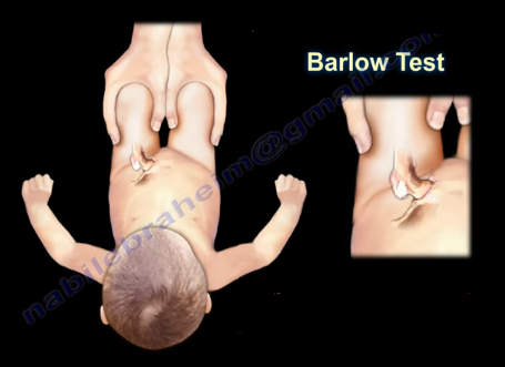
The Barlow maneuver identifies the unstable hip that is in a
reduced position that the clinician can passively dislocate. When performing
the Barlow Test, the examiner will flex the hip and knees to 90 degrees. The
maneuver is performed by bringing the thigh towards the midline (adducting the
hip). Mild pressure is then placed on the knee while directing the force
posteriorly. The femoral head will be pushed out of the socket. The Barlow Test
is considered positive if the hip can be popped out of socket with this
maneuver. The dislocation will be palpable.
The Ortolani Test is used to confirm the findings of the
Barlow test. The Ortolani maneuver is performed following the Barlow test to
determine if the hip is actually dislocated. The Ortolani test is performed by
the examiner flexing the hips and knees to 90 degrees. Reduction is done by
abduction of the hip and pushing the thigh anteriorly. The test is positive is
a palpable and audible clunk is heard from the hip being reduced. A hip click
is a nonspecific finding.
In summary, The Barlow Test is performed when the hip is
reduced and is used to dislocate the hip. The Ortolani Test is performed only
after the Barlow Test has been performed and the hip has been dislocated. The
Ortolani Test will reduce the hip.
Both of these tests are used for screening newborns during
the neonatal period. The hips are examined one at a time and usually the hips
are flexed during these maneuvers. Early diagnosis by these tests and
preferably with ultrasound is essential to detect hip instability and
dislocation in the neonatal period.
Treatment is directed at stabilizing the
hip that has positive Barlow and Ortolani Signs. The first born female with a
breach presentation and a positive family history are at risk of developing
developmental dysplasia of the hip (DDH). If the hip remains dislocated for weeks,
these two tests are usually not reliable. Barlow and Ortolani are not positive
after 3 months due to the soft tissue contracture around the hip region.
Limitation of abduction becomes the most consistent clinical findings. Children
older than 12 months will have other findings which include asymmetry of hip
abduction, a positive Trendelenburg gait and a positive Galeazzi sign.
The Froment’s sign occurs due to weakness of the adductor
pollicis muscle in ulnar nerve palsy. The adductor pollicis muscle has two
heads:
Transverse Head
Originiates from the anterior body of the third
metacarpal
Oblique Head
Originates from the base of the second and the
third metacarpals as well as the trapezoid and capitate bones
The two heads of the adductor pollicis muscle then insert
into the base of the proximal phalanx of the thumb and the ulnar sesamoid
bones. The muscle is innervated by the deep branch of the ulnar nerve. The
function of the adductor pollicis muscle is to adduct the thumb. It is
important in pinch strength. When the ulnar nerve is injured, the adductor
pollicis function is lost and thumb adduction will not occur.
The Foment’s Sign is used to test the function of the
adductor pollicis muscle. When pinching a piece of paper between the thumb and
index finger against resistance, the thumb IP joint will flex if the adductor
pollicis muscle is weak. The flexion of the thumb occurs by the flexor pollicis
longus, which is innervated by the median nerve. The flexor pollicis longus,
which is innervated by the median nerve, substitutes the function of the
adductor pollicis which is innervated by the ulnar nerve.
The bulbocavernosus reflex indicates the absence or presence
of spinal shock. Spinal shock usually occurs between 24-72 hours after a spinal
injury. Spinal shock is manifested by the absence of the bulbocavernosus
reflex, hypotension, bradycardia, and complete loss of motor sensation and
reflexes. When the reflex is absent, this means that the patient is in spinal
shock because the anal sphincter will not contract when the reflex is absent.
When the reflex is present, this signals the end of spinal
shock; the anal sphincter will contract when the reflex is present. The reflex
is check by monitoring anal sphincter contraction in response to squeezing of
the penis of clitoris, or by pulling on an indwelling Foley catheter. It is a
polysynaptic response mediated by S2-S4.
What is Spinal Shock?
Spinal shock is the loss of sensation and motor power
following a spinal cord injury. Spinal shock is the loss of sensation and motor
power following a spinal cord injury. After an injury to the spine, if the
patient has no motor or sensory below the level of the lesion, the physician
must determine if the patient is in spinal shock by checking the
bulbocavernosus reflex.
If there is no anal contraction (absent bulbocavernosus
reflex), this indicates that the patient is still in shock and the prognosis
cannot be determined. If anal contraction is present (positive bulbocavernosis
reflex), this indicates the end of spinal shock. The patient’s condition and
prognosis at this point can be determined by examining sacral sparing (positive
sacral sparing indicates an incomplete lesion).
Loss of sensation and motor power below the level of injury
indicates complete spinal cord injury. Once the diagnosis of neurogenic shock
is established, the blood pressure should be managed with vasopressors to prevent
fluid overload. With the end of spinal shock, the prognosis can be determined. Examine
the patient thoroughly, including sacral sparing. The patient may have normal,
partial, or complete indications.
Platelet Rich Plasma, or PRP, is a volume of the plasma of
autologous blood having a platelet concentration above the baseline. Platelets
facilitate healing by stimulating the release of different growth factors. The
growth factors recruit stem cells that assist with healing, repair, or
regeneration of the injured tissue. PRP is injected directly into the injured
tissue, stimulating a healing response in a more powerful form.
Red Blood Cell
In order to understand PRP, you must know that platelets are
components of blood cells. The development of PRP focuses on the concentration
of the patient’s own blood platelets to create an active mixture of growth
factors. The other components of blood are the red and white blood cells. Red blood
cells carry oxygen from the lungs and deliver it to the body tissues. White
blood cells form our immune system and help the body to fight against infection
and foreign materials.
Active Platelet
Platelets aid in hemostasis (stop the bleeding) and in
building new tissues—they act as a scaffold for tissue regeneration. Platelets
aid in the attraction and binding of stem cells. Platelets act as the directors
while stem cells work. Platelets divide, multiply and differentiate to become
the healing cells for injured tissue. The platelets become activated by
thrombin and other factors which cause a change in their morphology and the
release of multiple growth factors. These growth factors bind themselves to the
receptors of the cell causing intracellular changes down to the nucleus and affecting
its DNA. The result is a change in the
performance and function of the cell.
In order to produce PRP, 30-60mLs of the patient’s own
venous blood is drawn from the antecubital vein. The blood is then placed into
a device to be centrifuged which separates the blood into platelet poor plasma
(PPP), red blood cells (RBC), and platelet rich plasma (PRP). The blood is then
placed in a centrifuge for 15 minutes at 3,200 rpm. The centrifuge spins and
separates the platelets from the rest of the blood components and increases the
concentration of platelets and growth factors. The more platelet concentration,
the greater the healing power. After the centrifuge process is complete, the
plasma has been separated from the blood producing the PRP and the platelet
poor plasma is withdrawn to be discarded. Platelet rich plasma is withdrawn for
injection. Sodium Bicarbonate is used to neutralize the acidity of the sample.
The more platelet concentration, the more growth factors and healing power the
sample has.
Ultrasounds help deliver a concentrated sample of platelets
to the injured tissue. Ultrasound increases the accuracy of delivering the
sample to the injured tissue. Preferably, injections should be performed with
the aid of ultrasound imaging. Needling induced injury releases thrombin which
activates the platelets. Platelets help in hemostasis and produce growth
factors as well as chemotactic factors. Platelets act as a scaffold for
mesenchymal stem cells which start the process of tissue regeneration. Patients
will typically experience minimal to moderate discomfort which may last up to
one week following the injection. Avoid the use of anti-inflammatory medications
for 6 weeks after the injection.
The hangman’s fracture refers to the bilateral fracture of
the pars interarticularis. When this fracture occurs, the spinal canal is
widened and there is a low risk for spinal cord injury. This fracture usually
occurs due to motor vehicle accidents.
Hyperextension will fracture the pars interarticularis with
secondary flexion, injuring the disc and posterior ligament. The patient may
have other associated spine fractures.
Hangman’s fractures are identified by types using the Levine and Edwards
Classification.
Type I
Type I:
Stable fracture with less than 3mm displacement
No angulation
Treatment: Cervical Orthosis
Type II
Type II:
Most common type
Significant translation and some angulation
Unstable fracture
Treatment: Cervical traction to improve the displacement and immobilization in halo vest
Type IIa
Type IIa:
Slight translation but severe anglulation seen in flexion distraction injuries with tearing of the posterior longitudinal ligament and the disc.
The fracture is unstable
Treatment: reduction in extension and compression in a halo
Do NOT use traction when there is severe angulation of the fracture
Type III
Type III:
C2-C3 facet dislocation
Rare fracture that results from initial anterior facet dislocation of C2 on C3, followed by an extension injury fracturing the neural arch.
Results in translation with unilateral or bilateral facet dislocation of C2-C3.
Unstable fracture
Treatment: surgery for reduction of the facet dislocation and stabilization of the injury
Typical and Atypical fractures
A typical hangman’s fracture displaces the vertebral body
anteriorly and its posterior element posteriorly. This creates increased space
for the spinal cord.
Atypical Hangman's Fracture
An atypical hangman’s fracture line leave the canal
circumferentially intact, which puts the spinal cord at risk of injury if
displacement occurs.
 A patient with a complete anterior interosseous nerve injury
or a high medial nerve injury should be asked to make a fist. The first and
second digits will have difficulty in flexing, while the other digits will
flex. The third digit will be weak, while the fourth and fifth digits are
normal. This position of the hand is similar to the position taken during a
hand blessing. The Benedictine sign is different from an “ulnar claw hand”.
Ulnar claw hand refers to damage to the ULNAR nerve and is seen when attempting
to extend all the digits (leaving the 4th and 5th digits
flexed). The O.K. sign is used to check for paralysis of the anterior
interosseous nerve due to entrapment or compression injury. A patient with
paralysis of the anterior interosseous nerve will be unable to make the O.K.
sign. This is due to weakness of the flexor pollicis longus and flexor
digitorum profundus muscles. A typical pinch attitude is associated with
anterior interosseous nerve injury.
 The anterior interosseous nerve arises from the median nerve
about 4-6cm distal to the elbow, which is about 1/3 of the way down the
forearm. It exits from the anterolateral aspect of the median nerve and it runs
between the radius and the ulna on the interosseous membrane between and below
the muscles of the flexor digitorum profundus and the flexor pollicis longus.