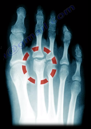
Freiberg’s Disease is caused by avascular necrosis of the
head of the 2nd metatarsal. This condition is more common in
patients who have a longer 2nd metatarsal bone relative to the 1st
metatarsal. This leads to the transfer of excessive loads onto the 2nd
metatarsal, which may interfere with the blood supply. This disease tends to
occur more commonly in young females during growth spurts.
Freiberg’s Disease usually presents itself as pain and
swelling at the 2nd metatarsophalangeal joint that is related to
activities and walking in high heels. There may be point tenderness and
swelling over the head of the 2nd metatarsal. There may also be
limited range of motion in the 2nd metatarsophalangeal joint. Early
in the disease, x-rays may only show minimal changes. Radiological evidence of
the condition may only be clearly visible on an MRI and bone scan. In more
severe cases, sclerosis, fragmentation, collapse of the metatarsal head, and
severe arthritis, may make the condition easily visible on an x-ray.
Treatment
Conservative treatment consists of nonsteroidal
anti-inflammatory medications, activity modification, orthotics, and immobilization
with a short leg cast. Surgical intervention involving a joint debridement is
indicated only after the failure of all conservative measures. Other procedures
may be indicated depending on the complexity and severity of the case.
Pain located at the heel can have several different causes.
It is important to make the correct diagnosis for the cause of the heel pain,
so that appropriate treatment can be given to the patient. Common causes of
heel pain include:
Baxter’s Nerve Compression
Plantar Fasciitis
Fat Pad Atrophy
Achilles Tendonitis
Stress Fractures of the Calcaneus
Lumbar Spine Radiculopathy
The sites and locations from different causes of the pain are
all very close to each other, making it difficult to determine the source of
the pain and to diagnose injuries.
In plantar fasciitis, irritation and swelling will occur at
the thick tissue on the bottom of the foot. This fascia can become inflamed and
painful, making walking more difficult. Plantar fasciitis is most severe in the
morning when patients first stand on their feet. Stretching exercises or
walking, often helps in relieving the painful tightening associated with
plantar fasciitis. Pain symptoms will intensify with prolonged exercise or
standing. Plantar fasciitis is usually
associated with a tight heel cord.
During fat pad atrophy, the fat that cushions the calcaneus
is thinned. This condition is common in elderly people and can cause
significant pain while walking. It also commonly occurs in patients with a
history of steroid injections.
Achilles Tendonitis is a chronic injury to the tendon that
joins the heel to the muscles of the lower leg, primarily occurring from
overuse. The Achilles tendon gives us the ability to rise up on our toes,
allowing for the act of walking. If a patient has Achilles tendonitis, walking
can be painful and difficult.
Stress fractures of the calcaneus can occur due to overuse
injuries. The patient will experience weight bearing pain. Stress fracture
injuries of the heel are typical in patients who engage in running sports, due
to the repetitive shock being placed on the heel. Side to side compression of
the calcaneus is painful.
Pain on the lateral side of the foot may be a result of
L5-S1 radiculopathy.
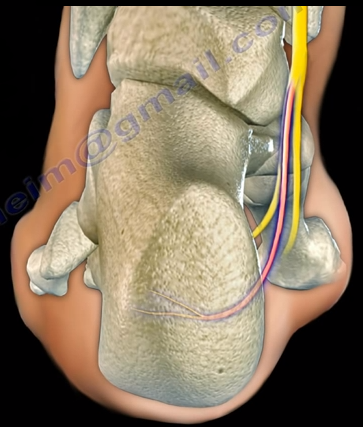
The Baxter’s nerve is the first branch of the lateral
plantar nerve. It also contributes to 20% of all heel pain causes. The Baxter’s
nerve provides motor innervation to the abductor digiti minimi muscle. When the
nerve is affected by compression, the symptoms are similar to planter
fasciitis. Entrapment of the Baxter’s nerve may cause up to 20% of heel pain,
but is still overlooked. The first branch of the lateral planter nerve is the
inferior calcaneal nerve (Baxter’s Nerve).
The nerve courses vertically between
the abductor hallucis and the quadratus plantae muscles, then makes a 90°
horizontal turn, coursing laterally beneath the calcaneus to innervate the
abductor digiti minimi muscle. The Baxter’s nerve can become entrapped within
the medial heel. The entrapment will result in heel pain, paresthesia, abnormal
sensations on the plantar aspect (bottom) of the heel, and medial heel
tenderness. There are two sites of entrapment that are typical with Baxter’s
nerve impingements. The first site is located between the fascia of the
abductor hallucis and quadratus plantae muscles. The second site is where the
nerve passes along the anterior aspect of the medial calcaneal tuberosity.
Treatment
Management is usually conservative and consists of heel
stretching exercises, rest, NSAIDs, and Orthotics. Surgery is done as a last
resort with neurolysis of the nerve after release of the deep fascia of the abductor
hallucis muscle. There is a good-excellent result in about 90% of patients
after decompression of the nerve. Involvement of the Baxter’s nerve may affect
running athletes causing pain on the medial plantar aspect of the foot.
Wednesday, December 21, 2016
Stress Fractures:
Common for Athletes and Those with
Active
Lifestyles
Figure 1
Fractures
are typically caused by a single traumatic event such as a car accident or
fall. Here, bones are exposed to forces so extreme they are unable to support
energy that is placed upon them. In contrast, there are fractures caused by a
repetitive application of force. These fractures, called stress fractures,
occur when muscles become so fatigued they lose their ability to absorb
additional shock. These fractures are categorized as overuse injuries and are characterized
by tiny cracks in the bone.
Bone
is incredibly resilient. Each day, bone broken down by daily living is replaced
by the body in a necessary balancing act. However, this balance is disturbed with
excessive physical training.
Stress fractures often present in athletes such as
tennis players, basketball players, gymnasts and track and field participants.
Typical stress fracture causes include improper equipment, increased physical
stress, increasing the amount or intensity of an activity too rapidly and the
impact of an unfamiliar surface.
Figure 2
Stress
fractures are usually categorized one of two ways: a fatigue fracture or an
insufficiency fracture. Fatigue fractures are the result of bone being overused
or being exposed to repetitive stress beyond its ability to repair itself. In
contrast, insufficiency fractures are the result of bone being deficient in
minerals or vitamins.
A typical cause of an insufficiency fracture is if a bone
has been weakened by osteoporosis.
Figure 3
While
stress fractures can occur anywhere on the body, more than 50 percent occur in
the lower leg. (Figure 1) (Figure 2). Stress fractures rarely occur in the
upper extremities because they are not weight bearing bones, like the tibia and
bones that run from the mid-foot to the toes. (Figure 3).
There
are certain red flags that may signal stress fractures. These red flags
include: pain that increases over time, pain that increases with activity and
decreases with rest, pain that persists
Figure 5
Figure 4
even
at rest, pain that occurs earlier in each successive workout and swelling. A
runner with groin pain and a negative x-ray must have a back scan or an MRI to
rule out hip stress fractures.
Early
diagnosis of hip fractures is necessary to avoid displacement of the fracture.
The best treatment for stress fractures is rest. After about 6 to 8
weeks, patients should be able to return to their regular activities. It’s important to do your best to safeguard against stress fractures. This includes implementing a
prevention plan that includes the following: alternating activities that accomplish the same fitness goals (crosstraining); setting
incremental goals when participating in a new sports activity; and
maintaining a healthy diet(Figure 4) with bone strengthening vitamins and
minerals such as calcium and vitamin D (Figure 5). In some situatoins, however, surgery is needed to treat stress
fractues.(Figure 6)
 Freiberg’s Disease is caused by avascular necrosis of the
head of the 2nd metatarsal. This condition is more common in
patients who have a longer 2nd metatarsal bone relative to the 1st
metatarsal. This leads to the transfer of excessive loads onto the 2nd
metatarsal, which may interfere with the blood supply. This disease tends to
occur more commonly in young females during growth spurts.
 Freiberg’s Disease usually presents itself as pain and
swelling at the 2nd metatarsophalangeal joint that is related to
activities and walking in high heels. There may be point tenderness and
swelling over the head of the 2nd metatarsal. There may also be
limited range of motion in the 2nd metatarsophalangeal joint. Early
in the disease, x-rays may only show minimal changes. Radiological evidence of
the condition may only be clearly visible on an MRI and bone scan. In more
severe cases, sclerosis, fragmentation, collapse of the metatarsal head, and
severe arthritis, may make the condition easily visible on an x-ray.