Showing posts with label University of Toledo Medical Center. Show all posts

Tuesday, September 4, 2018

SLAP Tear- Symptoms, Diagnosis, and Treatment


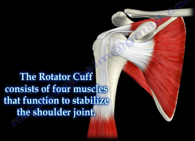
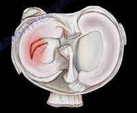
A SLAP tear is a tear that occurs where the biceps tendon inserts into the superior labrum. A SLAP tear is different from a Bankart lesion. SLAP tears are not common and can be hard to diagnose. Symptoms of a SLAP tear include: pain deep within the shoulder or in the back of the shoulder, as well as catching, popping, or clicking sensations. The patient may also experience pain when throwing a ball with a decrease in velocity and the feeling of having a dead arm after pitching. Patients will also experience pain with overhead activity which mimics impingement syndrome. This typically affects throwing athletes. When the biceps tendon is involved, pain may also be located at the front of the shoulder. A SLAP tear can be an isolated lesion or it can be associated with internal impingement, articular sided cuff tear, or instability.
A SLAP tear is diagnosed with a clinical examination and testing. The O’Brien’s test is the most commonly used test. Multiple tests are usually used including the anterior slide test and the clunk test. An MRI with contrast is the best imaging technique. When performing the O’Brien’s test, the patient is standing or sitting with the arm at 90° of flexion, 10° of adduction, and full internal rotation with the forearm pronated.  The examiner applies pressure to the forearm and instructs the patient to resist the applied downward force. Pain at the shoulder joint suggests a SLAP lesion. Decrease in pain of the shoulder joint on supination of the arm is suggestive of a SLAP tear.
Treatment consists of physical therapy, anti-inflammatory medications, injections, and surgery (when conservative treatment fails). If surgery is necessary, a labral debridement will be performed for minor tearing and fraying. Biceps Tenodesis is becoming popular, as it is a procedure that cuts the biceps tendon where it attaches to the labrum and reinserts it in another area, usually in front of the shoulder. A biceps tenotomy is a procedure that cuts the biceps tendon from the glenoid, releasing the long head of the biceps tendon from its attachment allowing it to fall into the upper arm out of the shoulder joint. A biceps tenotomy is probably best suitable for some elderly patients. A SLAP repair is a procedure which uses sutures to anchor the torn labrum to the glenoid. This repair is usually done for athletes and patients under the age of 40 years.

Tuesday, August 28, 2018

Massive Rotator Cuff Tear


To view my Youtube video, 'Massive Rotator Cuff Tear- Classic', click here.


Massive rotator cuff tears are a disabling problem. These tears can cause pain, weakness, and sometimes swelling of the shoulder. The rotator cuff consists of four muscles that function to stabilize the shoulder joint: supraspinatus rotator cuff tendon, subscapularis tendon, infraspinatus rotator cuff tendon, and the teres minor rotator cuff tendon. The supraspinatus tendon is the most common of the rotator cuff tendons to become ruptured. Massive tears of the rotator cuff that are greater than 5cm usually involving both the supraspinatus and infraspinatus tendons.


Massive tears of the rotator cuff are defined as tears greater than 5cm, usually involving both the supraspinatus and infraspinatus tendons. Retraction of the rotator cuff tendons along with muscle atrophy and fatty infiltration can occur. This makes surgical reconstruction difficult with the surgical outcome being unpredictable and less than satisfactory.

Treatment varies from physiotherapy to replacement of the humeral head. Arthroscopic or open repair is usually the selected treatment. Reconstruction can be done in selected cases. A rotator cuff arthropathy is performed on massive cuff tears that are associated with superior migration of the humeral head as well as instability and arthritis of the shoulder. The patient will have pseudoparalysis and an x-ray will show shift of the humerus proximally. An MRI will show massive cuff tear with retraction at the level of the glenoid with atrophy of the muscle and fatty infiltration. A reverse shoulder is the treatment of choice for the elderly with rotator cuff arthropathy as it improves the pain and function. Hemiarthroplasty is the treatment for younger patients. A standard head or a big humeral head can be selected.


A patient with a massive tear of the cuff usually develops weakness of the shoulder and becomes unable to actively lift the arm without assistance. Fluid collection within the shoulder may occur with a massive tear of the rotator cuff.

Tuesday, August 21, 2018

Toe Deformities


Deformities of the toes are not uncommon and can occur from muscle imbalance, or other causes such as rheumatoid arthritis, diabetes, compartment syndrome, synovitis, or neurological disorders. Hammer toe occurs as flexion of the proximal interphalangeal (PIP) joint. Hammer toe is similar to the Boutonniere deformity of the finger. Claw Toe is a hyperextension deformity of the MTP joint and flexion of the PIP and DIP, resembling a pirate hook. Claw toe is similar to an intrinsic minus deformity of the hand, or “claw hand”. Mallet toe is similar in appearance to mallet finger, and is a flexion deformity of the DIP joint.

Tuesday, July 10, 2018

McMurray's Test- Meniscal Tear


Meniscal injuries are very common. The McMurray’s Test is a rotational maneuver of the knee that is frequently used to aid in the diagnosis of meniscal tears. With a meniscal tear, the patient usually complains of knee pain localized to the lateral or medial side of the knee joint. The patient will have locking, clicking, pain, or effusion.


During the physical examination, joint line tenderness is the most sensitive finding. Swelling of the knee and a possible extension lag (locked knee) is also a common finding. Pain at a higher level is usually associated with the medial collateral ligament. Pain at a lower level is usually associated with the pes anserine bursa.


What is the McMurrays test?             


The McMurray’s test is a knee examination test that provokes pain or a painful click as the knee is brought from flexion to extension with either internal or external rotation. The McMurray’s test uses the tibia to trap the meniscus between the femoral condyles of the femur and the tibia. When performing the test, the patient should be lying supine with the knee hyperflexed. The examiner then grasps the patient’s heel with one hand and places the other hand over the knee joint. To test the medial meniscus, the knee is fully flexed, and the examiner then passively externally rotates the tibia and places a valgus force. The knee is then extended in order to test the medial meniscus. To test the lateral meniscus, the examiner passively internally rotates the tibia and places a varus force. The knee is then extended in order to test the lateral meniscus. A positive test is indicated by pain, clicking or popping within the joint and may signal a tear of either the medial or lateral meniscus when the knee is brought from flexion to extension.


How reliable is the McMurray’s test?


There are mixed reviews for the validity of this test. An MRI is a very sensitive exam and makes the diagnosis easier, while excluding other associated injuries.


Tuesday, June 26, 2018

Distal Femur Fractures & Nonunion- Use of Fibular Graft


The physician may be faced with some complex distal femur fractures or nonunion where the bony stock is not adequate or the fixation may have failed. It may also be a situation where a bone graft cannot be obtained from the patient. In some of these cases, I use an intramedually fibular graft in addition to plate fixation. The intramedullary fibular graft technique can be used in: complicated cases, comminuted fractures with osteoporosis, failure of traditional method of fixation, complex nonunion, and complex supracondylar periprothetic fractures.


How do you perform the technique?


The physician must find the starting point, which is the center of the intercondylar notch just
superior to the Blumensaat’s Line. The physician will insert a guide wire after reduction of the fracture. Then, the physician will ream over the guide wire to the appropriate size of the fibular graft, which you may need to fashion slightly. The physician needs to be sure to change the beaded guide wire to a smooth one and put the fibular graft through the guide wire into the medullary canal across the fracture of the nonunion. If the medullary canal of the fibula is small and it will not go through the guide wire, then place the fibular graft free hand. The physician should be sure that the fibular graft is not prominent through the joint. Next, fix the fracture or nonunion with a plate preferably a locking plate. You can augment the fixation with bone graft, allograft, or bone graft substitute. This procedure can also be helpful in periprosthetic fractures of the distal femur. If the prosthesisi is stable, you will do fixation of the fracture of the nonunion. It will be ideal to use a plate fixation after insertion of an intramedullary fibular graft, especially if the bony stock is very poor and if you can pass the fibular graft through the femoral component.

Tuesday, June 12, 2018

Congenital Dislocation of the Knee


Congenital Dislocation of the knee is rare and may occur due to a contracture of the quadriceps. This condition usually occurs in patients with myelo, arthrogryposis, or Larsen’s syndrome. The patient with a congenital dislocation of the knee may have developmental dysplasia of the hip (DDH) and club foot. On examination, the patient will have a hyperextended knee at birth. They may have their foot placed against their face and there will be limited flexion at the knee. The patient may have a dimple or skin crease at the anterior aspect of the knee. You must examine the hip to rule out ipsilateral hip dislocation. 50% or more patients will have hip dysplasia. The etiology is not known; however, it could be due to fetal positioning or congenital absence of the cruciate.
There are grades, or a spectrum, for this deformity. Grade I deformities are referred to as Severe Genu Recurvatum, and the knee is hyperextended. If the range of passive flexion is more than 90°, it is considered to be a simple recurvatum. Grade II deformities are identified by subluxation with a range of 30-90° in passive flexion. Grade III deformities are complete dislocations with a range of passive flexion being less than 30°.

Congenital dislocation of the knee will take priority over treatment of hip dysplasia or club foot. The Pavlik harness and club foot cast will require knee flexion, so the physician will need to treat the knee dislocation first. With Grade I deformities, the initial treatment will be stretching of the knee and serial casting with the knee in flexion. In serial stretching and casting, the goal is to obtain at least 90° of flexion and reduction of the deformity over the course of several weeks. The physician should avoid pseudo-correction through an iatrogenic fracture of the proximal tibial physis. The prognosis is usually good if reduction is achieved without surgery. With Grade II deformities, if the infant is less than 1 month old, you will do serial casting first followed by percutaneous quadriceps recession, especially if the flexion is less than 90°. In Grade III deformities, a V-Y quadricepsplasty with above the knee cast is done in Grade III (frank dislocation), especially if nonsurgical treatment fails to reduce the tibia on the femur.   The result of open surgery is better when it is done in children younger than 6 months. In general, open reduction is reserved for children who did not respond to stretching and cast immobilization. It is important that the hip dysplasia is recognized and the knee dislocation is corrected early. This will help in early reduction of the hip.

Tuesday, April 24, 2018

Tibial Bowing


Tibial bowing is not uncommon. The direction and the apex of the tibial bow can alert the clinician to the type of the deformity, its treatment, and its prognosis. Tibial bowing can occur anteriorly, laterally, anterolaterally, and posteromedially.
Anterior bowing is often associated with fibular hemimelia. This bowing may be associated with the loss of the lateral rays of the foot, equinovalgus foot deformity, tarsal coalition, and significant leg length discrepancy.


Lateral bowing is a common variation, which occurs bilaterally. This condition is mild and not associated with other problems.


Anterolateral bowing is a serious tibial bowing that may increase and lead to a fracture as well as pseudoarthrosis of the tibia. This type of bowing occurs early in infancy. Pseudoarthrosis is usually associated with neurofibromatosis. 10% of patients with neurofibromatosis will have anterolateral tibial bowing. Neurofibromatosis is found in 50% of the patients with ALB. The patient should be carefully examined for café-au-lait spots. In this type of bowing, bone ends are usually thin and the fibula may also be involved. Treatment of anterolateral bowing is bracing with total contact orthosis. In order to treat pseudoarthosis in the tibia, surgery is usually needed. Multiple options are available for surgery, and none of these options are perfect. 50% of patients may undergo amputation due to the inability to achieve healing of pseudoarthrosis.

Posteromedial bowing is a rare calcaneovalgus deformity of the foot plus leg length discrepancy. This condition usually resolves, but may have residual leg length discrepancy.

Tuesday, April 10, 2018

Pes Anserine Bursitis


Several bursa are seen around the knee area. These bursa include the suprapatellar, prepatellar, infrapatellar, and pes anserine. The pes answerine bursa is a small fluid filled sac located between the tibia and the three tendons of the Sartorius, Gracilis, and Semi-tendinosus.
These muscles are innervated by three separate nerves, the femoral, obturator, and the tibial branch of the sciatic nerve, respectively. Pes Anserine bursitis, or “breast stroke knee”, is an inflammatory condition of the medial knee at the pes anserine bursa that is common in swimmers.

What is the pes anserine?

The pes anserine is the common area of insertion for the three tendons along the proximal medial aspect of the tibia. This condition is also sometimes referred to as a “goosefoot” because the pes anserinus tendons resemble the shape of a goose foot. Pes Anserine bursitis is usually seen as causing pain, tenderness, and localized swelling after trauma or total knee replacement. The pain is seen below the joint line on the medial part of the proximal tibial with the bursa being deep to the tendons.


Treatment

Treatment consists of physical therapy, nonsteroidal anti-inflammatory medications, and injections. The physician will need to rule out meniscal tears, stress fractures, or osteonecrosis of the tibia, as these are all differential diagnosis.


Tuesday, April 3, 2018

Cauda Equina—Central Disc Herniation


Disc herniations usually occurs posterolaterally, but it may also be central. The cauda equina is composed of several nerves within the lower end of the spinal canal. The top of the spinal cord is a tubular bundle of nervous tissue extending from the brain.

The following section of the spinal cord is called the Conus Medullaris and is the lower end of the spinal cord. The Cauda Equina is made up of multiple nerve roots beginning at the level of L1.


The most common disc herniation is the Posterolateral Disc Herniation. This type of herniation is a nerve root injury, which will cause changes to both the sensory and motor skills as well as the reflexes. A posterolateral disc herniation usually affects the foot and ankle, and may cause unilateral leg pain and weakness. Observe for a positive straight leg raising test. These herniations are usually initially treated with conservative methods.  


A central disc herniation will cause cauda equina syndrome, which is a compression over the lumbosacral nerve roots. This compression will cause more back pain than leg pain, and bladder and bowel symptoms will be evident. This herniation is considered a surgical emergency.

Tuesday, March 27, 2018

Heel Pain & The Baxter's Nerve


Pain located at the heel can have several different causes. It is important to make the correct diagnosis for the cause of the heel pain, so that appropriate treatment can be given to the patient. Common causes of heel pain include:


  1. Baxter’s Nerve Compression
  2. Plantar Fasciitis
  3. Fat Pad Atrophy
  4. Achilles Tendonitis
  5. Stress Fractures of the Calcaneus
  6. Lumbar Spine Radiculopathy
The sites and locations from different causes of the pain are all very close to each other, making it difficult to determine the source of the pain and to diagnose injuries.
In plantar fasciitis, irritation and swelling will occur at the thick tissue on the bottom of the foot. This fascia can become inflamed and painful, making walking more difficult. Plantar fasciitis is most severe in the morning when patients first stand on their feet. Stretching exercises or walking, often helps in relieving the painful tightening associated with plantar fasciitis. Pain symptoms will intensify with prolonged exercise or standing.  Plantar fasciitis is usually associated with a tight heel cord.


During fat pad atrophy, the fat that cushions the calcaneus is thinned. This condition is common in elderly people and can cause significant pain while walking. It also commonly occurs in patients with a history of steroid injections.
Achilles Tendonitis is a chronic injury to the tendon that joins the heel to the muscles of the lower leg, primarily occurring from overuse. The Achilles tendon gives us the ability to rise up on our toes, allowing for the act of walking. If a patient has Achilles tendonitis, walking can be painful and difficult.


Stress fractures of the calcaneus can occur due to overuse injuries. The patient will experience weight bearing pain. Stress fracture injuries of the heel are typical in patients who engage in running sports, due to the repetitive shock being placed on the heel. Side to side compression of the calcaneus is painful.
Pain on the lateral side of the foot may be a result of L5-S1 radiculopathy.
The Baxter’s nerve is the first branch of the lateral plantar nerve. It also contributes to 20% of all heel pain causes. The Baxter’s nerve provides motor innervation to the abductor digiti minimi muscle. When the nerve is affected by compression, the symptoms are similar to planter fasciitis. Entrapment of the Baxter’s nerve may cause up to 20% of heel pain, but is still overlooked. The first branch of the lateral planter nerve is the inferior calcaneal nerve (Baxter’s Nerve).


The nerve courses vertically between the abductor hallucis and the quadratus plantae muscles, then makes a 90° horizontal turn, coursing laterally beneath the calcaneus to innervate the abductor digiti minimi muscle. The Baxter’s nerve can become entrapped within the medial heel. The entrapment will result in heel pain, paresthesia, abnormal sensations on the plantar aspect (bottom) of the heel, and medial heel tenderness. There are two sites of entrapment that are typical with Baxter’s nerve impingements. The first site is located between the fascia of the abductor hallucis and quadratus plantae muscles. The second site is where the nerve passes along the anterior aspect of the medial calcaneal tuberosity.


Treatment


Management is usually conservative and consists of heel stretching exercises, rest, NSAIDs, and Orthotics. Surgery is done as a last resort with neurolysis of the nerve after release of the deep fascia of the abductor hallucis muscle. There is a good-excellent result in about 90% of patients after decompression of the nerve. Involvement of the Baxter’s nerve may affect running athletes causing pain on the medial plantar aspect of the foot.

Wednesday, March 21, 2018

Malunions of the Clavicle


It is difficult to reduce and maintain the reduction of clavicle fractures as there will be deforming forces in the midshaft area of the clavicle. The clavicle will heal regardless as to if a sling or figure 8 strap is used. Despite the presence of a bump, fracture displacement, and deformity, healing of the fracture still occurs rapidly. Healing occurs in about 85% of cases, however, the clavicle will not look aligned due to the difficulty in reducing the fracture. It is hard to achieve a reduction of these fracture without surgery. Without surgical reduction, the fracture may end with some degree of malunion and possible shortening of the clavicle. The fragments will not line up with the distal fragment appearing to be downward and anteriorly rotated. Shortening is clinically significant because it alters the dynamics of the muscles around the shoulder. It also narrows the costoclavicular space. The patient may complain of decreased shoulder strength and endurance if the patient had a displaced midshaft clavicle fracture that healed with more than 2 cm of shortening.


What are the symptoms of clavicle malunion?


Pain, easy fatigability, cosmetic problems (especially in females), neurological dysfunction (possible involvement of the brachial plexus—especially the ulnar nerve), and the patient may have thoracic outlet syndrome.
X-rays should be done of both shoulders. Bilateral panoramic views are beneficial in order to measure the shortening. The physician will also want to check the amount of overlap at the ends of the clavicle.


Treatment


Surgical treatment of the malunion may be successful in restoring the function and relieving the pain. A clavicle osteotomy is done in the plane of the healed fracture. The fracture is recreated with correction of the deformity and the length of the clavicle is restored. A local or iliac crest bone graft can be used if needed.

If an osteotomy is done with reduction of the clavicle to its preinjury position and leaves a significant bone defect, the physician will need a tricortical piece of bone at the osteotomy area. It is probably better to use a precontoured clavicle plate in the superior position with six cortices, three placed on each side of the osteotomy.


Tuesday, March 6, 2018

Tibial Spine Fractures in Children


Tibial Spine fractures are similar to ACL injuries in adults. It occurs due to hyperextension of the knee and is commonly seen during falls from a bicycle. An injured child with a swollen knee and who has fallen from a bicycle should alert the clinician to the possibility of a tibial spine fracture. A meniscal injury may also occur, especially with the medial meniscus. The interposition of the meniscus or rotation of the fracture may prevent a closed reduction.
Tibial spine fractures are classified using the Meyers and McKeever Classification and are separated into three types. Type I classifications are nondisplaced, Type II fractures are identified as being minimally displaced with an intact posterior hinge, and Type III fractures are classified as being completely displaced.
The presentation and examination is similar to an ACL tear with immediate swelling as well as a positive Lachman’s Test or Anterior Drawer Test. An x-ray will show the fracture and a CT scan will help in planning for surgery. An MRI may be required to show a trapped or a meniscal injury.

Treatment


Treatment will consist of an aspiration of the large hematoma. Nonoperative treatment is used for Type I fractures and reducible Type II fractures; a closed reduction and immobilization in 0-20 of flexion. Surgery is performed in Type II fractures and unreducible Type II fractures. An ORIF or arthroscopic reduction and fixation will be performed. During surgery, the trapped meniscus will be moved out of the way and the surgeon will use sutures or screws for fixation. It is important for the surgeon to remember to avoid the physis.

Complication


ACL laxity is common but not clinically significant. Stiffness or arthrofibrosis occurs with surgical fixation. Growth arrest is rare.

Tuesday, February 27, 2018

Sciatica, Disc Herniation, and Piriformis Syndrome


The sciatic nerve is a large nerve that comes from the lumbosacral plexus. The sciatic nerve has five nerve roots, L4, L5, S1, S2, and S3. The sciatic nerve runs from the lower spine, through the buttock to the lower leg and foot. The sciatic nerve initially emerges from the pelvis and exits the greater sciatic notch anteriorly and deep to the piriformis muscle, exiting below the piriformis muscle.
The sciatic nerve then enters the thigh between the ischial tuberosity and the greater trochanter of the femur. In about 10% of patients, the sciatic nerve is separated by all or part of the piriformis muscle. The sciatic nerve enters the thigh beneath the lower border of the gluteus maximus muscle. The nerve then runs down and branches out within the posterior aspect of the thigh, down to the leg and foot. The sciatic nerve gives multiple sensory and motor branches to specific areas and muscles in the leg and foot. The two main branches of the sciatic nerve are the posterior tibial nerve and the common peroneal nerve.
Irritation of the sciatic nerve may occur at multiple sites. The first site that we need to look at is the spine—which is where irritation may occur, usually from lumbar disc herniation. This is considered true sciatica or lumbar radiculopathy. Another site for irritation of the sciatic nerve is at the piriformis muscle. The sciatic nerve may become compressed by the piriformis muscle (piriformis syndrome). Piriformis Syndrome is a diagnosis of exclusion! If the patient has symptoms of sciatica, then there must be an MRI of the spine that is negative, proving that the symptoms are not associated with a possible disc problem. Once the MRI is negative, then you can say that the condition of sciatica may come from piriformis syndrome.
Disc Herniation

Tuesday, February 20, 2018

Jefferson Fractures


Fifty percent of patients with Jefferson fractures will have associated spine injuries. The canal is wide with a low risk of spinal cord injuries unless the transverse ligament is disrupted. It is difficult to view Jefferson Fractures on an x-ray (usually seen on the lateral side”. This fracture is considered a “Junctional Fracture” and could be missed. The classic Jefferson fracture is a burst fracture that results from an axial load. It could be a four part fracture with bilateral fractures of the anterior and posterior arch. There are variations which include two and three part fractures and incomplete formations of the posterior arch can be mistaken as a fracture.
When speaking of Jefferson fractures, it is important to be familiar with the structures that may be involved. These bony structures include: The Atlas (C1), Axis (C2), and the odontoid process. C1 and C2 are stabilized together by the transverse ligament and C1 and C2 provide a 50% of rotation of the neck. The C1 is a ring. At the upper cervical region, the spinal canal is 2.5 times larger than the cord size. The stability and treatment of Jefferson fractures depends on the integrity of the transverse ligament and the displacement of the fracture. You need to know about the important ligaments related to the Jefferson fracture. These ligaments include: the transverse ligament, the apical ligament, and the Alar ligament.

Diagnosing ligamentous injury


In order to determine a ligamentous injury, the physician will want to check the Atlanto-dens interval (A.D.I). Normally, this interval should be less than 3mm in adults and less than 5mm in children. If the ADI is between 3-5mm, this indicates an injury to the transverse ligament; the transverse ligament holds the odontoid and C1 together, alar and apical ligaments will be intact. If the A.D.I measures greater than 5mm, then there is an injury to the transverse, alar, and apical ligaments.


Fracture Types


A bony injury with the intact transverse ligament and a lateral mass displacement less than 7mm and the A.D.I is less than 3mm is considered a stable fracture. Nondisplaced fractures of this nature should be treated with a rigid orthosis. If the fracture is displaced, a halo will need to be used.
Another type of fracture can occur at C1 with a transverse ligament tear. The Atlanto-dens interval will be more than 3 mm in adults. The treatment will depend on the type of injury to the transverse ligament. With bony avulsions of the transverse ligament, the halo will need to be used cautiously. However, some surgeons prefer to do a fusion of C1 and C2. If there is an intrasubstance tear of the transverse ligament, the surgeon will perform a fusion at C1-C2. The surgeon will need to do early surgery as this is a significant injury with a risk of spinal cord compression.


In regards to “Open Mouth Views”, the normal overhang is visible during an “Open Mouth View”. If it is just a bony injury Jefferson fracture, the combined overhang will be less than 7mm and the transverse ligament is intact and it is a stable fracture. If a Jefferson fracture has a combined overhang of more than 7mm, then the transverse ligament is probably torn and there is an unstable fracture present.

Radiological Studies


A CT scan is probably the best study in diagnosing the characteristics of the bony injury. An MRI is the best study in diagnosing any associated transverse ligament injuries.

Tuesday, February 13, 2018

Pronator Teres Syndrome


The nerve that is involved in pronator teres syndrome is the median nerve. Pronator Teres Syndrome is caused by a compression of the median nerve at the level of the elbow which occurs more in women. In the forearm, the median nerve runs between the two heads of the pronator teres muscle and then it lies between the flexor digitorum superficialis and flexor digitorum profundus muscles. This syndrome may be associated with medial epicondylitis. The principle symptoms of numbness in the radial 3 ½ fingers as well as thenar weakness which may be mistakenly attributed to carpal tunnel syndrome.
The most common cause of entrapment is due to compression of the median nerve between the two heads of the pronator teres muscle. This commonly occurs in people who perform repetitive forceful pronation of the forearm. Compression may be due to the thickening of the bicipital aponeurosis. The aponeurosis crosses from lateral to medial over the antecubital fossa and may irritate the median nerve. Compression of the nerve may also occur due to the fibrous arch of the origin of the flexor digitorum superficialis (FDS).


The median nerve runs down the medial side of the arm and passes 2 ½ to 4 cm below the level of the medial epicondyle before it enters between the two heads of the pronator teres. About 1% of
patients have a medial supracondylar humeral spur about 5cm proximally to the medial epicondyle. The ligament of Struthers is attached to this bony projection which connects the process to the medial epicondyle. The bony process points towards the elbow joint and the median nerve can become compressed by the supracondylar spur. The median nerve can also become trapped by the ligament of Struthers that extends from the supracondylar process to the medial epicondyle. The ligament of Struthers is different from the arcade of Struthers, which deals with the compression of the ulnar nerve around the elbow.


Paresthesia in these lateral 3 ½ fingers may occur with the compression of the median nerve at the elbow region or at the carpal tunnel region. These symptoms are similar to carpal tunnel syndrome but the symptoms are worse with rotation of the forearm. The patient will complain of dull aching pain over the proximal forearm with no nighttime symptoms. The pain is usually worsened by repetitive or forceful pronation. Tenderness of palpation to the pronator teres muscle will be detected. The median nerve gives off a palmar cutaneous branch before entering the carpal tunnel. Sensory disturbances over the palm of the hand occur due to involvement of the palmar cutaneous branch of the medial nerve and this occurs proximal to the carpal tunnel. Sensory disturbances in this area indicates median nerve problems proximal to the carpal tunnel. This differentiates between carpal tunnel syndrome and pronator teres syndrome.

There are specific provocative tests that produce the pain and distal paresthesia that are used to localize the site of compression. The Tinel’s sign at the wrist and the Phalen’s test will be negative. The Median nerve compression tests are negative at the carpal tunnel; however, there will be a positive Tinel’s sign at the proximal forearm. There will be abnormal sensation in the “palmar triangle”. When compression of the nerve involves the supracondylar process, the test is considered positive if symptoms of tingling worsen while tapping on the spur.
Occassionally, the spur can be felt. The pronator teres muscle can be assessed as the cause of the median nerve compression in different ways. Resisted forearm pronation with elbow flexion will test for compression at the two heads of the pronator teres muscle. During this test, the patient’s forearm is held in resisted pronation and flexion. While remaining in a pronated position, the forearm is gradually extended. Compression of the median nerve may also be tested by: resisted elbow flexion with forearm supination (compression at the bicipital aponeurosis) and resisted contraction of the FDS to the middle finger (compression at the FDS arch).


Differential Diagnosis

C6/C7 Radiculopathy occurs due to involvement of the nerves at these levels which will cause numbness of the thumb, index, and long fingers, as well as weakness of the muscles of the forearm that are innervated by the median nerve. The radial nerve part of C6-C7 will show normal function of the wrist extensors and the triceps.

X-rays, imaging and nerve conduction studies may be helpful in the diagnosis.

Treatment typically consists of rest, splints, and NSAIDs. Surgical decompression of the median nerve through all 4 or 5 possible sites of compression when non-operative management fails for 3-6 months. The results of surgery are variable. Full recovery is not always seen in all patients as only about 80% of patients improve from surgery. The skin incision may leave an unsatisfactory scar.