Showing posts with label education. Show all posts

Monday, July 25, 2022

Ankle fracture Maisonneuve Fracture – Everything You Need to Know


      Ankle fracture Maisonneuve Fracture – Everything You Need to Know


                         https://www.youtube.com/watch?v=m35lMOtMJHQ

Maisonneuve fracture involves fracture of the proximal fibula associated with an occult and unstable injury of the ankle. The problem in these patients occur when the ankle injury is presented without a fracture of the lateral malleolus, or the medial malleolus and the injury is mistakenly diagnosed as an ankle sprain and the proximal fibular fracture is missed. Examine the leg for tenderness in the proximal fibula to diagnose a proximal fibula fracture. The patient could be mistakenly treated for having an isolated proximal fibular fracture alone and the ankle injury is missed.

 High index of suspicion is necessary to diagnose and treat this injury. Maisonneuve fracture equals syndesmotic injury. Syndesmotic Injury equals Syndesmotic Reduction and Fixation. If ankle x-rays show medial or posterior malleolus fracture, or a medial clear space widening with no fracture of the lateral malleolus, then you must obtain a long-leg films to assess possible proximal fibular fracture. Clinical examination of their entire leg for pain and tenderness in addition to long leg films of the entire leg that includes the ankle, and the knee is mandatory in case of the patient with approximate fibular fracture to exclude the presence of an additional ankle injury, or if the patient has an unexplained increase in the medial clear space of the ankle joint. You should be searching for the presence of a high fibular fracture. Look for signs of syndesmotic injury such as an unexplained increase in medial clear space or tibiofibular clear space is widened and it should be less than 5 millimeters.

So how do you explain this injury? It is explained by the presence of rotation force to the ankle with transmission of the force through the interosseous membrane, which exits through a proximal fibular fracture. Maisonneuve fracture occurs from external rotation of the foot, most often with pronation mechanism. This force has to go somewhere! If you don't see a fracture of the fibula then do the squeeze test or the external rotation stress test (both will 
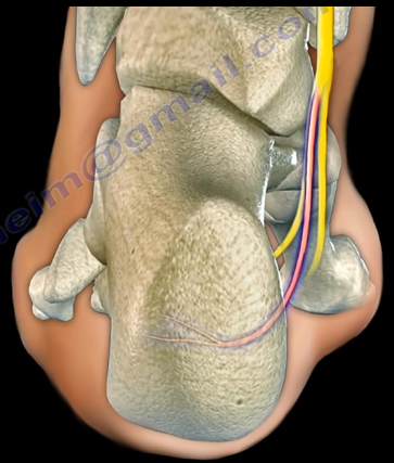
show syndesmotic). The injury can involve the deltoid ligament injury or medial malleolar fracture medially and a fibular fracture proximally. Additionally, the tibiofibular ligaments are also involved, which can be the anterior tibiofibular ligament, interosseous ligament, the posterior tibiofibular ligament or posterior malleolar fracture. This looks like a very unstable ankle injury that may not be very obvious at presentation and you have to look out for it.

So how do you treat an Maisonneuve Fracture? This treated by fixation of the tibiofibular syndesmotic injury (key of treatment) or syndesmotic screws. if you have a medial site injury and there is a tear of the deltoid ligament, leave it alone. if there's a medial malleolus fracture you should fix that of the lateral side if there's approximate fibular fracture leave it alone. If there is a medial malleolar fracture, it should be fixed. If there is a proximal fibular fracture on the lateral side, leave it alone. As for the Syndesmotic Injury, the fixation has to be stable and adequate. Because of the magnitude of the injury, the Maisonneuve fracture may require more syndesmotic screws than with a routine ankle fracture with syndesmotic injury. After the fixation you will give a short leg non-weight bearing splint for six to eight weeks. Here is a patient taste example: the proximal fibular fracture and you can see increase in the medial clear space and you can see that the syndesmosis is widened. You can see that in the posterior malleolar fracture the patient is fixed with syndesmotic screws.

Tuesday, October 30, 2018

Olecranon Fractures


Fractures of the olecranon may occur due to a direct blow to the elbow or from a fall onto an outstretched hand. Nondisplaced fractures have less than 2mm of separation and are considered stable. Fracture separation will not increase with elbow flexion. Extensor mechanism is intact and the patient will be able to extend the elbow against gravity. Displaced fractures could be an avulsion, oblique, transverse, comminuted, or dislocated.
Olecranon fracture dislocations can be anterior (transolecranon) or posterior (similar to monteggia fracture dislocation). An examination will show that the patient is unable to extend the elbow with these displaced fracture types. A true lateral view x-ray will clearly show the olecranon fracture. Usually, these fractures are followed by stiffness of the elbow in about 50% of the patients. However, this does not affect the function.
The goal of treatment for olecranon fractures should be restoration of the articular surface, preservation of the continuity of extensor mechanisms, maintain elbow stability, and avoid stiffness of the elbow. Nonoperative treatment is used for nondisplaced fractures and it may be used for some displaced fractures in elderly patients (treat elbow in some flexion with a splint). I would personally use minimally invasive techniques in these patients unless the skin is very bad, or the fracture is very comminuted.
There are three techniques used for surgical treatment: the tension band technique, detach olecranon and reattach triceps, and plate and screw fixation. The tension band technique is only used for transverse fractures with no comminution. K-wires and screws are used, and the surgeon may use either a 6.5mm screw or Kwires for the tension band. When doing the tension band technique, you want to engage the anterior cortex of the ulna. The surgeon should avoid over penetration to avoid affecting the forearm rotation or injuring the anterior interosseous nerve. The surgeon needs to be sure that the pins are not fixing the radius and that after the operation, the patient can perform pronation and supination of the forearm (pull the pins out slightly if needed). The distractive force of the triceps is converted to compression force at the articular surgace, especially when bending the elbow. The drill hole for the K-wire should be positioned about 4-5cm from the fracture which gives enough safe distance so that the fracture will not propagate. Place the tension band wire through the drilled holes before application of the K wires. The surgeon should be sure that the hook to the K-wire is posterior. Make sure that the tension band wire is close to the bone so there is no laxity in the fixation and instability. An intramedullary screw could be used. This screw fixation may need a washer to capture the tension band wire. Intramedullary 6.5mm screw fixation is a reasonable option for fixation but it may need to be supplemented with tension band wires. Never use cancellous screw alone. The tension band technique are for transverse fractures of the olecranon. If fractures comminution is present, change the plan of fixation.

When detaching the olecranon and reattaching the triceps, an excision of the fracture fragment and triceps advancement is used:

  • If the fracture is less than 50%
  • To treat elderly patients (especially if fracture is comminuted)
  • For some nonunions when the fracture is small and cannot be fixed

The surgeon must be sure that the procedure is done with the elbow is stable. If the elbow has ligamentous instability and excision of the fracture fragment is done, this will make the elbow very unstable. The triceps should be attached closer to the articular surface.
Special olecranon plates are available when using a plate and screw fixation technique. The bridge plate and screw fixation technique is used in comminuted, Monteggia, oblique fractures extending to the coranoid, and fracture dislocation. The plate is placed on the tension side of the olecranon (dorsal side). Sometimes, an opening is made through the triceps and the plate is placed against the bone, then suturing the triceps tendon over the plate to avoid hardware prominence.
In summary, if the patient is elderly with a small, comminuted fracture fragment less than 50% of the joint space, excise the fragment, and reattach the triceps tendon to the olecranon. If the olecranon fracture is transverse and proximal to the base of the coranoid process, then use the tension band technique. Use plate fixation for all olecranon fracture scenerios, such comminuted fractures, oblique fractures, unstable fractures, dislocation, or fractures distal to the coranoid process. The typical exam question scenario will discuss a comminuted fracture that should be treated with a plate. You probably need to remove above 20% of the plate fixations due to hardware irritation. Hardware irritation is worse with the tension band surgical treatment (may need to remove in more than 50% of cases).

Tuesday, October 2, 2018

Tests Orthopaedic Surgeons Should Think About


There are some important tests that every Orthopaedic Surgeon should think about. This doesn’t mean the tests are needed for every patient. It just means that the physician needs to think about these tests to see if it will benefit the patient or not. Some tests commonly ordered are hemoglobin A1C (HbA1c), Vitamin D25, and C-reactive protein (CRP) & sedimentation rate.


Hemoglobin A1C test are ordered for diabetic patients. HbA1c is a good test for monitoring long-term glucose (sugar) control on patients with diabetes. HbA1c is a percentage of the glycanated hemoglobin relative to the total hemoglobin in the blood. The normal range of HbA1c is 4-6%. More than 7% is high. Another test, the 25-Hydroxy Vitamin D blood test is ordered for patients with osteoporosis, nonunions, fragility fractures, and occasionally in patients with infections. If infection is suspected and the physician needs to monitor the progress of treatment, C-reactive protein (CRP) & sedimentation rate tests should be ordered. A Methicillin-resistant staphylococcus aureus (MRSA) screening should be ordered for patients who could be carriers. Nutritional assessments may be necessary for other patients.

Joints should be aspirated prior to injecting of the joint. The physician should additionally be sure that there is no infection when injecting the joint. A fluid analysis from the joint should be completed. Important vascular studies that can be ordered include: A.B.I., CTA, or a Doppler. Some radiological studies are performed with a dye injection. For example, an MRI of the spine will require gadolinium, while an MRI arthrogram may be used for the hip or shoulder. Tests rarely ordered include: alpha-defensin test (infection); Nicotine/Cotinine test (smoking); Protein S, Protein C, or Factor 5 leiden tests. There are some special tests and precautions that must be taken for patients with epilepsy. It is important that the physician does not perform a procedure if the epilepsy is not controlled. It is important to know that anti-epileptic medication can interfere with vitamin D metabolism in the liver.
Patients on anticoagulation medications should be monitored, especially patients with atrial fibrillation, which makes the orthopaedic procedure more complicated. You want to give the patient anticoagulation, but not encroaching on the management of atrial fibrillation. Patients with a short or thick neck, or a history of sleep apnea, may need additional sleep studies before surgery and may need special precautions after surgery. Sleep apnea will affect the post-operative care of the patient. The physician should avoid ordering unnecessary tests and focus on ordering the most important tests. Orthopaedics deal with concepts and every condition will have a reasonable way of diagnosing it and a reasonable way of treating it.  

Wednesday, September 26, 2018

Adhesive Capsulitis, Frozen Shoulder


Adhesive Capsulitis, or frozen shoulder, is a painful progressive loss of shoulder motion. It affects both active and passive movement of the shoulder joint. The shoulder will be stiff and painful and occurs due to inflammation, fibrosis, scarring, and contraction of the capsule. A normal shoulder joint capsule is elastic and allows great range of motion. Inflammation and thickening of the shoulder capsule and may lead to adhesive capsulitis. Frozen shoulder may occur without any specific cause, however it may be triggered by a mild trauma to the shoulder.


This condition develops slowly and goes through three phases:

  1. Pain and freezing
  2. Stiffness or frozen
  3. Resolution

During the pain and freezing phase, the pain is worse at night and increases with any movement. This phase will last several months. During the second phase, range of motion is limited as pain is diminishing. This may last up to one year. The resolution phase may begin overtime and may last up to three years.


Conditions associated with frozen shoulder include:

  • Diabetes
  • Thyroid problems
  • Auto immune disease
  • Stroke
  • Rheumatoid arthritis
  • Trauma or post-surgery

A patient with frozen shoulder will have loss of both active (movement without assistance) and passive (movement with assistance) motion. External rotation of the shoulder is very limited and the condition is self-limiting and may resolve on its own. X-rays are needed to rule out degenerative arthritis.  An MRI or arthrogram will show small fluid in joint cavity. Rotator cuff may be normal and synovitis and narrowing of the rotator cuff interval is usually seen.

Treatment consists of anti-inflammatory medications, physical therapy, injections, and manipulation under anesthesia. Surgery will be done in the form of a release of the capsule when nonoperative methods fail. The physician should always check the patient for diabetes.  

Tuesday, August 28, 2018

Massive Rotator Cuff Tear


To view my Youtube video, 'Massive Rotator Cuff Tear- Classic', click here.


Massive rotator cuff tears are a disabling problem. These tears can cause pain, weakness, and sometimes swelling of the shoulder. The rotator cuff consists of four muscles that function to stabilize the shoulder joint: supraspinatus rotator cuff tendon, subscapularis tendon, infraspinatus rotator cuff tendon, and the teres minor rotator cuff tendon. The supraspinatus tendon is the most common of the rotator cuff tendons to become ruptured. Massive tears of the rotator cuff that are greater than 5cm usually involving both the supraspinatus and infraspinatus tendons.


Massive tears of the rotator cuff are defined as tears greater than 5cm, usually involving both the supraspinatus and infraspinatus tendons. Retraction of the rotator cuff tendons along with muscle atrophy and fatty infiltration can occur. This makes surgical reconstruction difficult with the surgical outcome being unpredictable and less than satisfactory.

Treatment varies from physiotherapy to replacement of the humeral head. Arthroscopic or open repair is usually the selected treatment. Reconstruction can be done in selected cases. A rotator cuff arthropathy is performed on massive cuff tears that are associated with superior migration of the humeral head as well as instability and arthritis of the shoulder. The patient will have pseudoparalysis and an x-ray will show shift of the humerus proximally. An MRI will show massive cuff tear with retraction at the level of the glenoid with atrophy of the muscle and fatty infiltration. A reverse shoulder is the treatment of choice for the elderly with rotator cuff arthropathy as it improves the pain and function. Hemiarthroplasty is the treatment for younger patients. A standard head or a big humeral head can be selected.


A patient with a massive tear of the cuff usually develops weakness of the shoulder and becomes unable to actively lift the arm without assistance. Fluid collection within the shoulder may occur with a massive tear of the rotator cuff.

Wednesday, August 8, 2018

Discoid Meniscus


The meniscus is a cushion structure made of cartilage which fits within the knee joint between the tibia and the femur. The medial meniscus is C-shaped and the lateral meniscus in the more circular. The meniscus is made up of type I collagen that provides shock absorption and stability to the knee joint. The meniscus helps to protect the knee joint, allowing the bones to slide freely on each other. Discoid meniscus is a rare variation of the meniscus that usually affects the lateral meniscus of the knee in less than 5% of the population and could be bilateral in about 25% of the cases.
Discoid meniscus is a large meniscus with abnormal attachment causing increased mobility of the meniscus. It causes a pop, click, or snapping with locking and pain. There will be loss of full knee extension with tenderness on the lateral joint space. Symptoms occur more during extension of the knee. The discoid meniscus occurs due to the abnormal development and increase in size of the meniscus. An x-ray could show increased widening of the joint space. An MRI will show the “bow tie” sign in three or more sagittal continuous cuts. The coronal MRI will show a thick and flat meniscus extending beyond the halfway point of the condyle.


Watanabe Classification of Discoid Lateral Meniscus


  • Type I: Block-shaped stable
  • Type II: Block-shaped, stable, partial meniscus (has good peripheral attachment)
  • Type III: Unstable meniscus with stability arising only form the ligament of Wrisberg. (no posterior meniscal tibial attachment).

Treatment


An asymptomatic patient will be treated with observation. A symptomatic patient may receive a partial meniscectomy and saucerization with repair of type III (no posterior tibial meniscal attachment)

Tuesday, July 3, 2018

Hip Dislocation Following Total Hip Surgery


There are two types of hip joint dislocations: posterior and anterior. The position of the leg is important in determining the type of hip dislocation. When the hip is dislocated, the leg is usually shortened and it assumes a different position than the normal leg (the other leg). If the dislocation is posterior, the leg will be in adduction and internal rotation. If the dislocation is anterior, the leg will be in abduction and external rotation. Notice that the affected extremity is shortened and externally rotated. Leg shortening can also be seen in hip fractures and the leg will be shortened and externally rotated.


Dislocation of the hip following total hip surgery may require revision surgery, but it is rare. The majority of hip dislocations after total hip dislocations are posterior, and they are usually treated without surgery. Most occur within the first month of THA; 1-4% in primary, 16% in revision. There is more incidence of dislocation in revision hip replacement.


Causes & Risk Factors:

  • Posterior Approach (try to repair the capsule adequately)
  • Malposition of the component
    • Ideally, the normal cup component will be in 20° of anteversion and 40° of abduction
    • When the hip dislocates posterior, always check for retroversion of the cup.

  • Prior hip fracture surgery, especially in the elderly
  • Weakness of the abductor muscle—must achieve soft tissue tension and function
  • Alcohol abuse
  • Improper neck length—looseness of the hip

The patient should be careful to avoid all activities that cause dislocation after total hip surgery. The patient should use a pillow between the legs while sleeping on their back and they should be careful to not cross their legs in their sleep. Patients cannot sleep on their sides as well. The patient should not bend the body at the waist farther than 90°. When sitting, the patient must avoid chairs that make it difficult to stand up, and sit at more than a 90° angle. The patient must not sit with their legs crossed in the chair. The patient must be made aware that if the leg is changed from its usual position, or becomes shortened, then the hip is probably dislocated and their doctor should be consulted.


X-rays of the dislocated total hip should include AP and lateral views. Look for eccentric wear and look for the position of the prosthesis. CT scans may be needed before or after reduction of the dislocation to check the version of the components. Treatment is variable and depends on the situation. The treatment should be tailored for each case. The majority of these cases with early dislocations can be treated successfully with closed reduction and immobilization.

The treatment should start with closed reduction of the total hip and immobilization. Hip stability is checked after reduction of the dislocation. Immobilization can be done by a brace or a hip spica. Trochanteric osteotomy and advancement of the trochanter and tensioning the abductor muscle. Screws or wires can be used. The prosthesis must be in good alignment for this procedure to work. Constrained acetabular components are used when the abductor muscle is deficient and the component position is good. Revision total hip is done in recurrent dislocation with malposition of the component or polyethylene wear.

Tuesday, June 19, 2018

Isolated Fibular Fractures


Fibular fractures are usually associated with a complex injury, however they can be an isolated fracture. Complex injuries where a fibula fracture can occur include: fracture of the fibula and tibia, ankle fracture, pilon fracture, and Maisonneuve fractures.

Maisonneuve fractures involve a fracture of the proximal fibula associated with an occult injury of the ankle. Isolated fibular fractures are rare and usually the result of direct trauma. The fibula carries about 15% of the axial load and is the site of muscle attachment for the peroneus muscles and the flexor hallucis longus muscle. Check the patient who has a fibular fracture and no other fracture involving the tibia to rule out a possible Maisonneuve fracture, especially if there is no history of direct trauma to the leg. A high index of suspicion is necessary to diagnose and treat this injury. For high fibular fractures, the physician should look for signs of syndesmotic injury. Syndesmotic injury may include an unexplained increase in the medial clear space or the tibiofibular clear space is widened (should be less than 5mm). The x-ray will show the fracture to be rotational or oblique. Maisonneuve fractures require surgery to fix the syndesmosis.


Treatment will consist of reduction and fixation. It is important to determine if the injury is a Maisonneuve fracture or an isolated fibular fracture. An isolated fibular fracture will not need surgery.

Tuesday, June 12, 2018

Congenital Dislocation of the Knee


Congenital Dislocation of the knee is rare and may occur due to a contracture of the quadriceps. This condition usually occurs in patients with myelo, arthrogryposis, or Larsen’s syndrome. The patient with a congenital dislocation of the knee may have developmental dysplasia of the hip (DDH) and club foot. On examination, the patient will have a hyperextended knee at birth. They may have their foot placed against their face and there will be limited flexion at the knee. The patient may have a dimple or skin crease at the anterior aspect of the knee. You must examine the hip to rule out ipsilateral hip dislocation. 50% or more patients will have hip dysplasia. The etiology is not known; however, it could be due to fetal positioning or congenital absence of the cruciate.
There are grades, or a spectrum, for this deformity. Grade I deformities are referred to as Severe Genu Recurvatum, and the knee is hyperextended. If the range of passive flexion is more than 90°, it is considered to be a simple recurvatum. Grade II deformities are identified by subluxation with a range of 30-90° in passive flexion. Grade III deformities are complete dislocations with a range of passive flexion being less than 30°.

Congenital dislocation of the knee will take priority over treatment of hip dysplasia or club foot. The Pavlik harness and club foot cast will require knee flexion, so the physician will need to treat the knee dislocation first. With Grade I deformities, the initial treatment will be stretching of the knee and serial casting with the knee in flexion. In serial stretching and casting, the goal is to obtain at least 90° of flexion and reduction of the deformity over the course of several weeks. The physician should avoid pseudo-correction through an iatrogenic fracture of the proximal tibial physis. The prognosis is usually good if reduction is achieved without surgery. With Grade II deformities, if the infant is less than 1 month old, you will do serial casting first followed by percutaneous quadriceps recession, especially if the flexion is less than 90°. In Grade III deformities, a V-Y quadricepsplasty with above the knee cast is done in Grade III (frank dislocation), especially if nonsurgical treatment fails to reduce the tibia on the femur.   The result of open surgery is better when it is done in children younger than 6 months. In general, open reduction is reserved for children who did not respond to stretching and cast immobilization. It is important that the hip dysplasia is recognized and the knee dislocation is corrected early. This will help in early reduction of the hip.

Tuesday, April 24, 2018

Tibial Bowing


Tibial bowing is not uncommon. The direction and the apex of the tibial bow can alert the clinician to the type of the deformity, its treatment, and its prognosis. Tibial bowing can occur anteriorly, laterally, anterolaterally, and posteromedially.
Anterior bowing is often associated with fibular hemimelia. This bowing may be associated with the loss of the lateral rays of the foot, equinovalgus foot deformity, tarsal coalition, and significant leg length discrepancy.


Lateral bowing is a common variation, which occurs bilaterally. This condition is mild and not associated with other problems.


Anterolateral bowing is a serious tibial bowing that may increase and lead to a fracture as well as pseudoarthrosis of the tibia. This type of bowing occurs early in infancy. Pseudoarthrosis is usually associated with neurofibromatosis. 10% of patients with neurofibromatosis will have anterolateral tibial bowing. Neurofibromatosis is found in 50% of the patients with ALB. The patient should be carefully examined for café-au-lait spots. In this type of bowing, bone ends are usually thin and the fibula may also be involved. Treatment of anterolateral bowing is bracing with total contact orthosis. In order to treat pseudoarthosis in the tibia, surgery is usually needed. Multiple options are available for surgery, and none of these options are perfect. 50% of patients may undergo amputation due to the inability to achieve healing of pseudoarthrosis.

Posteromedial bowing is a rare calcaneovalgus deformity of the foot plus leg length discrepancy. This condition usually resolves, but may have residual leg length discrepancy.

Tuesday, April 17, 2018

Freiberg's Disease


Freiberg’s Disease is caused by avascular necrosis of the head of the 2nd metatarsal. This condition is more common in patients who have a longer 2nd metatarsal bone relative to the 1st metatarsal. This leads to the transfer of excessive loads onto the 2nd metatarsal, which may interfere with the blood supply. This disease tends to occur more commonly in young females during growth spurts.

Freiberg’s Disease usually presents itself as pain and swelling at the 2nd metatarsophalangeal joint that is related to activities and walking in high heels. There may be point tenderness and swelling over the head of the 2nd metatarsal. There may also be limited range of motion in the 2nd metatarsophalangeal joint. Early in the disease, x-rays may only show minimal changes. Radiological evidence of the condition may only be clearly visible on an MRI and bone scan. In more severe cases, sclerosis, fragmentation, collapse of the metatarsal head, and severe arthritis, may make the condition easily visible on an x-ray.

Treatment


Conservative treatment consists of nonsteroidal anti-inflammatory medications, activity modification, orthotics, and immobilization with a short leg cast. Surgical intervention involving a joint debridement is indicated only after the failure of all conservative measures. Other procedures may be indicated depending on the complexity and severity of the case.

Tuesday, April 10, 2018

Pes Anserine Bursitis


Several bursa are seen around the knee area. These bursa include the suprapatellar, prepatellar, infrapatellar, and pes anserine. The pes answerine bursa is a small fluid filled sac located between the tibia and the three tendons of the Sartorius, Gracilis, and Semi-tendinosus.
These muscles are innervated by three separate nerves, the femoral, obturator, and the tibial branch of the sciatic nerve, respectively. Pes Anserine bursitis, or “breast stroke knee”, is an inflammatory condition of the medial knee at the pes anserine bursa that is common in swimmers.

What is the pes anserine?

The pes anserine is the common area of insertion for the three tendons along the proximal medial aspect of the tibia. This condition is also sometimes referred to as a “goosefoot” because the pes anserinus tendons resemble the shape of a goose foot. Pes Anserine bursitis is usually seen as causing pain, tenderness, and localized swelling after trauma or total knee replacement. The pain is seen below the joint line on the medial part of the proximal tibial with the bursa being deep to the tendons.


Treatment

Treatment consists of physical therapy, nonsteroidal anti-inflammatory medications, and injections. The physician will need to rule out meniscal tears, stress fractures, or osteonecrosis of the tibia, as these are all differential diagnosis.


Tuesday, April 3, 2018

Cauda Equina—Central Disc Herniation


Disc herniations usually occurs posterolaterally, but it may also be central. The cauda equina is composed of several nerves within the lower end of the spinal canal. The top of the spinal cord is a tubular bundle of nervous tissue extending from the brain.

The following section of the spinal cord is called the Conus Medullaris and is the lower end of the spinal cord. The Cauda Equina is made up of multiple nerve roots beginning at the level of L1.


The most common disc herniation is the Posterolateral Disc Herniation. This type of herniation is a nerve root injury, which will cause changes to both the sensory and motor skills as well as the reflexes. A posterolateral disc herniation usually affects the foot and ankle, and may cause unilateral leg pain and weakness. Observe for a positive straight leg raising test. These herniations are usually initially treated with conservative methods.  


A central disc herniation will cause cauda equina syndrome, which is a compression over the lumbosacral nerve roots. This compression will cause more back pain than leg pain, and bladder and bowel symptoms will be evident. This herniation is considered a surgical emergency.

Tuesday, March 27, 2018

Heel Pain & The Baxter's Nerve


Pain located at the heel can have several different causes. It is important to make the correct diagnosis for the cause of the heel pain, so that appropriate treatment can be given to the patient. Common causes of heel pain include:


  1. Baxter’s Nerve Compression
  2. Plantar Fasciitis
  3. Fat Pad Atrophy
  4. Achilles Tendonitis
  5. Stress Fractures of the Calcaneus
  6. Lumbar Spine Radiculopathy
The sites and locations from different causes of the pain are all very close to each other, making it difficult to determine the source of the pain and to diagnose injuries.
In plantar fasciitis, irritation and swelling will occur at the thick tissue on the bottom of the foot. This fascia can become inflamed and painful, making walking more difficult. Plantar fasciitis is most severe in the morning when patients first stand on their feet. Stretching exercises or walking, often helps in relieving the painful tightening associated with plantar fasciitis. Pain symptoms will intensify with prolonged exercise or standing.  Plantar fasciitis is usually associated with a tight heel cord.


During fat pad atrophy, the fat that cushions the calcaneus is thinned. This condition is common in elderly people and can cause significant pain while walking. It also commonly occurs in patients with a history of steroid injections.
Achilles Tendonitis is a chronic injury to the tendon that joins the heel to the muscles of the lower leg, primarily occurring from overuse. The Achilles tendon gives us the ability to rise up on our toes, allowing for the act of walking. If a patient has Achilles tendonitis, walking can be painful and difficult.


Stress fractures of the calcaneus can occur due to overuse injuries. The patient will experience weight bearing pain. Stress fracture injuries of the heel are typical in patients who engage in running sports, due to the repetitive shock being placed on the heel. Side to side compression of the calcaneus is painful.
Pain on the lateral side of the foot may be a result of L5-S1 radiculopathy.
The Baxter’s nerve is the first branch of the lateral plantar nerve. It also contributes to 20% of all heel pain causes. The Baxter’s nerve provides motor innervation to the abductor digiti minimi muscle. When the nerve is affected by compression, the symptoms are similar to planter fasciitis. Entrapment of the Baxter’s nerve may cause up to 20% of heel pain, but is still overlooked. The first branch of the lateral planter nerve is the inferior calcaneal nerve (Baxter’s Nerve).


The nerve courses vertically between the abductor hallucis and the quadratus plantae muscles, then makes a 90° horizontal turn, coursing laterally beneath the calcaneus to innervate the abductor digiti minimi muscle. The Baxter’s nerve can become entrapped within the medial heel. The entrapment will result in heel pain, paresthesia, abnormal sensations on the plantar aspect (bottom) of the heel, and medial heel tenderness. There are two sites of entrapment that are typical with Baxter’s nerve impingements. The first site is located between the fascia of the abductor hallucis and quadratus plantae muscles. The second site is where the nerve passes along the anterior aspect of the medial calcaneal tuberosity.


Treatment


Management is usually conservative and consists of heel stretching exercises, rest, NSAIDs, and Orthotics. Surgery is done as a last resort with neurolysis of the nerve after release of the deep fascia of the abductor hallucis muscle. There is a good-excellent result in about 90% of patients after decompression of the nerve. Involvement of the Baxter’s nerve may affect running athletes causing pain on the medial plantar aspect of the foot.

Wednesday, March 21, 2018

Malunions of the Clavicle


It is difficult to reduce and maintain the reduction of clavicle fractures as there will be deforming forces in the midshaft area of the clavicle. The clavicle will heal regardless as to if a sling or figure 8 strap is used. Despite the presence of a bump, fracture displacement, and deformity, healing of the fracture still occurs rapidly. Healing occurs in about 85% of cases, however, the clavicle will not look aligned due to the difficulty in reducing the fracture. It is hard to achieve a reduction of these fracture without surgery. Without surgical reduction, the fracture may end with some degree of malunion and possible shortening of the clavicle. The fragments will not line up with the distal fragment appearing to be downward and anteriorly rotated. Shortening is clinically significant because it alters the dynamics of the muscles around the shoulder. It also narrows the costoclavicular space. The patient may complain of decreased shoulder strength and endurance if the patient had a displaced midshaft clavicle fracture that healed with more than 2 cm of shortening.


What are the symptoms of clavicle malunion?


Pain, easy fatigability, cosmetic problems (especially in females), neurological dysfunction (possible involvement of the brachial plexus—especially the ulnar nerve), and the patient may have thoracic outlet syndrome.
X-rays should be done of both shoulders. Bilateral panoramic views are beneficial in order to measure the shortening. The physician will also want to check the amount of overlap at the ends of the clavicle.


Treatment


Surgical treatment of the malunion may be successful in restoring the function and relieving the pain. A clavicle osteotomy is done in the plane of the healed fracture. The fracture is recreated with correction of the deformity and the length of the clavicle is restored. A local or iliac crest bone graft can be used if needed.

If an osteotomy is done with reduction of the clavicle to its preinjury position and leaves a significant bone defect, the physician will need a tricortical piece of bone at the osteotomy area. It is probably better to use a precontoured clavicle plate in the superior position with six cortices, three placed on each side of the osteotomy.


Wednesday, March 14, 2018

Elbow Joint Dislocations


An Elbow dislocation occurs when the radius and ulna bones of the forearm move out of place from the humerus bone of the upper arm. There are two basic types of elbow dislocations:

  1. Simple

    No fracture of the bones around the elbow joint

    Usually ligamentous injury

  2. Complex

    Fracture has occurred along with ligamentous injury


Simple elbow dislocations typically occur when the patient falls onto an outstretched hand. Injury progression from lateral to medial in most patients. Posterolateral simple dislocations are the most common, occurring approximately 90% of the time.
The proximal ulna and radius are displaced posterolaterally relative to the distal humerus. Postemedial dislocations occur at the proximal ulna and radius and are displaced posteromedially relative to the distal humerus. In medial dislocations, the proximal ulna and radius are displaced medially relative to the distal humerus. With lateral dislocations, the proximal ulna and radius are displaced laterally relative to the distal humerus. Anterior dislocations are rare, as they result from a direct force applied to the posterior aspect of the forearm with the elbow in a flexed position. Anterior dislocations occur when the proximal ulna and radius are displaced anteriorly relative to the distal humerus. If stable, simple acute fractures can be treated with a closed reduction and a splint for two to three days (no more than two weeks) in addition to range-of-motion exercises and physical therapy. Unstable simple fractures are rare, but can be stabilized by ligament repair and/or by the use of an external fixator or cross pinning of the joint in the elderly.

Chronic Elbow dislocations will need to be treated with an open reduction and external fixator and is usually hinged. Recurrent elbow dislocations (diagnosed by pivot shift) occurs due to a deficiency of the lateral collateral ligament and is treated by a reconstruction of the ulnohumeral ligament with a tendon graft.