Showing posts with label Spinal Cord. Show all posts

Monday, February 18, 2019

Acute Low Back Pain Lumbar Disc Herniation


Acute Low Back Pain Lumbar Disc Herniation

Low back pain is a common condition. 90% of patients with low back pain will improve without surgery. Usually they get better with spontaneous resolution of the symptoms within 12 weeks. We usually advise the patient for early return to activity and function as the symptoms and the pain permits. The risk factors for development of low back pain are numerous, some include: vibration exposure, poor physical fitness, smoking and obesity, anxiety and depression, job dissatisfaction, or repetitive bending or “stooping” on the job. In summary, if the patient has no red flags and has a normal neurological exam, there is no reason to get early radiological studies. Getting early x-rays and early MRIs leads to a better patient satisfaction but does not give a better patient outcome. If there is no specific pain pattern, then there is no need for further workup. MRIs are good studies, but they give false positives. There is degeneration or a bulge of a disc in 35% of all asymptomatic subjects between 25-39 years of age. In patients 60 years old or older, the majority of the patients will have changes in the MRI. MRI abnormalities are common and must be correlated with the age and the clinical signs and symptoms of the patient. An MRI is good for diagnosing the lumbar disc herniation, which is sometimes called a ruptured disc, a slipped disc, or a herniated disc. The most common location of a disc herniation is a posterolateral herniation involving one nerve root. A foramninal L4-L5 herniation occurs in about 8%-10% of the cases. It involves the exiting nerve. A central herniation involves multiple nerve roots. It predominantly causes low back pain more than leg pain. It may cause bladder and bowel symptoms. This type of disc herniation causes Cauda Equina Syndrome which needs urgent diagnosis and surgical treatment. Clinical evaluation for a herniated disc examines sensory and motor reflexes. The Straight Leg Raising Test is the most important finding. It can be done in either the sitting or supine position. The test is positive as indicated by pain in the leg when the patient’s leg is raised to flex the hip with the knee extended. A positive straight leg test means a tension sign, something is putting tension or stress on the sciatic nerve. When the test is positive, it indicates possible disc herniation.
Treatment is typically non-operative. First, reassure the patient. Let the patient take some rest (no more than a few days), give the patient anti-inflammatory medication, and instruct them to attend physical therapy. Indications for surgery include progressive neurological deficits, Cauda Equina Syndrome, the patient is not getting better with time and treatment or if the symptoms are not getting better with conservative treatment, or the patient has a positive tension sign with persistent sever pain. Patients with sciatica and positive tension signs or patients with positive neurological findings on clinical exam with positive MRI findings make ideal surgical candidates. Surgery results in relief of leg pain in the majority of patients. Back pain may persist in some patients. Surgery results in neurological improvement, 50 % motor and sensory and 25% reflexes. In patients with discogenic back pain, they may need fusion which is a major procedure.The worst pressure on the disc occurs with prolonged sitting and bending over. This is the position that produces the highest pressure on the disc. If a patient has back pain but no radiation, by the patient’s history or physical examination and there are no red flags, then there is no reason to get x-rays or MRI early in the treatment of the patient. Red flags include a history of trauma, a tumor, infection, or Cauda Equina Syndrome symptoms. To rule out a history of trauma you should rule out fractures with x-rays, MRI, or CT scans. Tumors are a risk if the patient is older than 50 years old, if the patient had weight loss, or if the patient has pain at rest or at night. An infection may be present if the patient has fever and chills, if the patient has a history of diabetes, or if the patient has a history of IV drug abuse. Cauda Equina Symptoms may be present if the patient has back pain more than leg pain or if the patient also has bladder and bowel symptoms. Cauda Equina Syndrome needs to be diagnosed and surgically treated early. An MRI needs to be ordered urgently in the course of treatment. The MRI should be ordered STAT. There may need to be a wet read; a wet read is an early preliminary read of the radiographs. A wet read needs to be communicated with the physician and can be done while the patient is still on the table of the MRI.

Monday, February 4, 2019

Cervical Radiculopathy


Cervical Radiculopathy

Cervical radiculopathy is caused by cervical nerve root compression. The patient will have pain and/or progressive neurological deficit that results from conditions such as disc herniation that irritates a nerve in the cervical spine. Cervical radiculopathy is an irritation of the cervical nerve root. Cervical spine and shoulder problems overlap. The condition is of cervical spine etiology if the patient’s symptoms are relieved by shoulder abduction, by placing the hand over the head. The relief of the symptoms occurs due to decreased tension on the nerve roots. In cervical disc problems, be aware of false positive MRIs especially if the patient is above the age of 40 years old. Nerve conduction studies are not useful; they have a high false negative rate. EMG and nerve studies may differentiate radiculopathy from peripheral nerve entrapment. Cervical disc problems usually affect the lower numbered nerve root.
When you see the middle finger numbness, then this is C7. When compression of the C7 nerve root, there will be middle finger numbness, triceps weakness, and the triceps reflex will be affected. The cervical nerve roots are horizontal in orientation. It does not matter if cervical disc herniation is central or foraminal, it will compress the same nerve root. C7 nerve root runs above the pedicle of the C7 vertebra. C5-C6 is the most commonly affected disc and that will compress the C6 nerve root. The patient will come to the doctor with unilateral arm pain that is relieved by arm elevation. The numbness and paresthesia will occur in specific dermatomes. The patient may also have upper trapezius pain or interscapular pain. The patient may complain of occipital headache. When you examine the patient, do provocative tests such as the spurling’s test and the shoulder abduction test. The Spurling’s test is done by extending and rotating the neck towards the involved side. It reproduces the symptoms by narrowing the neuroforamen. The Spurling’s test differentiates cervical radiculopathy from peripheral nerve entrapment. Lifting the arm above the head relieves the symptoms if the cervical nerve roots are irritated. The Shoulder Abduction test differentiates cervical pathology from other causes of painful shoulder etiology. Make sure that you do not have a double crush syndrome, one in the neck and one in the peripheral nerve. Make sure that you differentiate radiculopathy from myelopathy. Make sure that you exclude a coexisting myelopathy. Examine the patient for upper motor neuron signs or cervical
myelopathy. Test the patient for gait instability. Test the patient for Hoffman’s sign. Test the patient for Babinski reflex. Test the patient for ankle Clonus. Check to see if the patient has hyperflexia in the upper and lower extremities (triceps/quadriceps). Even if there is a bad cervical spine disc problem on the MRI, treat it conservatively for about 3 months. Give the patient therapy and nonsteroidal anti-inflammatory medication (NSAIDS). 75% of the patients will improve with nonoperative treatment. Cervical radiculopathy is generally treated nonoperatively, in contrast to cervical myelopathy. Do surgery when there is persistent, severe pain for 6-12 weeks and/or progressive neurological deficit such as weakness or numbness. The procedure to treat cervical radiculopathy surgically is usually done anteriorly with direct removal of the lesion that causes the radiculopathy such as a herniated disc or spurs. When you place the anterior bone graft or the allograft in the disc space, you open the nueroforamen, and that will indirectly relieve the nerve. Then you will add the anterior plate. Some surgeons prefer to do a posterior approach.

Tuesday, August 21, 2018

Toe Deformities


Deformities of the toes are not uncommon and can occur from muscle imbalance, or other causes such as rheumatoid arthritis, diabetes, compartment syndrome, synovitis, or neurological disorders. Hammer toe occurs as flexion of the proximal interphalangeal (PIP) joint. Hammer toe is similar to the Boutonniere deformity of the finger. Claw Toe is a hyperextension deformity of the MTP joint and flexion of the PIP and DIP, resembling a pirate hook. Claw toe is similar to an intrinsic minus deformity of the hand, or “claw hand”. Mallet toe is similar in appearance to mallet finger, and is a flexion deformity of the DIP joint.

Tuesday, April 3, 2018

Cauda Equina—Central Disc Herniation


Disc herniations usually occurs posterolaterally, but it may also be central. The cauda equina is composed of several nerves within the lower end of the spinal canal. The top of the spinal cord is a tubular bundle of nervous tissue extending from the brain.

The following section of the spinal cord is called the Conus Medullaris and is the lower end of the spinal cord. The Cauda Equina is made up of multiple nerve roots beginning at the level of L1.


The most common disc herniation is the Posterolateral Disc Herniation. This type of herniation is a nerve root injury, which will cause changes to both the sensory and motor skills as well as the reflexes. A posterolateral disc herniation usually affects the foot and ankle, and may cause unilateral leg pain and weakness. Observe for a positive straight leg raising test. These herniations are usually initially treated with conservative methods.  


A central disc herniation will cause cauda equina syndrome, which is a compression over the lumbosacral nerve roots. This compression will cause more back pain than leg pain, and bladder and bowel symptoms will be evident. This herniation is considered a surgical emergency.

Tuesday, February 20, 2018

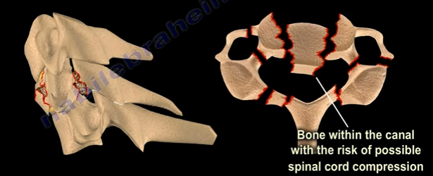
Jefferson Fractures


Fifty percent of patients with Jefferson fractures will have associated spine injuries. The canal is wide with a low risk of spinal cord injuries unless the transverse ligament is disrupted. It is difficult to view Jefferson Fractures on an x-ray (usually seen on the lateral side”. This fracture is considered a “Junctional Fracture” and could be missed. The classic Jefferson fracture is a burst fracture that results from an axial load. It could be a four part fracture with bilateral fractures of the anterior and posterior arch. There are variations which include two and three part fractures and incomplete formations of the posterior arch can be mistaken as a fracture.
When speaking of Jefferson fractures, it is important to be familiar with the structures that may be involved. These bony structures include: The Atlas (C1), Axis (C2), and the odontoid process. C1 and C2 are stabilized together by the transverse ligament and C1 and C2 provide a 50% of rotation of the neck. The C1 is a ring. At the upper cervical region, the spinal canal is 2.5 times larger than the cord size. The stability and treatment of Jefferson fractures depends on the integrity of the transverse ligament and the displacement of the fracture. You need to know about the important ligaments related to the Jefferson fracture. These ligaments include: the transverse ligament, the apical ligament, and the Alar ligament.

Diagnosing ligamentous injury


In order to determine a ligamentous injury, the physician will want to check the Atlanto-dens interval (A.D.I). Normally, this interval should be less than 3mm in adults and less than 5mm in children. If the ADI is between 3-5mm, this indicates an injury to the transverse ligament; the transverse ligament holds the odontoid and C1 together, alar and apical ligaments will be intact. If the A.D.I measures greater than 5mm, then there is an injury to the transverse, alar, and apical ligaments.


Fracture Types


A bony injury with the intact transverse ligament and a lateral mass displacement less than 7mm and the A.D.I is less than 3mm is considered a stable fracture. Nondisplaced fractures of this nature should be treated with a rigid orthosis. If the fracture is displaced, a halo will need to be used.
Another type of fracture can occur at C1 with a transverse ligament tear. The Atlanto-dens interval will be more than 3 mm in adults. The treatment will depend on the type of injury to the transverse ligament. With bony avulsions of the transverse ligament, the halo will need to be used cautiously. However, some surgeons prefer to do a fusion of C1 and C2. If there is an intrasubstance tear of the transverse ligament, the surgeon will perform a fusion at C1-C2. The surgeon will need to do early surgery as this is a significant injury with a risk of spinal cord compression.


In regards to “Open Mouth Views”, the normal overhang is visible during an “Open Mouth View”. If it is just a bony injury Jefferson fracture, the combined overhang will be less than 7mm and the transverse ligament is intact and it is a stable fracture. If a Jefferson fracture has a combined overhang of more than 7mm, then the transverse ligament is probably torn and there is an unstable fracture present.

Radiological Studies


A CT scan is probably the best study in diagnosing the characteristics of the bony injury. An MRI is the best study in diagnosing any associated transverse ligament injuries.

Tuesday, October 24, 2017

Hangman's Fracture


The hangman’s fracture refers to the bilateral fracture of the pars interarticularis. When this fracture occurs, the spinal canal is widened and there is a low risk for spinal cord injury. This fracture usually occurs due to motor vehicle accidents.

Hyperextension will fracture the pars interarticularis with secondary flexion, injuring the disc and posterior ligament. The patient may have other associated spine fractures.


Hangman’s fractures are identified by types using the Levine and Edwards Classification.
Type I


Type I:


  • Stable fracture with less than 3mm displacement
  • No angulation
  • Treatment: Cervical Orthosis
Type II

Type II:

  • Most common type
  • Significant translation and some angulation
  • Unstable fracture
  • Treatment: Cervical traction to improve the displacement and immobilization in halo vest
Type IIa

Type IIa:

  • Slight translation but severe anglulation seen in flexion distraction injuries with tearing of the posterior longitudinal ligament and the disc.
  • The fracture is unstable
  • Treatment: reduction in extension and compression in a halo
  • Do NOT use traction when there is severe angulation of the fracture  

 
Type III

Type III:

  • C2-C3 facet dislocation
  • Rare fracture that results from initial anterior facet dislocation of C2 on C3, followed by an extension injury fracturing the neural arch.
  • Results in translation with unilateral or bilateral facet dislocation of C2-C3.
  • Unstable fracture
  • Treatment: surgery for reduction of the facet dislocation and stabilization of the injury
 

Typical and Atypical fractures

A typical hangman’s fracture displaces the vertebral body anteriorly and its posterior element posteriorly. This creates increased space for the spinal cord.

Atypical Hangman's Fracture
An atypical hangman’s fracture line leave the canal circumferentially intact, which puts the spinal cord at risk of injury if displacement occurs.


Friday, September 8, 2017

Orthopaedic Emergencies Part IV


In our final blog post regarding Orthopaedic Emergencies, we will review:

  1. Transverse Atlantal Ligament Rupture
  2. Bilateral Cervical Facet Dislocation
  3. Spinal Cord Compression
  4. Cauda Equina Syndrome


Transverse Atlantal Ligament Rupture


The normal Atlanto-Dental Interval is less than 3mm. An A.D.I measuring greater than 3mm will be translationally unstable in the sagittal plane due to transverse atlantal ligament rupture. This is usually apparent on x-rays or CT scan. If the condition is not diagnosed, it can result in spinal cord compression, respiratory arrest, and a catastrophic outcome. Treatment typically requires a posterior atlanto-axial arthrodesis.


Bilateral Cervical Facet Dislocation


Facet dislocations of the cervical spine:

  1. Unilateral Facet Dislocation
    1. Displacement is less than 50% of the vertebral body width
    2. May need surgery
  2. Bilateral Facet Dislocation
    1. Displacement greater than 50% of the vertebral body width
    2. Usually needs surgery
    3. Exclude disc herniation

Obtain a preoperative MRI to rule our disc herniation associated with facet dislocations.


Spinal Cord Compression


Spinal cord compression is more common with cervical spine injuries and thoracic spine injuries. Neurogenic shock resulting from spinal cord injury may complicate resuscitation of the patient and should be differentiated from hypovolemic shock. It is important to look for hypotension and bradycardia as well as thoracolumbar fractures which could be missed. Treatment consists of emergency management involving resuscitation and hemodynamic stabilization with concurrent neurologic examination. Protocol requires steroids given early. Definitive treatment consists of stabilization of unstable spinal injuries.


Cauda Equina Syndrome


Central disc herniation compressing the cauda equine. It results from injury to the lumbosacral nerve roots within the spinal canal. This syndrome presents with involvement of the bladder, bowel, and lower limbs and usually results from central disc herniation or fractures. Central disc herniation or bony fragments results in the compression of the nerve roots. Early diagnosis is imperative to find the cause of the compression on the nerve roots. Urgent decompression by the removal of the central disc herniation or stabilization of the fracture is necessary for treatment.