A patient with a complete anterior interosseous nerve injury
or a high medial nerve injury should be asked to make a fist. The first and
second digits will have difficulty in flexing, while the other digits will
flex. The third digit will be weak, while the fourth and fifth digits are
normal. This position of the hand is similar to the position taken during a
hand blessing. The Benedictine sign is different from an “ulnar claw hand”.
Ulnar claw hand refers to damage to the ULNAR nerve and is seen when attempting
to extend all the digits (leaving the 4th and 5th digits
flexed). The O.K. sign is used to check for paralysis of the anterior
interosseous nerve due to entrapment or compression injury. A patient with
paralysis of the anterior interosseous nerve will be unable to make the O.K.
sign. This is due to weakness of the flexor pollicis longus and flexor
digitorum profundus muscles. A typical pinch attitude is associated with
anterior interosseous nerve injury.
The anterior interosseous nerve arises from the median nerve
about 4-6cm distal to the elbow, which is about 1/3 of the way down the
forearm. It exits from the anterolateral aspect of the median nerve and it runs
between the radius and the ulna on the interosseous membrane between and below
the muscles of the flexor digitorum profundus and the flexor pollicis longus.
The anterior interosseous nerve supplies the flexor digitorum profundus muscle
for the index and long fingers. It also supplies the flexor pollicis longus and
the pronator quadratus muscles. The flexor digitorum profundus muscle for the
index and long fingers is supplied by the anterior interosseous nerve. The
medial part of the FDP is supplied by the ulnar nerve (FDP has dual
innervation). The anterior interosseous nerve passes dorsal to the pronator
quadratus with the anterior interosseous artery and provides innervation to the
volar wrist capsule. The terminal branch of the anterior interosseous nerve
innervates the carpal joint capsule.
In patients with Martin-Gruber Connection, the median nerve,
or anterior interosseous nerve to the ulnar nerve in the forearm may present
with intrinsic muscle weakness. It may be differentiated also from
Parsonage-Turner Synrome (acute brachial plexus neuritis) and patient may have
pain in the affected extremity. In anterior interosseous nerve entrapment, the
median nerve conduction study result will be normal, however the needle EMG of
the anterior interosseous innervated muscles will be abnormal.
Ganglion cysts can be important when they are located around
the shoulder, especially when they are located in the suprascapular notch and
the spinoglenoid notch. The suprascapular nerve passes under the transverse
scapular ligament at the suprascapular notch. The transverse scapular artery
runs above the transverse scapular ligament. The artery and nerve joint and
then pass through the spinoglenoid notch under the inferior scapular ligament.
The suprascapular nerve gives branches to the supraspinatus muscle and branches
to the infraspinatus muscle.
Nerve compression from a ganglion cyst at the suprascapular
notch affects both the supraspinatus and infraspinatus muscles, causing a
decrease in abduction and loss of external rotation of the shoulder. Nerve
compression at the spinoglenoid notch affects only infraspinatus muscle,
causing loss of external rotation of the shoulder with the arm to the side.
Spinoglenoid notch compression is usually associated with cysts and ganglia. In
addition to compression of the suprascapular nerve, these patients may also
have associated posterior labral tears.
The posterior cutaneous nerve of the thigh (small sciatic
nerve) arises from the sacral plexus from S1-S3. The posterior cutaneous nerve
of the thigh exits from the pelvis through the greater sciatic notch below the
piriformis muscle. The nerve descends below the gluteus maximus muscle along
with the inferior gluteal artery. It runs into the back of the thigh beneath
the fascia lata and over the long head of the biceps femoris muscle to the back
of the knee. The nerve then pierces the deep fascia and accompanies the short
saphenous vein to the middle of the back of the leg. The posterior cutaneous
nerve of the thigh innervates the distal part of the gluteal region, the skin
of the perineum and the posterior part of the thigh.
The nerve can become compressed
when passing through the tunnel below the piriformis muscle and under the
gluteus maximus muscle. This may result in sensitivity disturbances of the
innervation area of the nerve. Causes of the syndrome may be hypertrophy or
abnormality of the piriformis muscle such as entrapment below the piriformis
which compresses the nerve. Compression of the nerve can also occur due to
prolonged sitting. During the clinical examination, pain and sensitivity will
be evident. Pain and sensitivity disturbances are characteristic of the nerve
distribution site in the posterior part of the thigh down the knee. This
disturbance can be from hyperesthesia to hypoesthesia or burning sensation
similar to meralgia paresthetica of the lateral cutaneous nerve of the thigh.
Differential diagnosis include piriformis syndrome. The
patient should avoid sitting for long periods of time, especially on a hard
base. Treatment consists of physical therapy, massage, and injection. Surgery
is rarely needed.
Deformities of the toes are not uncommon and can occur from
muscle imbalance, or other causes such as rheumatoid arthritis, diabetes,
compartment syndrome, synovitis, or neurological disorders. Hammer toe occurs
as flexion of the proximal interphalangeal (PIP) joint. Hammer toe is similar
to the Boutonniere deformity of the finger. Claw Toe is a hyperextension deformity
of the MTP joint and flexion of the PIP and DIP, resembling a pirate hook. Claw
toe is similar to an intrinsic minus deformity of the hand, or “claw hand”.
Mallet toe is similar in appearance to mallet finger, and is a flexion
deformity of the DIP joint.
Disc herniations usually occurs posterolaterally, but it may
also be central. The cauda equina is composed of several nerves within the
lower end of the spinal canal. The top of the spinal cord is a tubular bundle
of nervous tissue extending from the brain.
The following section of the spinal
cord is called the Conus Medullaris and is the lower end of the spinal cord.
The Cauda Equina is made up of multiple nerve roots beginning at the level of
L1.
The most common disc herniation is the Posterolateral Disc
Herniation. This type of herniation is a nerve root injury, which will cause
changes to both the sensory and motor skills as well as the reflexes. A
posterolateral disc herniation usually affects the foot and ankle, and may
cause unilateral leg pain and weakness. Observe for a positive straight leg
raising test. These herniations are usually initially treated with conservative
methods.
A central disc herniation will cause cauda equina syndrome,
which is a compression over the lumbosacral nerve roots. This compression will
cause more back pain than leg pain, and bladder and bowel symptoms will be
evident. This herniation is considered a surgical emergency.
Pain located at the heel can have several different causes.
It is important to make the correct diagnosis for the cause of the heel pain,
so that appropriate treatment can be given to the patient. Common causes of
heel pain include:
Baxter’s Nerve Compression
Plantar Fasciitis
Fat Pad Atrophy
Achilles Tendonitis
Stress Fractures of the Calcaneus
Lumbar Spine Radiculopathy
The sites and locations from different causes of the pain are
all very close to each other, making it difficult to determine the source of
the pain and to diagnose injuries.
In plantar fasciitis, irritation and swelling will occur at
the thick tissue on the bottom of the foot. This fascia can become inflamed and
painful, making walking more difficult. Plantar fasciitis is most severe in the
morning when patients first stand on their feet. Stretching exercises or
walking, often helps in relieving the painful tightening associated with
plantar fasciitis. Pain symptoms will intensify with prolonged exercise or
standing. Plantar fasciitis is usually
associated with a tight heel cord.
During fat pad atrophy, the fat that cushions the calcaneus
is thinned. This condition is common in elderly people and can cause
significant pain while walking. It also commonly occurs in patients with a
history of steroid injections.
Achilles Tendonitis is a chronic injury to the tendon that
joins the heel to the muscles of the lower leg, primarily occurring from
overuse. The Achilles tendon gives us the ability to rise up on our toes,
allowing for the act of walking. If a patient has Achilles tendonitis, walking
can be painful and difficult.
Stress fractures of the calcaneus can occur due to overuse
injuries. The patient will experience weight bearing pain. Stress fracture
injuries of the heel are typical in patients who engage in running sports, due
to the repetitive shock being placed on the heel. Side to side compression of
the calcaneus is painful.
Pain on the lateral side of the foot may be a result of
L5-S1 radiculopathy.
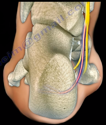
The Baxter’s nerve is the first branch of the lateral
plantar nerve. It also contributes to 20% of all heel pain causes. The Baxter’s
nerve provides motor innervation to the abductor digiti minimi muscle. When the
nerve is affected by compression, the symptoms are similar to planter
fasciitis. Entrapment of the Baxter’s nerve may cause up to 20% of heel pain,
but is still overlooked. The first branch of the lateral planter nerve is the
inferior calcaneal nerve (Baxter’s Nerve).
The nerve courses vertically between
the abductor hallucis and the quadratus plantae muscles, then makes a 90°
horizontal turn, coursing laterally beneath the calcaneus to innervate the
abductor digiti minimi muscle. The Baxter’s nerve can become entrapped within
the medial heel. The entrapment will result in heel pain, paresthesia, abnormal
sensations on the plantar aspect (bottom) of the heel, and medial heel
tenderness. There are two sites of entrapment that are typical with Baxter’s
nerve impingements. The first site is located between the fascia of the
abductor hallucis and quadratus plantae muscles. The second site is where the
nerve passes along the anterior aspect of the medial calcaneal tuberosity.
Treatment
Management is usually conservative and consists of heel
stretching exercises, rest, NSAIDs, and Orthotics. Surgery is done as a last
resort with neurolysis of the nerve after release of the deep fascia of the abductor
hallucis muscle. There is a good-excellent result in about 90% of patients
after decompression of the nerve. Involvement of the Baxter’s nerve may affect
running athletes causing pain on the medial plantar aspect of the foot.
 A patient with a complete anterior interosseous nerve injury
or a high medial nerve injury should be asked to make a fist. The first and
second digits will have difficulty in flexing, while the other digits will
flex. The third digit will be weak, while the fourth and fifth digits are
normal. This position of the hand is similar to the position taken during a
hand blessing. The Benedictine sign is different from an “ulnar claw hand”.
Ulnar claw hand refers to damage to the ULNAR nerve and is seen when attempting
to extend all the digits (leaving the 4th and 5th digits
flexed). The O.K. sign is used to check for paralysis of the anterior
interosseous nerve due to entrapment or compression injury. A patient with
paralysis of the anterior interosseous nerve will be unable to make the O.K.
sign. This is due to weakness of the flexor pollicis longus and flexor
digitorum profundus muscles. A typical pinch attitude is associated with
anterior interosseous nerve injury.
 The anterior interosseous nerve arises from the median nerve
about 4-6cm distal to the elbow, which is about 1/3 of the way down the
forearm. It exits from the anterolateral aspect of the median nerve and it runs
between the radius and the ulna on the interosseous membrane between and below
the muscles of the flexor digitorum profundus and the flexor pollicis longus.