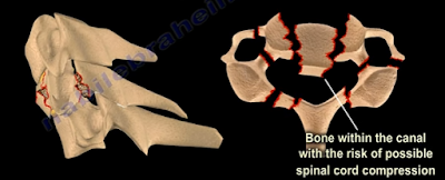
In our final blog post regarding Orthopaedic Emergencies, we will
review:
- Transverse Atlantal Ligament Rupture
- Bilateral Cervical Facet Dislocation
- Spinal Cord Compression
- Cauda Equina Syndrome
Transverse Atlantal Ligament Rupture
The normal Atlanto-Dental Interval is less than 3mm. An
A.D.I measuring greater than 3mm will be translationally unstable in the sagittal
plane due to transverse atlantal ligament rupture. This is usually apparent on
x-rays or CT scan. If the condition is not diagnosed, it can result in spinal
cord compression, respiratory arrest, and a catastrophic outcome. Treatment
typically requires a posterior atlanto-axial arthrodesis.
Bilateral Cervical Facet Dislocation
Facet dislocations of the cervical spine:
- Unilateral Facet Dislocation
- Displacement is less than 50% of the vertebral body width
- May need surgery
- Bilateral Facet Dislocation
- Displacement greater than 50% of the vertebral body width
- Usually needs surgery
- Exclude disc herniation
Obtain a preoperative MRI to rule our disc herniation
associated with facet dislocations.
Spinal Cord Compression
Spinal cord compression is more common with cervical spine
injuries and thoracic spine injuries. Neurogenic shock resulting from spinal cord
injury may complicate resuscitation of the patient and should be differentiated
from hypovolemic shock. It is important to look for hypotension and bradycardia
as well as thoracolumbar fractures which could be missed. Treatment consists of
emergency management involving resuscitation and hemodynamic stabilization with
concurrent neurologic examination. Protocol requires steroids given early.
Definitive treatment consists of stabilization of unstable spinal injuries.
Cauda Equina Syndrome
Central disc herniation compressing the cauda equine. It results
from injury to the lumbosacral nerve roots within the spinal canal. This
syndrome presents with involvement of the bladder, bowel, and lower limbs and
usually results from central disc herniation or fractures. Central disc
herniation or bony fragments results in the compression of the nerve roots.
Early diagnosis is imperative to find the cause of the compression on the nerve
roots. Urgent decompression by the removal of the central disc herniation or
stabilization of the fracture is necessary for treatment.


