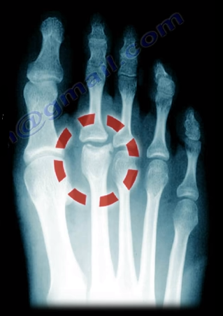
Freiberg’s Disease is caused by avascular necrosis of the
head of the 2nd metatarsal. This condition is more common in
patients who have a longer 2nd metatarsal bone relative to the 1st
metatarsal. This leads to the transfer of excessive loads onto the 2nd
metatarsal, which may interfere with the blood supply. This disease tends to
occur more commonly in young females during growth spurts.
Freiberg’s Disease usually presents itself as pain and
swelling at the 2nd metatarsophalangeal joint that is related to
activities and walking in high heels. There may be point tenderness and
swelling over the head of the 2nd metatarsal. There may also be
limited range of motion in the 2nd metatarsophalangeal joint. Early
in the disease, x-rays may only show minimal changes. Radiological evidence of
the condition may only be clearly visible on an MRI and bone scan. In more
severe cases, sclerosis, fragmentation, collapse of the metatarsal head, and
severe arthritis, may make the condition easily visible on an x-ray.
Treatment
Conservative treatment consists of nonsteroidal
anti-inflammatory medications, activity modification, orthotics, and immobilization
with a short leg cast. Surgical intervention involving a joint debridement is
indicated only after the failure of all conservative measures. Other procedures
may be indicated depending on the complexity and severity of the case.
Stem cells may help tissues that are injured or damaged to
renew and regenerate themselves. Depending on the treatment and medium, stem
cells have the ability to become different types of cells such as bone,
cartilage, and blood vessels. There are several conditions in which stem cells
are used as treatment, including: avascular necrosis, arthritis, and nonunion.
When Avascular Necrosis of the femoral head occurs due to
the diminished blood supply, there is a death of a segment of bone, which is
considered necrotic. The surgeon can inject stem cells into this area to revive
this area by drilling into the bone. When using stem cells to treat AVN, the
surgeon will need to create a channel for new blood vessels to form into the
area that lacks blood supply. After the channel is created, the stem cells are
injected into the necrotic femoral head.
Stem cell treatments for joint pain and arthritis is not
proven to be effective. However, there is some use in knee arthritis for
cartilage regeneration.
The best use of stem cells in Orthopaedics is its treatment
for nonunion fractures. A nonunion fracture is classified as a fracture that
does not heal after a reasonable period of time or a fixation failure. Nonunion
may also be due to motion of the bony ends and incomplete healing of the
fracture; fractures of this nature will need a lot of assistance. Two elements
are needed for treatment of nonunions: vascularity—which improve the local
conditions to facilitate healing; and stability—in the form of fixation such as
a rod or plate.
The most common causes of nonunion are smoking (5 times more
common), diabetes, obesity, osteoporosis, unstable fixation, infection (most
common), open fractures, and the severe displacement of the fracture.
Options available for treatment:
Bone Morphogenetic Protein—very expensive
Bone Graft—donor site morbidity
Stem Cells
Stem cells must be extracted from the bone marrow and are
aspirated and harvested from the anterior iliac crest. This procedure is
performed with an outlet view under fluoroscopy. Once extracted, the bone
marrow is prepared to be centrifuged. After centrifuging the bone marrow, a
good sample is extracted for injection.
The surgeon will mark and localize the
area for injection and the trocar is placed. The sample will then be injected
into the fracture area—occasionally, two areas of nonunion are treated. Adult
mesenchymal stem cells are special cells that can copy themselves, divide, and
multiply. They can differentiate into bone cells that heal the nonunion and lay
down new bone. This process can be monitored by alkaline phosphatase activity
or by the genes of the stem cells. The whole cellular mechanism can help increase
the vascularity of the nonunion.
It is important to note that adult mesenchymal stem cells
are not embryonic stem cells. There is a large amount of information in regards
to stem cells that is lacking or misleading. Cells should probably be combined
with some type of matrix. Additionally, surgeons need a better delivery system
and localization during the injection of the stem cells due to the fact that
the dye kills the cells. It is beneficial to allow the cells to expand and grow
in the culture prior to injection. Moreover, the effect of certain medications
such as aspirin, Plavix, and Coumadin, should be studied further.
Hip dislocations can occur posteriorly or anteriorly;
however, any type of hip dislocation is considered an emergency.
Posterior hip dislocations are more common and the lower
limb will be flexed, adducted, and internally rotated. Posterior dislocations
are frequently caused by dashboard injuries. The impact with the car dashboard
drives the femoral head backwards out of the acetabulum. The physician will
want to observe sciatic nerve function and examine the knee to rule out a PCL
injury as well. Weakness of the ankle and toe dorsiflexion due to an injury to
the peroneal division of the sciatic nerve may result in foot drop. The patient
will also be unable to dorsiflex the ankle.
Anterior hip dislocation is rare. Superior Anterior hip
dislocation results from the lower limb being extended, abducted, and
externally rotated. Inferior Anterior Hip Dislocations (obturator type) results
from the lower limb being flexed, abducted, and externally rotated.
An emergency reduction of dislocations is needed in less
than 8 hours of the injury. An urgent reduction is mandatory to avoid avascular
necrosis and interruption of the blood supply, which leads to a collapse of the
femoral head. AVN is the death of a segment of bone.
Treatment
A CT scan should be obtained after reduction to evaluate the
presence of fragments in the joint and access stability of the joint. Hip joint
dislocations may be associated with acetabular or femoral head fractures (Pipkin).
An urgent closed reduction of the hip dislocation followed by stabilization of
either of the fractures if needed according to the protocols.
 Freiberg’s Disease is caused by avascular necrosis of the
head of the 2nd metatarsal. This condition is more common in
patients who have a longer 2nd metatarsal bone relative to the 1st
metatarsal. This leads to the transfer of excessive loads onto the 2nd
metatarsal, which may interfere with the blood supply. This disease tends to
occur more commonly in young females during growth spurts.
Freiberg’s Disease is caused by avascular necrosis of the
head of the 2nd metatarsal. This condition is more common in
patients who have a longer 2nd metatarsal bone relative to the 1st
metatarsal. This leads to the transfer of excessive loads onto the 2nd
metatarsal, which may interfere with the blood supply. This disease tends to
occur more commonly in young females during growth spurts.  Freiberg’s Disease usually presents itself as pain and
swelling at the 2nd metatarsophalangeal joint that is related to
activities and walking in high heels. There may be point tenderness and
swelling over the head of the 2nd metatarsal. There may also be
limited range of motion in the 2nd metatarsophalangeal joint. Early
in the disease, x-rays may only show minimal changes. Radiological evidence of
the condition may only be clearly visible on an MRI and bone scan. In more
severe cases, sclerosis, fragmentation, collapse of the metatarsal head, and
severe arthritis, may make the condition easily visible on an x-ray.
Freiberg’s Disease usually presents itself as pain and
swelling at the 2nd metatarsophalangeal joint that is related to
activities and walking in high heels. There may be point tenderness and
swelling over the head of the 2nd metatarsal. There may also be
limited range of motion in the 2nd metatarsophalangeal joint. Early
in the disease, x-rays may only show minimal changes. Radiological evidence of
the condition may only be clearly visible on an MRI and bone scan. In more
severe cases, sclerosis, fragmentation, collapse of the metatarsal head, and
severe arthritis, may make the condition easily visible on an x-ray.