Showing posts with label growth plates. Show all posts

Tuesday, August 14, 2018

Triplane Fracture of the Distal Tibia


A triplane fracture of the distal tibia usually occurs during adolescence and occurs before complete closure of the distal tibial physis. The distal tibial physis (growth plate) is a weak area which closes from central to medial, with the lateral side being the last part to close. A Triplane fracture is a Salter-Harris Type IV Fracture, involving all three planes, the coronal (metaphysis), transverse (growth plate), and sagittal (epiphysis). The fracture has several variations and occurs due to external rotation forces. This fracture typically occurs in patients between 12-15 years of age.


Triplane fractures are complicated three-dimensional fractures. A two-part fracture is a Salter-Harris Type IV. A Three-part fracture is a combination of Salter-Harris Type III in an AP view and a Type II in a lateral view. CT scans are helpful.


An ORIF is necessary if there is displacement of the fragments of more than 2 mm.

Tuesday, April 17, 2018

Freiberg's Disease


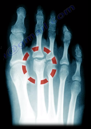
Freiberg’s Disease is caused by avascular necrosis of the head of the 2nd metatarsal. This condition is more common in patients who have a longer 2nd metatarsal bone relative to the 1st metatarsal. This leads to the transfer of excessive loads onto the 2nd metatarsal, which may interfere with the blood supply. This disease tends to occur more commonly in young females during growth spurts.

Freiberg’s Disease usually presents itself as pain and swelling at the 2nd metatarsophalangeal joint that is related to activities and walking in high heels. There may be point tenderness and swelling over the head of the 2nd metatarsal. There may also be limited range of motion in the 2nd metatarsophalangeal joint. Early in the disease, x-rays may only show minimal changes. Radiological evidence of the condition may only be clearly visible on an MRI and bone scan. In more severe cases, sclerosis, fragmentation, collapse of the metatarsal head, and severe arthritis, may make the condition easily visible on an x-ray.

Treatment


Conservative treatment consists of nonsteroidal anti-inflammatory medications, activity modification, orthotics, and immobilization with a short leg cast. Surgical intervention involving a joint debridement is indicated only after the failure of all conservative measures. Other procedures may be indicated depending on the complexity and severity of the case.

Tuesday, March 6, 2018

Tibial Spine Fractures in Children


Tibial Spine fractures are similar to ACL injuries in adults. It occurs due to hyperextension of the knee and is commonly seen during falls from a bicycle. An injured child with a swollen knee and who has fallen from a bicycle should alert the clinician to the possibility of a tibial spine fracture. A meniscal injury may also occur, especially with the medial meniscus. The interposition of the meniscus or rotation of the fracture may prevent a closed reduction.
Tibial spine fractures are classified using the Meyers and McKeever Classification and are separated into three types. Type I classifications are nondisplaced, Type II fractures are identified as being minimally displaced with an intact posterior hinge, and Type III fractures are classified as being completely displaced.
The presentation and examination is similar to an ACL tear with immediate swelling as well as a positive Lachman’s Test or Anterior Drawer Test. An x-ray will show the fracture and a CT scan will help in planning for surgery. An MRI may be required to show a trapped or a meniscal injury.

Treatment


Treatment will consist of an aspiration of the large hematoma. Nonoperative treatment is used for Type I fractures and reducible Type II fractures; a closed reduction and immobilization in 0-20 of flexion. Surgery is performed in Type II fractures and unreducible Type II fractures. An ORIF or arthroscopic reduction and fixation will be performed. During surgery, the trapped meniscus will be moved out of the way and the surgeon will use sutures or screws for fixation. It is important for the surgeon to remember to avoid the physis.

Complication


ACL laxity is common but not clinically significant. Stiffness or arthrofibrosis occurs with surgical fixation. Growth arrest is rare.

Thursday, February 8, 2018

Bone Growth in Children


A Special Thanks to Miranda Ebraheim for assisting with this article


There are growth plates within the long bones which contributes to the development of the bones in children.

The growth distribution in the humerus is about 80% in the proximal and 20% in the distal area. Displaced fractures of the proximal humerus in children are usually treated without surgery.

In regards to the ulna, the growth distribution is about 80% proximal and 20% distal. Growth arrest is common in fractures involving the distal ulna and occur approximately 50% of the time. Within the radius, it is about 25% proximal and 75% distal. Fractures at the distal radius usually heals and corrects its angulation after a closed reduction. Surgery is rarely necessary. Fractures involving the growth plate of the distal radius rarely involves growth arrest.

Within the femur, the growth rate distribution is about 30% proximal and 70% distal. Fractures involving the growth plate of the distal femur may cause major growth disturbances. It is expected that a child grows 1 cm per year from the distal femur growth plate. Boys will continue to grow up until 16 years old, while girls stop growing at the age of 14.


Finally, there are the bones of the tibia and fibula. The growth distribution in the tibia is about 55% proximal and 45% distal. Within the fibula, it is about 60% proximal and 40% distal.

A growth spur occurs at the time of puberty. Puberty typically occurs in females around 8-13 years of age, and at 10-15 years of age in males. Fractures near the growth plate remodel well.