The knee jerk reflex or patellar reflex, is a deep tendon
reflex seen as a sudden kicking movement of the lower leg in response to a
sharp tap on the patellar tendon. Tapping the patellar ligament stimulates the
muscle spindles in the quadriceps. Impulses travel from the muscle spindles to the
spinal cord. In the spinal cord, synapses occur with motor neurons and
interneurons. The motor (efferent) neurons send activating impulses to the
quadriceps causing the muscles to contract and extend the knee. The interneuron
(relay neuron) forms a connection between the other neurons and interneurons. Interneurons
are neither motor nor sensory. Interneurons transmit impulses that inhibit the
antagonistic muscles (hamstrings). An abnormality of the reaction suggests that
there may be damage to the central nervous system.
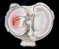
The meniscus is a cushion structure made of cartilage which
fits within the knee joint between the tibia and the femur. The medial meniscus
is C-shaped and the lateral meniscus in the more circular. The meniscus is made
up of type I collagen that provides shock absorption and stability to the knee
joint. The meniscus helps to protect the knee joint, allowing the bones to
slide freely on each other. Discoid meniscus is a rare variation of the
meniscus that usually affects the lateral meniscus of the knee in less than 5%
of the population and could be bilateral in about 25% of the cases.
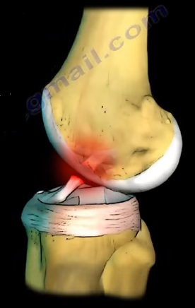
Discoid
meniscus is a large meniscus with abnormal attachment causing increased
mobility of the meniscus. It causes a pop, click, or snapping with locking and
pain. There will be loss of full knee extension with tenderness on the lateral
joint space. Symptoms occur more during extension of the knee. The discoid
meniscus occurs due to the abnormal development and increase in size of the
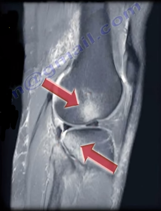
meniscus. An x-ray could show increased widening of the joint space. An MRI
will show the “bow tie” sign in three or more sagittal continuous cuts. The
coronal MRI will show a thick and flat meniscus extending beyond the halfway
point of the condyle.
Watanabe Classification of Discoid Lateral Meniscus
Type I: Block-shaped stable
Type II: Block-shaped, stable, partial meniscus (has good peripheral attachment)
Type III: Unstable meniscus with stability arising only form the ligament of Wrisberg. (no posterior meniscal tibial attachment).
Treatment
An asymptomatic patient will be treated with observation. A
symptomatic patient may receive a partial meniscectomy and saucerization with
repair of type III (no posterior tibial meniscal attachment)
Several types of tibial plateau fractures are a complex
management problem. The knee joint may have a significant comminution and
depression, and the physician may need to take an extensile approach for
reduction and fixation of this fracture. Personally, I use the intra-articular
extensile approach for tibial plateau fracture reduction and fixation. In
general, fracture of the tibial plateau is a complicated problem.
A vascular evaluation is necessary. The ankle-brachial index
(ABI) is needed in some types, such as in medial plateau fractures or in severe
types, such as Schatzker Type V or Type VI. The ABI should be more than 0.9.
Usually, medial tibial plateau fractures are considered to be a knee
dislocation. A fasciotomy may be needed if compartment syndrome occurs. The soft
tissue condition may be bad, and an external fixator may be initially used
until the soft tissue condition improves.
The association between tibial plateau fractures and meniscal
tear is not uncommon. A lateral plateau fracture will create a lateral meniscal
tear, while the medial plateau fracture will cause a medial meniscal tear. A
tear of the meniscus is usually peripheral. It should be recognized and dealt
with. The physician may want to look at the x-ray and see if there is a
depression or separation of more than 6mm, as this indicates a high chance of
meniscal tear.
The posteromedial fragment is another problem with tibial
plateau fractures which needs to be fixed separately. When an extensive
comminuted displaced tibial plateau fracture occurs, the physician may need
excellent exposure of the articular surface to allow for anatomic reduction of
the joint and visualization and repair or debridement of the meniscus if it is
torn. This extensile exposure is important, especially if the posterior part of
the plateau is involved. The traditional way to see the articular cartilage of
the tibial plateau is to use the submeniscal approach by cutting the coronary
ligament, but the exposure is limited. Other extensile approaches are also
developed; however, we use the extensile intra-articular approach for complex,
comminuted tibial plateau fractures. This involves anterior detachment and
retraction of the meniscus to improve visualization of the tibial articular
surface. This approach can be utilized for lateral or medial tibial plateau
fractures and it is especially helpful in diagnosing and repairing the torn
meniscus. This allows for inspection of the meniscus pathology in fractures of
the articular surface. This improves reduction of the fracture and the torn
meniscus is repaired and reattached to the coronary ligament. Incision and
reflection of the meniscus allows great exposure and inspection of the joint
which is followed by reattachment and suturing of the anterior horn of the
meniscus to its normal position which is followed by reattachment of the
meniscotibial (coronary) ligament. The sutures are tied to the sides of the
patellar tendon on the opposite side of the meniscus.
Meniscal injuries are very common. The McMurray’s Test is a
rotational maneuver of the knee that is frequently used to aid in the diagnosis
of meniscal tears. With a meniscal tear, the patient usually complains of knee
pain localized to the lateral or medial side of the knee joint. The patient
will have locking, clicking, pain, or effusion.
During the physical examination, joint line tenderness is
the most sensitive finding. Swelling of the knee and a possible extension lag
(locked knee) is also a common finding. Pain at a higher level is usually
associated with the medial collateral ligament. Pain at a lower level is
usually associated with the pes anserine bursa.
What is the McMurrays test?
The McMurray’s test is a knee examination test that provokes
pain or a painful click as the knee is brought from flexion to extension with
either internal or external rotation. The McMurray’s test uses the tibia to
trap the meniscus between the femoral condyles of the femur and the tibia. When
performing the test, the patient should be lying supine with the knee
hyperflexed. The examiner then grasps the patient’s heel with one hand and
places the other hand over the knee joint. To test the medial meniscus, the
knee is fully flexed, and the examiner then passively externally rotates the
tibia and places a valgus force. The knee is then extended in order to test the
medial meniscus. To test the lateral meniscus, the examiner passively
internally rotates the tibia and places a varus force. The knee is then
extended in order to test the lateral meniscus. A positive test is indicated by
pain, clicking or popping within the joint and may signal a tear of either the
medial or lateral meniscus when the knee is brought from flexion to extension.
How reliable is the McMurray’s test?
There are mixed reviews for the validity of this test. An
MRI is a very sensitive exam and makes the diagnosis easier, while excluding
other associated injuries.
A patellar tendon rupture is a rupture of the tendon that
connects the patella to the tibia. Rupture often occurs at the lower pole
insertion site of the patella and it could be associated with degenerative
changes. Rupture most often occurs in patients younger than 40 years of age.
When the tendon is ruptured, the quadriceps muscle pulls the patella upward.
One way to measure the height of the patella is by measuring the Blumensaat’s
line. The knee needs to be flexed at least 30 degrees, then a line can be drawn
through the roof of the intercondylar notch and usually touches the tip of the
patella. The patella moves upward with the patellar tendon rupture (patella
alta).
Associated Risk Factors
Rheumatoid Arthritis
Diabetes
Chronic Renal Failure
Systemic Corticosteroid Therapy
Chronic Patellar Tendonitis
Degenerative Changes
During the radiographic evaluation, an AP and Lateral x-ray
is necessary. The patella alta is seen on the lateral view (*patella superior
to Blumensaat’s line). An MRI is effective in assessing the patellar tendon,
especially if other intraarticular or soft tissue injuries are suspected.
Treatment consists of a surgical reattachment of the tendon.
The patient will need to keep their knee in extension and in a knee immobilizer
for about 4-6 weeks.
Several bursa are seen around the knee area. These bursa
include the suprapatellar, prepatellar, infrapatellar, and pes anserine. The
pes answerine bursa is a small fluid filled sac located between the tibia and
the three tendons of the Sartorius, Gracilis, and Semi-tendinosus.
These
muscles are innervated by three separate nerves, the femoral, obturator, and
the tibial branch of the sciatic nerve, respectively. Pes Anserine bursitis, or
“breast stroke knee”, is an inflammatory condition of the medial knee at the
pes anserine bursa that is common in swimmers.
What is the pes anserine?
The pes anserine is the common area of insertion for the three
tendons along the proximal medial aspect of the tibia. This condition is also
sometimes referred to as a “goosefoot” because the pes anserinus tendons
resemble the shape of a goose foot. Pes Anserine bursitis is usually seen as
causing pain, tenderness, and localized swelling after trauma or total knee
replacement. The pain is seen below the joint line on the medial part of the
proximal tibial with the bursa being deep to the tendons.
Treatment
Treatment consists of physical therapy, nonsteroidal anti-inflammatory
medications, and injections. The physician will need to rule out meniscal
tears, stress fractures, or osteonecrosis of the tibia, as these are all
differential diagnosis.
Stem cells may help tissues that are injured or damaged to
renew and regenerate themselves. Depending on the treatment and medium, stem
cells have the ability to become different types of cells such as bone,
cartilage, and blood vessels. There are several conditions in which stem cells
are used as treatment, including: avascular necrosis, arthritis, and nonunion.
When Avascular Necrosis of the femoral head occurs due to
the diminished blood supply, there is a death of a segment of bone, which is
considered necrotic. The surgeon can inject stem cells into this area to revive
this area by drilling into the bone. When using stem cells to treat AVN, the
surgeon will need to create a channel for new blood vessels to form into the
area that lacks blood supply. After the channel is created, the stem cells are
injected into the necrotic femoral head.
Stem cell treatments for joint pain and arthritis is not
proven to be effective. However, there is some use in knee arthritis for
cartilage regeneration.
The best use of stem cells in Orthopaedics is its treatment
for nonunion fractures. A nonunion fracture is classified as a fracture that
does not heal after a reasonable period of time or a fixation failure. Nonunion
may also be due to motion of the bony ends and incomplete healing of the
fracture; fractures of this nature will need a lot of assistance. Two elements
are needed for treatment of nonunions: vascularity—which improve the local
conditions to facilitate healing; and stability—in the form of fixation such as
a rod or plate.
The most common causes of nonunion are smoking (5 times more
common), diabetes, obesity, osteoporosis, unstable fixation, infection (most
common), open fractures, and the severe displacement of the fracture.
Options available for treatment:
Bone Morphogenetic Protein—very expensive
Bone Graft—donor site morbidity
Stem Cells
Stem cells must be extracted from the bone marrow and are
aspirated and harvested from the anterior iliac crest. This procedure is
performed with an outlet view under fluoroscopy. Once extracted, the bone
marrow is prepared to be centrifuged. After centrifuging the bone marrow, a
good sample is extracted for injection.
The surgeon will mark and localize the
area for injection and the trocar is placed. The sample will then be injected
into the fracture area—occasionally, two areas of nonunion are treated. Adult
mesenchymal stem cells are special cells that can copy themselves, divide, and
multiply. They can differentiate into bone cells that heal the nonunion and lay
down new bone. This process can be monitored by alkaline phosphatase activity
or by the genes of the stem cells. The whole cellular mechanism can help increase
the vascularity of the nonunion.
It is important to note that adult mesenchymal stem cells
are not embryonic stem cells. There is a large amount of information in regards
to stem cells that is lacking or misleading. Cells should probably be combined
with some type of matrix. Additionally, surgeons need a better delivery system
and localization during the injection of the stem cells due to the fact that
the dye kills the cells. It is beneficial to allow the cells to expand and grow
in the culture prior to injection. Moreover, the effect of certain medications
such as aspirin, Plavix, and Coumadin, should be studied further.
The anterior cruciate ligament is located at the front of
the knee. Rupture of the anterior cruciate ligament (ACL) is a condition
commonly seen in sports, usually due to a non-contact pivoting injury. The
Pivot Shift test is a specific test for an ACL deficient knee (ACL injury).
A pivot shift is pathognomonic for an ACL tear and is best demonstrated in a
chronic setting. The Lachman’s test is the most sensitive examination test for
an ACL injury.
The ACL keeps the tibia from sliding out in front of the femur
and provides rotational stability to the knee. Rupture of the ACL causes
anterolateral rotatory instability. The tibia moves anterolaterally in
extension; however, when you flex the knee the IT band becomes a flexor of the
knee and pulls back, reducing the tibia. The Pivot Shift Test goes
from extension (tibia subluxed) to flexion, with the tibia reduced by the
iliotibial band.
Both the Lachman’s test and the Pivot Shift test are
associated with 20-30 degrees of knee flexion. The Lachman’s test starts at
20-30 degrees of flexion, but with the Pivot Shift test, you will feel the clunk at
20-30 degrees of flexion. Remember: 20-30 degrees of flexion is important for
examination of the ACL. The femur is stabilized with one hand and the other
hand pulls the tibia anteriorly and posteriorly against the femur. The tibia
can be pulled forward more than normal (anterior translation). The examiner
will have a sense of increased movement and lack of a solid end point.
When performing the Pivot Shift test, the patient should be
totally relaxed and lying supine. The knee is in the
subluxed position when in full extension. The pivot shift starts
with extension of the knee and you can feel a “clunk” at 20-30 degrees of
flexion. The physician will hold the knee in full extension, then add valgus force, and internal
rotation of the tibia to increase the rotational instability of the knee. Then
the physician will take the knee into flexion. A palpable clunk is very specific of an
ACL tear. The iliotibial band will reduce the tibia and create the clunk on the
outside of the knee. The physician should always compare the results with the
other side.
The ACL prevents anterior translation of the tibia. It is a
secondary restraint to tibial rotation and varus and valgus. The ACL consists
of two bundles:
The Posterolateral Bundle
Anteromedial Bundle
The Posterolateral bundle prevents the pivot shift and
contributes to rotational stability. This bundle also prevents internal
rotation of the tibia with the knee in near extension (tight in extension,
loose in flexion). If it is sectioned, it increases the anterior translation
and tibial rotation at 30° of flexion. The Anteromedial bundle is tight in
flexion and if sectioned, it increases the anterior translation at 90° of
flexion.
The Lachman’s test is the most sensitive test, especially in
acute settings. The examiner will find no end point with anterior translation
of the knee and the physical examination can be difficult or limited due to
pain. With the Pivot Shift test, the patient must be completely relaxed. The
test is helpful in chronic situations, especially if the patient complains of
the knee giving way.
During the Pivot Shift, the knee subluxes in extension and reduces
at 20-30 degrees of flexion. The Pivot Shift correlates closely with patient
satisfaction of their reconstructed knee. It is also a measure of functional
instability following ACL reconstruction. Verticle femoral tunnel placement
will cause rotational instability seen as a positive pivot shift, and the
malposition of the bone tunnel will be seen in an AP view x-ray of the knee. The
9 or 10 o’clock position is better than the 12 o’clock. A vertical position is
bad.
The patient with an ACL injury usually has a non-contact
pivoting injury even with:
Awkward landing
Feeling a “Pop” sensation
Immediate swelling
Aspiration usually shows blood in the knee (75%
chance of ACL tear with hemorrhage in the knee)
Positive Lachman’s Test (may be hard to examine
due to pain)
An MRI is going to be the best imaging technique. An MRI of
the knee joint will show bone lesions or bruising in the typical location
associated with tears of the ACL. These injuries are typically located at the
middle of the femoral condyle and posterior part of the tibia laterally. It is
also possible to find a triple injury within the MRI (O’Donoghue’s Unhappy
Triad).
O’Donoghue’s Unhappy Triad consists of:
Tear of the Lateral Meniscus
Anterior Cruciate Ligament Injury (ACL tear)
Medial Collateral Ligament Injury
In chronic ACL tears, the posterior horn of the medial
meniscus is the most commonly injured structure. In acute ACL tears, send the
patient for therapy for range of motion, brace the patient, and allow the MCL
to heal and reconstruct the ACL later if needed. It is important to stress
hamstring therapy in ACL tears. The patient will probably complain of
instability immediately or later on.
A tibial plateau depression fracture occurs when axial
forces from the femoral condyles against the articular surface of the tibia
cause the injury. The compression fracture may also be associated with other
fractures. Several techniques are used for the treatment of this fracture.
Balloon osteoplasty is proposed as a useful tool for reduction and elevation of
the tibial plateau depression fracture.
The technique of balloon osteoplasty can be done with or
without the supplementation of screws and plate fixation. Trajectory lines are
drawn on the medial side of the knee for access to the fracture area. A small
incision is made at the point where the lines cross. A cannula and trocar tip
stylet is used to access the fractured area under radiographic guidance. The
cannula and trocar is advanced under the area of depression using a mallet or
drill when needed. Once inserted, the trocar is then removed from the cannula.
The precision drill is then inserted into the cannula and advanced until it
passes under the depressed area of bone. Then, the precision drill is removed
from the cannula and the inflatable bone tamp is inserted through the cannula
to the area of the depressed fragments. The inflatable bone tamp is centered
under the area of depression and then inflated to reduce the depression
fracture. Once the depression has been adequately reduced, the inflatable bone
tamp can be deflated and removed from the cannula.
The void that has been left by the inflatable bone tamp is
now ready to be replaced with bone filler. The device loaded with injectable
material is inserted and used until the void is completely filled. Finally, you
will remove the injectable material device and the cannula. Plate fixation may
be added before or after the injectable material is inserted.