Showing posts with label medicine. Show all posts

Tuesday, October 30, 2018

Olecranon Fractures


Fractures of the olecranon may occur due to a direct blow to the elbow or from a fall onto an outstretched hand. Nondisplaced fractures have less than 2mm of separation and are considered stable. Fracture separation will not increase with elbow flexion. Extensor mechanism is intact and the patient will be able to extend the elbow against gravity. Displaced fractures could be an avulsion, oblique, transverse, comminuted, or dislocated.
Olecranon fracture dislocations can be anterior (transolecranon) or posterior (similar to monteggia fracture dislocation). An examination will show that the patient is unable to extend the elbow with these displaced fracture types. A true lateral view x-ray will clearly show the olecranon fracture. Usually, these fractures are followed by stiffness of the elbow in about 50% of the patients. However, this does not affect the function.
The goal of treatment for olecranon fractures should be restoration of the articular surface, preservation of the continuity of extensor mechanisms, maintain elbow stability, and avoid stiffness of the elbow. Nonoperative treatment is used for nondisplaced fractures and it may be used for some displaced fractures in elderly patients (treat elbow in some flexion with a splint). I would personally use minimally invasive techniques in these patients unless the skin is very bad, or the fracture is very comminuted.
There are three techniques used for surgical treatment: the tension band technique, detach olecranon and reattach triceps, and plate and screw fixation. The tension band technique is only used for transverse fractures with no comminution. K-wires and screws are used, and the surgeon may use either a 6.5mm screw or Kwires for the tension band. When doing the tension band technique, you want to engage the anterior cortex of the ulna. The surgeon should avoid over penetration to avoid affecting the forearm rotation or injuring the anterior interosseous nerve. The surgeon needs to be sure that the pins are not fixing the radius and that after the operation, the patient can perform pronation and supination of the forearm (pull the pins out slightly if needed). The distractive force of the triceps is converted to compression force at the articular surgace, especially when bending the elbow. The drill hole for the K-wire should be positioned about 4-5cm from the fracture which gives enough safe distance so that the fracture will not propagate. Place the tension band wire through the drilled holes before application of the K wires. The surgeon should be sure that the hook to the K-wire is posterior. Make sure that the tension band wire is close to the bone so there is no laxity in the fixation and instability. An intramedullary screw could be used. This screw fixation may need a washer to capture the tension band wire. Intramedullary 6.5mm screw fixation is a reasonable option for fixation but it may need to be supplemented with tension band wires. Never use cancellous screw alone. The tension band technique are for transverse fractures of the olecranon. If fractures comminution is present, change the plan of fixation.

When detaching the olecranon and reattaching the triceps, an excision of the fracture fragment and triceps advancement is used:

  • If the fracture is less than 50%
  • To treat elderly patients (especially if fracture is comminuted)
  • For some nonunions when the fracture is small and cannot be fixed

The surgeon must be sure that the procedure is done with the elbow is stable. If the elbow has ligamentous instability and excision of the fracture fragment is done, this will make the elbow very unstable. The triceps should be attached closer to the articular surface.
Special olecranon plates are available when using a plate and screw fixation technique. The bridge plate and screw fixation technique is used in comminuted, Monteggia, oblique fractures extending to the coranoid, and fracture dislocation. The plate is placed on the tension side of the olecranon (dorsal side). Sometimes, an opening is made through the triceps and the plate is placed against the bone, then suturing the triceps tendon over the plate to avoid hardware prominence.
In summary, if the patient is elderly with a small, comminuted fracture fragment less than 50% of the joint space, excise the fragment, and reattach the triceps tendon to the olecranon. If the olecranon fracture is transverse and proximal to the base of the coranoid process, then use the tension band technique. Use plate fixation for all olecranon fracture scenerios, such comminuted fractures, oblique fractures, unstable fractures, dislocation, or fractures distal to the coranoid process. The typical exam question scenario will discuss a comminuted fracture that should be treated with a plate. You probably need to remove above 20% of the plate fixations due to hardware irritation. Hardware irritation is worse with the tension band surgical treatment (may need to remove in more than 50% of cases).

Tuesday, October 9, 2018

Anterior Interosseous Nerve- The Benedictine and O.K. Sign


A patient with a complete anterior interosseous nerve injury or a high medial nerve injury should be asked to make a fist. The first and second digits will have difficulty in flexing, while the other digits will flex. The third digit will be weak, while the fourth and fifth digits are normal. This position of the hand is similar to the position taken during a hand blessing. The Benedictine sign is different from an “ulnar claw hand”. Ulnar claw hand refers to damage to the ULNAR nerve and is seen when attempting to extend all the digits (leaving the 4th and 5th digits flexed). The O.K. sign is used to check for paralysis of the anterior interosseous nerve due to entrapment or compression injury. A patient with paralysis of the anterior interosseous nerve will be unable to make the O.K. sign. This is due to weakness of the flexor pollicis longus and flexor digitorum profundus muscles. A typical pinch attitude is associated with anterior interosseous nerve injury.

The anterior interosseous nerve arises from the median nerve about 4-6cm distal to the elbow, which is about 1/3 of the way down the forearm. It exits from the anterolateral aspect of the median nerve and it runs between the radius and the ulna on the interosseous membrane between and below the muscles of the flexor digitorum profundus and the flexor pollicis longus.
The anterior interosseous nerve supplies the flexor digitorum profundus muscle for the index and long fingers. It also supplies the flexor pollicis longus and the pronator quadratus muscles. The flexor digitorum profundus muscle for the index and long fingers is supplied by the anterior interosseous nerve. The medial part of the FDP is supplied by the ulnar nerve (FDP has dual innervation). The anterior interosseous nerve passes dorsal to the pronator quadratus with the anterior interosseous artery and provides innervation to the volar wrist capsule. The terminal branch of the anterior interosseous nerve innervates the carpal joint capsule.


In patients with Martin-Gruber Connection, the median nerve, or anterior interosseous nerve to the ulnar nerve in the forearm may present with intrinsic muscle weakness. It may be differentiated also from Parsonage-Turner Synrome (acute brachial plexus neuritis) and patient may have pain in the affected extremity. In anterior interosseous nerve entrapment, the median nerve conduction study result will be normal, however the needle EMG of the anterior interosseous innervated muscles will be abnormal.

Wednesday, September 26, 2018

Adhesive Capsulitis, Frozen Shoulder


Adhesive Capsulitis, or frozen shoulder, is a painful progressive loss of shoulder motion. It affects both active and passive movement of the shoulder joint. The shoulder will be stiff and painful and occurs due to inflammation, fibrosis, scarring, and contraction of the capsule. A normal shoulder joint capsule is elastic and allows great range of motion. Inflammation and thickening of the shoulder capsule and may lead to adhesive capsulitis. Frozen shoulder may occur without any specific cause, however it may be triggered by a mild trauma to the shoulder.


This condition develops slowly and goes through three phases:

  1. Pain and freezing
  2. Stiffness or frozen
  3. Resolution

During the pain and freezing phase, the pain is worse at night and increases with any movement. This phase will last several months. During the second phase, range of motion is limited as pain is diminishing. This may last up to one year. The resolution phase may begin overtime and may last up to three years.


Conditions associated with frozen shoulder include:

  • Diabetes
  • Thyroid problems
  • Auto immune disease
  • Stroke
  • Rheumatoid arthritis
  • Trauma or post-surgery

A patient with frozen shoulder will have loss of both active (movement without assistance) and passive (movement with assistance) motion. External rotation of the shoulder is very limited and the condition is self-limiting and may resolve on its own. X-rays are needed to rule out degenerative arthritis.  An MRI or arthrogram will show small fluid in joint cavity. Rotator cuff may be normal and synovitis and narrowing of the rotator cuff interval is usually seen.

Treatment consists of anti-inflammatory medications, physical therapy, injections, and manipulation under anesthesia. Surgery will be done in the form of a release of the capsule when nonoperative methods fail. The physician should always check the patient for diabetes.  

Tuesday, September 11, 2018

Anatomy of the Posterior Cutaneous Nerve of the Thigh


The posterior cutaneous nerve of the thigh (small sciatic nerve) arises from the sacral plexus from S1-S3. The posterior cutaneous nerve of the thigh exits from the pelvis through the greater sciatic notch below the piriformis muscle. The nerve descends below the gluteus maximus muscle along with the inferior gluteal artery. It runs into the back of the thigh beneath the fascia lata and over the long head of the biceps femoris muscle to the back of the knee. The nerve then pierces the deep fascia and accompanies the short saphenous vein to the middle of the back of the leg. The posterior cutaneous nerve of the thigh innervates the distal part of the gluteal region, the skin of the perineum and the posterior part of the thigh.
The nerve can become compressed when passing through the tunnel below the piriformis muscle and under the gluteus maximus muscle. This may result in sensitivity disturbances of the innervation area of the nerve. Causes of the syndrome may be hypertrophy or abnormality of the piriformis muscle such as entrapment below the piriformis which compresses the nerve. Compression of the nerve can also occur due to prolonged sitting. During the clinical examination, pain and sensitivity will be evident. Pain and sensitivity disturbances are characteristic of the nerve distribution site in the posterior part of the thigh down the knee. This disturbance can be from hyperesthesia to hypoesthesia or burning sensation similar to meralgia paresthetica of the lateral cutaneous nerve of the thigh.

Differential diagnosis include piriformis syndrome. The patient should avoid sitting for long periods of time, especially on a hard base. Treatment consists of physical therapy, massage, and injection. Surgery is rarely needed.

Tuesday, September 4, 2018

SLAP Tear- Symptoms, Diagnosis, and Treatment


A SLAP tear is a tear that occurs where the biceps tendon inserts into the superior labrum. A SLAP tear is different from a Bankart lesion. SLAP tears are not common and can be hard to diagnose. Symptoms of a SLAP tear include: pain deep within the shoulder or in the back of the shoulder, as well as catching, popping, or clicking sensations. The patient may also experience pain when throwing a ball with a decrease in velocity and the feeling of having a dead arm after pitching. Patients will also experience pain with overhead activity which mimics impingement syndrome. This typically affects throwing athletes. When the biceps tendon is involved, pain may also be located at the front of the shoulder. A SLAP tear can be an isolated lesion or it can be associated with internal impingement, articular sided cuff tear, or instability.
A SLAP tear is diagnosed with a clinical examination and testing. The O’Brien’s test is the most commonly used test. Multiple tests are usually used including the anterior slide test and the clunk test. An MRI with contrast is the best imaging technique. When performing the O’Brien’s test, the patient is standing or sitting with the arm at 90° of flexion, 10° of adduction, and full internal rotation with the forearm pronated.  The examiner applies pressure to the forearm and instructs the patient to resist the applied downward force. Pain at the shoulder joint suggests a SLAP lesion. Decrease in pain of the shoulder joint on supination of the arm is suggestive of a SLAP tear.
Treatment consists of physical therapy, anti-inflammatory medications, injections, and surgery (when conservative treatment fails). If surgery is necessary, a labral debridement will be performed for minor tearing and fraying. Biceps Tenodesis is becoming popular, as it is a procedure that cuts the biceps tendon where it attaches to the labrum and reinserts it in another area, usually in front of the shoulder. A biceps tenotomy is a procedure that cuts the biceps tendon from the glenoid, releasing the long head of the biceps tendon from its attachment allowing it to fall into the upper arm out of the shoulder joint. A biceps tenotomy is probably best suitable for some elderly patients. A SLAP repair is a procedure which uses sutures to anchor the torn labrum to the glenoid. This repair is usually done for athletes and patients under the age of 40 years.

Tuesday, August 28, 2018

Massive Rotator Cuff Tear


To view my Youtube video, 'Massive Rotator Cuff Tear- Classic', click here.


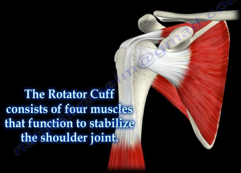
Massive rotator cuff tears are a disabling problem. These tears can cause pain, weakness, and sometimes swelling of the shoulder. The rotator cuff consists of four muscles that function to stabilize the shoulder joint: supraspinatus rotator cuff tendon, subscapularis tendon, infraspinatus rotator cuff tendon, and the teres minor rotator cuff tendon. The supraspinatus tendon is the most common of the rotator cuff tendons to become ruptured. Massive tears of the rotator cuff that are greater than 5cm usually involving both the supraspinatus and infraspinatus tendons.


Massive tears of the rotator cuff are defined as tears greater than 5cm, usually involving both the supraspinatus and infraspinatus tendons. Retraction of the rotator cuff tendons along with muscle atrophy and fatty infiltration can occur. This makes surgical reconstruction difficult with the surgical outcome being unpredictable and less than satisfactory.

Treatment varies from physiotherapy to replacement of the humeral head. Arthroscopic or open repair is usually the selected treatment. Reconstruction can be done in selected cases. A rotator cuff arthropathy is performed on massive cuff tears that are associated with superior migration of the humeral head as well as instability and arthritis of the shoulder. The patient will have pseudoparalysis and an x-ray will show shift of the humerus proximally. An MRI will show massive cuff tear with retraction at the level of the glenoid with atrophy of the muscle and fatty infiltration. A reverse shoulder is the treatment of choice for the elderly with rotator cuff arthropathy as it improves the pain and function. Hemiarthroplasty is the treatment for younger patients. A standard head or a big humeral head can be selected.


A patient with a massive tear of the cuff usually develops weakness of the shoulder and becomes unable to actively lift the arm without assistance. Fluid collection within the shoulder may occur with a massive tear of the rotator cuff.

Tuesday, August 21, 2018

Toe Deformities


Deformities of the toes are not uncommon and can occur from muscle imbalance, or other causes such as rheumatoid arthritis, diabetes, compartment syndrome, synovitis, or neurological disorders. Hammer toe occurs as flexion of the proximal interphalangeal (PIP) joint. Hammer toe is similar to the Boutonniere deformity of the finger. Claw Toe is a hyperextension deformity of the MTP joint and flexion of the PIP and DIP, resembling a pirate hook. Claw toe is similar to an intrinsic minus deformity of the hand, or “claw hand”. Mallet toe is similar in appearance to mallet finger, and is a flexion deformity of the DIP joint.

Wednesday, August 8, 2018

Discoid Meniscus


The meniscus is a cushion structure made of cartilage which fits within the knee joint between the tibia and the femur. The medial meniscus is C-shaped and the lateral meniscus in the more circular. The meniscus is made up of type I collagen that provides shock absorption and stability to the knee joint. The meniscus helps to protect the knee joint, allowing the bones to slide freely on each other. Discoid meniscus is a rare variation of the meniscus that usually affects the lateral meniscus of the knee in less than 5% of the population and could be bilateral in about 25% of the cases.
Discoid meniscus is a large meniscus with abnormal attachment causing increased mobility of the meniscus. It causes a pop, click, or snapping with locking and pain. There will be loss of full knee extension with tenderness on the lateral joint space. Symptoms occur more during extension of the knee. The discoid meniscus occurs due to the abnormal development and increase in size of the meniscus. An x-ray could show increased widening of the joint space. An MRI will show the “bow tie” sign in three or more sagittal continuous cuts. The coronal MRI will show a thick and flat meniscus extending beyond the halfway point of the condyle.


Watanabe Classification of Discoid Lateral Meniscus


  • Type I: Block-shaped stable
  • Type II: Block-shaped, stable, partial meniscus (has good peripheral attachment)
  • Type III: Unstable meniscus with stability arising only form the ligament of Wrisberg. (no posterior meniscal tibial attachment).

Treatment


An asymptomatic patient will be treated with observation. A symptomatic patient may receive a partial meniscectomy and saucerization with repair of type III (no posterior tibial meniscal attachment)

Tuesday, July 24, 2018

Lumbosacral Plexus


The sciatic nerve is the key nerve of the lumbosacral plexus. It arises from the spinal nerves of L4 through S3. The sciatic nerve is the cornerstone of the lumbosacral plexus. Knowledge of the lumbosacral plexus starts with the sciatic nerve because it is the most important nerve. The lumbosacral plexus is comprised of the sciatic nerve and S4. The branches of the lumbosacral plexus are the superior gluteal nerve (L4-S1), the inferior gluteal nerve (L5-S2), the posterior cutaneous nerve of the thigh (S1-S3), and the pudendal nerve (S2-S4) (SIPP).

Tuesday, April 10, 2018

Pes Anserine Bursitis


Several bursa are seen around the knee area. These bursa include the suprapatellar, prepatellar, infrapatellar, and pes anserine. The pes answerine bursa is a small fluid filled sac located between the tibia and the three tendons of the Sartorius, Gracilis, and Semi-tendinosus.
These muscles are innervated by three separate nerves, the femoral, obturator, and the tibial branch of the sciatic nerve, respectively. Pes Anserine bursitis, or “breast stroke knee”, is an inflammatory condition of the medial knee at the pes anserine bursa that is common in swimmers.

What is the pes anserine?

The pes anserine is the common area of insertion for the three tendons along the proximal medial aspect of the tibia. This condition is also sometimes referred to as a “goosefoot” because the pes anserinus tendons resemble the shape of a goose foot. Pes Anserine bursitis is usually seen as causing pain, tenderness, and localized swelling after trauma or total knee replacement. The pain is seen below the joint line on the medial part of the proximal tibial with the bursa being deep to the tendons.


Treatment

Treatment consists of physical therapy, nonsteroidal anti-inflammatory medications, and injections. The physician will need to rule out meniscal tears, stress fractures, or osteonecrosis of the tibia, as these are all differential diagnosis.


Wednesday, March 21, 2018

Malunions of the Clavicle


It is difficult to reduce and maintain the reduction of clavicle fractures as there will be deforming forces in the midshaft area of the clavicle. The clavicle will heal regardless as to if a sling or figure 8 strap is used. Despite the presence of a bump, fracture displacement, and deformity, healing of the fracture still occurs rapidly. Healing occurs in about 85% of cases, however, the clavicle will not look aligned due to the difficulty in reducing the fracture. It is hard to achieve a reduction of these fracture without surgery. Without surgical reduction, the fracture may end with some degree of malunion and possible shortening of the clavicle. The fragments will not line up with the distal fragment appearing to be downward and anteriorly rotated. Shortening is clinically significant because it alters the dynamics of the muscles around the shoulder. It also narrows the costoclavicular space. The patient may complain of decreased shoulder strength and endurance if the patient had a displaced midshaft clavicle fracture that healed with more than 2 cm of shortening.


What are the symptoms of clavicle malunion?


Pain, easy fatigability, cosmetic problems (especially in females), neurological dysfunction (possible involvement of the brachial plexus—especially the ulnar nerve), and the patient may have thoracic outlet syndrome.
X-rays should be done of both shoulders. Bilateral panoramic views are beneficial in order to measure the shortening. The physician will also want to check the amount of overlap at the ends of the clavicle.


Treatment


Surgical treatment of the malunion may be successful in restoring the function and relieving the pain. A clavicle osteotomy is done in the plane of the healed fracture. The fracture is recreated with correction of the deformity and the length of the clavicle is restored. A local or iliac crest bone graft can be used if needed.

If an osteotomy is done with reduction of the clavicle to its preinjury position and leaves a significant bone defect, the physician will need a tricortical piece of bone at the osteotomy area. It is probably better to use a precontoured clavicle plate in the superior position with six cortices, three placed on each side of the osteotomy.


Friday, January 12, 2018

PIP Dorsal Fracture Dislocation


Proximal interphalangeal (PIP) dorsal fracture dislocations can be challenging in management. In PIP dorsal fracture dislocations, there is an involvement of the articular surfaces of the joint. These injuries are the most disabling PIP joint injuries. A reduction of the middle phalanx on the condyles of the proximal phalanx is the primary goal of treatment. The patient frequently presents with a chronic dorsal subluxation due to delay in seeking treatment or from failed treatment.


85% of motion for grasping objects occurs at the PIP joint. The PIP joint has the largest arc of motion (120 degrees) of the three joints in each digit.

There are three different types of injury:

  1. Hyperextension
  2. Impact Shear
  3. Pylon


In cases of Hyperextension injuries, the size of the fragment can range from a small piece of bone, up to 30% of the joint surface with minimal comminution (usually stable).  In order to provide accurate treatment, the physician will want to test the stability of the fracture and apply a dorsal blocking splint. It is important to observe the lateral view for any subluxation.


Impaction/Shear injuries are produced by an axial load applied to a slightly extended or flexed PIP joint. The middle phalanx is driven over the head of the proximal phalanx with comminution and impaction of the base of the middle phalanx. These injuries are characterized by the loss of the volar plate tether. A 50% loss of the palmar of the middle phalanx base will make the PIP joint unstable. The extensor tendon and superficialis tendon will aggravate the dorsal subluxation. Splints will be inadequate if the injury is not stable. Treatment will include skeletal traction and early range of motion. An open reduction and internal fixation may be necessary if the fragment is large enough. A Palmar Plate Arthroplasty may be considered to advance the palmar plate into the defect. This procedure will support the palmar plate by filling the defect behind it with fracture chips, bone graft, or by a superficialis slip. Chronic impaction/shear cases are a difficult problem and can be treated with an arthrodesis or volar plate arthroplasty.


A pylon fracture results from an axial force that fractures the volar and dorsal articular surfaces with impaction on the central part. A pylon fracture is not a true fracture dislocation and will not have a good outcome. Dynamic external traction should be done in order to help in ligamentotaxis. Early range of motion is important for remodeling of the joint.


Classification of a PIP Dorsal Fracture Dislocation will depend on the degree of involvement of the articular surface.

  • Type I (stable) <30%
  • Type II (tenuous)= 30-50%
  • Type III (unstable) > 50%

Treatment will consist of a Dorsal Extension Blocking Splint if the PIP can be reduced in less than 30% of flexion. If less than 40%, the joint involved and stable. For Type I and Type II fractures, decrease the flexion gradually every week. Type III fractures will require ORIF, volar plate arthroplasty, or hemi-hamate graft (if >40% joint involved and unstable).


With a Hemi-Hamate Graft, the damaged palmar lip of the middle phalanx with a size matched portion of the hamate bone obtained from its distal dorsal articular surface between the 4th and 5th metacarpals. It restores both articular congruity and osseous stability. Extension block pinning is probably a helpful technique. Treatment must provide stable reduction to allow for early mobilization.


Stability of the reduction depends on the size of the avulsed fragment and the amount of ligament remaining attached to the middle phalanx. If less than 40% of the articular segment is avulsed, some of the collateral ligament will be intact. This will keep the reduction stable. If more than 40% of the articular segment has avulsed, only very little or none of the ligament will remain attached to the base of the middle phalanx, rendering the reduction unstable.

Recognizing Subluxation

AP and lateral x-rays are necessary for diagnosis. An AP view will help to recognize impaction fractures.  Subluxation can be missed on x-rays. The physician will want to look for the characteristic “V” sign of diverging joint surfaces, which indicates injury and subluxation.


If a reduction of the avulsion fracture is achieved with less than a 30 degree bend, nonoperative treatment is a good choice. However, if it takes more than 30 degrees of flexion to reduce the fragment, this may be an indication for surgery.