A patient with a complete anterior interosseous nerve injury
or a high medial nerve injury should be asked to make a fist. The first and
second digits will have difficulty in flexing, while the other digits will
flex. The third digit will be weak, while the fourth and fifth digits are
normal. This position of the hand is similar to the position taken during a
hand blessing. The Benedictine sign is different from an “ulnar claw hand”.
Ulnar claw hand refers to damage to the ULNAR nerve and is seen when attempting
to extend all the digits (leaving the 4th and 5th digits
flexed). The O.K. sign is used to check for paralysis of the anterior
interosseous nerve due to entrapment or compression injury. A patient with
paralysis of the anterior interosseous nerve will be unable to make the O.K.
sign. This is due to weakness of the flexor pollicis longus and flexor
digitorum profundus muscles. A typical pinch attitude is associated with
anterior interosseous nerve injury.
The anterior interosseous nerve arises from the median nerve
about 4-6cm distal to the elbow, which is about 1/3 of the way down the
forearm. It exits from the anterolateral aspect of the median nerve and it runs
between the radius and the ulna on the interosseous membrane between and below
the muscles of the flexor digitorum profundus and the flexor pollicis longus.
The anterior interosseous nerve supplies the flexor digitorum profundus muscle
for the index and long fingers. It also supplies the flexor pollicis longus and
the pronator quadratus muscles. The flexor digitorum profundus muscle for the
index and long fingers is supplied by the anterior interosseous nerve. The
medial part of the FDP is supplied by the ulnar nerve (FDP has dual
innervation). The anterior interosseous nerve passes dorsal to the pronator
quadratus with the anterior interosseous artery and provides innervation to the
volar wrist capsule. The terminal branch of the anterior interosseous nerve
innervates the carpal joint capsule.
In patients with Martin-Gruber Connection, the median nerve,
or anterior interosseous nerve to the ulnar nerve in the forearm may present
with intrinsic muscle weakness. It may be differentiated also from
Parsonage-Turner Synrome (acute brachial plexus neuritis) and patient may have
pain in the affected extremity. In anterior interosseous nerve entrapment, the
median nerve conduction study result will be normal, however the needle EMG of
the anterior interosseous innervated muscles will be abnormal.
Adhesive Capsulitis, or frozen shoulder, is a painful progressive
loss of shoulder motion. It affects both active and passive movement of the
shoulder joint. The shoulder will be stiff and painful and occurs due to
inflammation, fibrosis, scarring, and contraction of the capsule. A normal
shoulder joint capsule is elastic and allows great range of motion.
Inflammation and thickening of the shoulder capsule and may lead to adhesive
capsulitis. Frozen shoulder may occur without any specific cause, however it
may be triggered by a mild trauma to the shoulder.
This condition develops slowly and goes through three
phases:
Pain and freezing
Stiffness or frozen
Resolution
During the pain and freezing phase, the pain is worse at
night and increases with any movement. This phase will last several months.
During the second phase, range of motion is limited as pain is diminishing.
This may last up to one year. The resolution phase may begin overtime and may
last up to three years.
Conditions associated with frozen shoulder include:
Diabetes
Thyroid problems
Auto immune disease
Stroke
Rheumatoid arthritis
Trauma or post-surgery
A patient with frozen shoulder will have loss of both active
(movement without assistance) and passive (movement with assistance) motion.
External rotation of the shoulder is very limited and the condition is
self-limiting and may resolve on its own. X-rays are needed to rule out
degenerative arthritis. An MRI or
arthrogram will show small fluid in joint cavity. Rotator cuff may be normal
and synovitis and narrowing of the rotator cuff interval is usually seen.
Treatment consists of anti-inflammatory medications,
physical therapy, injections, and manipulation under anesthesia. Surgery will
be done in the form of a release of the capsule when nonoperative methods fail.
The physician should always check the patient for diabetes.
Ganglion cysts can be important when they are located around
the shoulder, especially when they are located in the suprascapular notch and
the spinoglenoid notch. The suprascapular nerve passes under the transverse
scapular ligament at the suprascapular notch. The transverse scapular artery
runs above the transverse scapular ligament. The artery and nerve joint and
then pass through the spinoglenoid notch under the inferior scapular ligament.
The suprascapular nerve gives branches to the supraspinatus muscle and branches
to the infraspinatus muscle.
Nerve compression from a ganglion cyst at the suprascapular
notch affects both the supraspinatus and infraspinatus muscles, causing a
decrease in abduction and loss of external rotation of the shoulder. Nerve
compression at the spinoglenoid notch affects only infraspinatus muscle,
causing loss of external rotation of the shoulder with the arm to the side.
Spinoglenoid notch compression is usually associated with cysts and ganglia. In
addition to compression of the suprascapular nerve, these patients may also
have associated posterior labral tears.
To view my Youtube video, 'Massive Rotator Cuff Tear- Classic', click here.
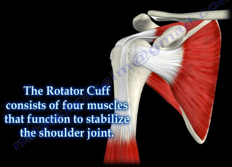
Massive rotator cuff tears are a disabling problem. These
tears can cause pain, weakness, and sometimes swelling of the shoulder. The
rotator cuff consists of four muscles that function to stabilize the shoulder
joint: supraspinatus rotator cuff tendon, subscapularis tendon, infraspinatus
rotator cuff tendon, and the teres minor rotator cuff tendon. The supraspinatus
tendon is the most common of the rotator cuff tendons to become ruptured. Massive
tears of the rotator cuff that are greater than 5cm usually involving both the
supraspinatus and infraspinatus tendons.
Massive tears of the rotator cuff are defined as tears
greater than 5cm, usually involving both the supraspinatus and infraspinatus
tendons. Retraction of the rotator cuff tendons along with muscle atrophy and
fatty infiltration can occur. This makes surgical reconstruction difficult with
the surgical outcome being unpredictable and less than satisfactory.
Treatment varies from physiotherapy to replacement of the
humeral head. Arthroscopic or open repair is usually the selected treatment.
Reconstruction can be done in selected cases. A rotator cuff arthropathy is
performed on massive cuff tears that are associated with superior migration of
the humeral head as well as instability and arthritis of the shoulder. The
patient will have pseudoparalysis and an x-ray will show shift of the humerus
proximally. An MRI will show massive cuff tear with retraction at the level of
the glenoid with atrophy of the muscle and fatty infiltration. A reverse shoulder
is the treatment of choice for the elderly with rotator cuff arthropathy as it
improves the pain and function. Hemiarthroplasty is the treatment for younger
patients. A standard head or a big humeral head can be selected.
A patient with a massive tear of the cuff usually develops
weakness of the shoulder and becomes unable to actively lift the arm without
assistance. Fluid collection within the shoulder may occur with a massive tear
of the rotator cuff.
The acromioclavicular joint is located at the top of the shoulder, where
the acromion of the scapula and the clavicle join together. The AC joint is a
small synovial gliding joint that can be affected by arthritis and
osteoarthritis. The oblique orientation of the joint’s articular surfaces may
allow the acromion to be driven underneath the clavicle when the AC joint is
injured. The condition could be subtle. Injuries of the acromioclavicular joint
most commonly occur due to separation of the AC joint. Falling directly onto
the shoulder can injure the ligaments that stabilize the AC joint. The AC
ligament provides anterior-posterior stability of the AC joint. The posterior
and superior AC ligaments are most important for stability. The
coracoclavicular ligaments provide superior-inferior stability. Activity
related pain with overhead activity and arm adduction.
During the physical examination, in order to test for injury
to the AC joint, the physician will begin by palpating the AC joint. They
should check to see if pain is present with direct palpation of the AC joint.
If pressing down onto the AC joint causes pain, this is a sign of an AC joint
problem such as distal clavicle osteolysis, arthritis, sprain of the AC
ligament, or separation. Osteolysis of the distal clavicle is a localized area
of inflammation, hyperemia, microfracture, bone resorption, and eventually
arthritis of the AC joint. When pulling down on the shoulder, if there is a
separation of the AC joint, the clavicle will rise and a bump will be seen in
the area of the joint. Sometimes, this is demonstrated by adding weights and
comparing both sides. The cross body adduction test can also be done by
bringing the shoulder across the body. This squeezes the acromion and clavicle
together, causing pain directly in the area of the joint if an AC joint
separation or arthritis is present.
The acromioclavicular joint is best evaluated using the
Zanca view radiograph. Using the Zanca view, the x-ray beam is directed with a cephalad
angle of 15 degrees. Clavicular osteolysis can be assessed using the Zanca
view. The acromion will be normal with the abnormality isolated to the distal
clavicle. The Zanca view is also used for diagnosis of arthritis of the AC
joint. It can show osteophytes and joint space narrowing. The patient’s
symptoms may not correlate with the x-ray findings. An MRI will show an
increased signal and edema in the AC joint.
An Elbow dislocation occurs when the radius and ulna bones
of the forearm move out of place from the humerus bone of the upper arm. There
are two basic types of elbow dislocations:
Simple
No fracture of the bones around the elbow
joint
Usually ligamentous injury
Complex
Fracture has occurred along with ligamentous
injury
Simple elbow dislocations typically occur when the patient
falls onto an outstretched hand. Injury progression from lateral to medial in
most patients. Posterolateral simple dislocations are the most common, occurring
approximately 90% of the time.
The proximal ulna and radius are displaced
posterolaterally relative to the distal humerus. Postemedial dislocations occur
at the proximal ulna and radius and are displaced posteromedially relative to
the distal humerus. In medial dislocations, the proximal ulna and radius are
displaced medially relative to the distal humerus. With lateral dislocations,
the proximal ulna and radius are displaced laterally relative to the distal
humerus. Anterior dislocations are rare, as they result from a direct force
applied to the posterior aspect of the forearm with the elbow in a flexed
position. Anterior dislocations occur when the proximal ulna and radius are
displaced anteriorly relative to the distal humerus. If stable, simple acute
fractures can be treated with a closed reduction and a splint for two to three
days (no more than two weeks) in addition to range-of-motion exercises and
physical therapy. Unstable simple fractures are rare, but can be stabilized by
ligament repair and/or by the use of an external fixator or cross pinning of
the joint in the elderly.
Chronic Elbow dislocations will need to be treated with an
open reduction and external fixator and is usually hinged. Recurrent elbow
dislocations (diagnosed by pivot shift) occurs due to a deficiency of the
lateral collateral ligament and is treated by a reconstruction of the
ulnohumeral ligament with a tendon graft.
 A patient with a complete anterior interosseous nerve injury
or a high medial nerve injury should be asked to make a fist. The first and
second digits will have difficulty in flexing, while the other digits will
flex. The third digit will be weak, while the fourth and fifth digits are
normal. This position of the hand is similar to the position taken during a
hand blessing. The Benedictine sign is different from an “ulnar claw hand”.
Ulnar claw hand refers to damage to the ULNAR nerve and is seen when attempting
to extend all the digits (leaving the 4th and 5th digits
flexed). The O.K. sign is used to check for paralysis of the anterior
interosseous nerve due to entrapment or compression injury. A patient with
paralysis of the anterior interosseous nerve will be unable to make the O.K.
sign. This is due to weakness of the flexor pollicis longus and flexor
digitorum profundus muscles. A typical pinch attitude is associated with
anterior interosseous nerve injury.
A patient with a complete anterior interosseous nerve injury
or a high medial nerve injury should be asked to make a fist. The first and
second digits will have difficulty in flexing, while the other digits will
flex. The third digit will be weak, while the fourth and fifth digits are
normal. This position of the hand is similar to the position taken during a
hand blessing. The Benedictine sign is different from an “ulnar claw hand”.
Ulnar claw hand refers to damage to the ULNAR nerve and is seen when attempting
to extend all the digits (leaving the 4th and 5th digits
flexed). The O.K. sign is used to check for paralysis of the anterior
interosseous nerve due to entrapment or compression injury. A patient with
paralysis of the anterior interosseous nerve will be unable to make the O.K.
sign. This is due to weakness of the flexor pollicis longus and flexor
digitorum profundus muscles. A typical pinch attitude is associated with
anterior interosseous nerve injury.  The anterior interosseous nerve arises from the median nerve
about 4-6cm distal to the elbow, which is about 1/3 of the way down the
forearm. It exits from the anterolateral aspect of the median nerve and it runs
between the radius and the ulna on the interosseous membrane between and below
the muscles of the flexor digitorum profundus and the flexor pollicis longus.
The anterior interosseous nerve arises from the median nerve
about 4-6cm distal to the elbow, which is about 1/3 of the way down the
forearm. It exits from the anterolateral aspect of the median nerve and it runs
between the radius and the ulna on the interosseous membrane between and below
the muscles of the flexor digitorum profundus and the flexor pollicis longus.