Tuesday, December 19, 2017

Chondroblastoma


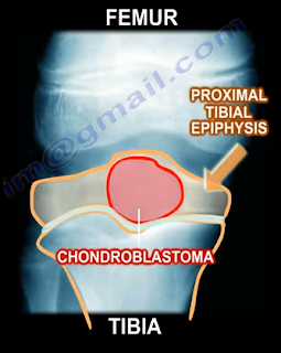
Chondroblastoma is a benign, aggressive cartilage tumor with an epiphyseal location. Chondroblastoma occurs more often in males, in younger patients between the ages of 10-25 years

of age, and in patients that may be skeletally immature. Chondroblastoma occurs commonly at the distal femur and the proximal tibia. 30% of chondroblastoma occur around the knee, followed by the proximal humerus, proximal femur, and the calcaneus.


Other epiphyseal lesions include:
  • Clear Cell Chondrosarcoma
    • Occurs in older age groups
    • Has a more aggressive histological pattern
    • Large cells with central nuclei
    • Occurs in the proximal humerus and proximal femur
  • Giant Cell Tumor
    • Occurs in older age groups
    • Has uniform cells and the nuclei of the stroma are similar to the nuclei of the giant cells
  • Osteomyelitis
    • Brodie’s abscess


During the clinical examination, the patient may complain of pain due to the tumor. The tumor abuts the joint which may cause joint symptoms and may cross the physis. 1% of these tumor metastasize to the lung. Radiologic images will show a lytic epiphyseal lesion with a sclerotic bony rim and sharp, well-defined borders. There may be calcification in the matrix. An MRI will show extensive surrounding edema.


You may find scattered giant cells and a cobblestone appearance of the cells with chicken wire calcifications. One-third of the lesions may have an aneurysmal bone cyst (ABC). The lesion is chondroid with polygonal cells. The lesion will have a defined cytoplasmic border and oval shaped nuclei with a prominent longitudinal groove (coffee bean appearance of the nuclei). Chondroblasts can be distinguied from giant cell tumors by staining for the S100 protein as the chondroblastoma will be reactive.
Chondroblastomas can be treated with an intralesional curettage and bone graft. The recurrence rate is less than 10%. Some surgeons may use adjuvants such as phenol or liquid nitrogen.
Tuesday, December 12, 2017

Wrist Pain Part I


There are eight carpal bones of the wrist that fit into a shallow socket formed by the bones of the forearm. Carpal Tunnel Syndrome occurs when there is pressure placed on the median nerve due to thickening of the transverse carpal ligament.
Thenar atrophy is commonly associated with severe carpal tunnel syndrome. Carpal tunnel syndrome can occur during pregnancy due to swelling, which compresses the median nerve in the hand, causing the fingers to feel numb. These symptoms typically go away after the delivery of the baby. Common causes of Carpal Tunnel syndrome are: obesity, hypothyroidism, arthritis, diabetes, trauma, and repetitive work (typing and lifting).  Carpal Tunnel Syndrome can lead to numbness, tingling, or weakness in the hand and fingers. The numbness usually occurs at night because we tend to sleep with our wrists flexed.
The Tinel’s test is used to determine symptoms of carpal tunnel syndrome. The test is considered positive if symptoms of tingling worsen while tapping on the median nerve at the wrist. The Phalen’s maneuver is a diagnostic test performed to determine if the patient has carpal tunnel syndrome. During this test, the back of the hands are pressed together, compressing the nerve, which may cause the symptoms of carpal tunnel syndrome.


Treatment for Carpal Tunnel Syndrome includes: anti-inflammatory medications, a splint, therapy, injections, and carpal tunnel release surgery. A carpal tunnel release is performed by cutting through the transverse carpal ligament. A carpal tunnel release allows the median nerve to pass freely through the tunnel to receive sensations from the thumb, index, and middle fingers of the hand. The idea is similar to a tunnel being widened so a large truck can pass through.

Tuesday, December 5, 2017

Pelvic Fractures with Bleeding


Pelvic fractures may cause significant bleeding. The superior gluteal artery is responsible for the majority of bleeding in pelvic fractures with an arterial injury. Most of the bleeding in pelvic fractures is from the veins and the fracture itself. Hemorrhage is the most life-threatening complication associated with pelvic injuries and will typically occur at the Superior Gluteal Artery. Hemorrhage of the Superior Gluteal Artery can be fatal. Approximately 10% of patients will have severe bleeding. Severe bleeding usually occurs in fracture patterns that are highly unstable to both rotational and vertical forces.


APC III (open book like type) is the complete disruption of anterior SI, sacrotuberous, and sacrospinous ligaments; disrupted posterior SI ligaments.
Vertical Shear Fractures are very bad fractures as they cause the complete disruption of the anterior and posterior ligaments; cephaloposterior displacement. Anteroposterior compression or vertical shear injuries are consistently associated with a higher risk of mortality from bleeding. The mortality rate is directly related to the amount of shock the patient is in at the time of presentation.
When treating patients with pelvic fractures and massive bleeding, it is important to remember that the patient will lose approximately 35% of their blood volume with acute hemorrhage before a sustained decrease in systolic blood pressure occurs. Immediate application of a pneumatic anti-shock garment is absolutely contraindicated in patients with a rupture of the diaphragm. Ringer’s lactate is the preferred initial fluid replacement used to resuscitate hypovolemic trauma patients in shock. A hypotensive blunt trauma patient will be given an initial fluid push with 2,000mL of Ringer’s lactate. A patient with bleeding and in shock will probably require O negative blood. If the patient is given 4 units of blood but remains hemodynamically unstable, then angiography and embolization is needed. Immediate application of an external fixator is another method to control bleeding, especially if the pelvis is unstable in external rotation. An abdominal and pelvic CT scan will clearly define the bony injury as well as the extent and source of the bleeding.
The best treatment for pelvic fractures with bleeding is a blood transfusion with correction of hypothermia and coagulopathy.