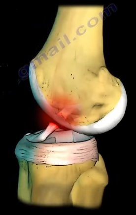
The anterior cruciate ligament is located in the front of
the knee. Rupture of the anterior cruciate ligament (ACL) is a condition
commonly seen in sports usually due to a non-contact pivoting injury. The Pivot
Shift test is a specific test for ACL deficient knee (ACL injury). Pivot shift
is pathognomonic for an ACL tear and is best demonstrated in a chronic setting.
Lachman’s test is the most sensitive examination test for ACL injury. o of flexion. The anteromedial bundle is
tight in flexion, and it increases anterior translation at 90o of
flexion. The Lachman’s test is the most sensitive test especially in acute
settings, and the examiner will find no end point with anterior translation of
the tibia. In an acute setting, physical examination can be difficult or
limited due to pain. With the Pivot shift test, the patient must be completely
relaxed, and the test is helpful in chronic situations especially if the
patient complains of the knee giving way.
In the Pivot shift, the knee subluxes
in extension and reduces at 20-30 degrees of flexion. The Pivot shift
correlates closely with patient satisfaction of their reconstructed knee. It is
a measure of functional instability following ACL reconstruction. Vertical
femoral tunnel placement will cause rotational instability seen as a positive
pivot shift, and the malposition of the bone tunnel will be seen in an AP view
x-ray of the knee. The 9 or 10 o’clock position is better than the 12 o’clock
position; the vertical position is bad. The patient with an ACL injury usually
has a non-contact pivoting injury event with an awkward landing, feeling a
“pop” sensation, or immediate swelling. Aspiration usually shows blood in the
knee which proves a 75% chance of ACL tear when you aspirate blood from the
knee. Patients will also exhibit a positive Lachman’s test which may be hard to
examine because of the pain. Aspiration of the knee may make the examination
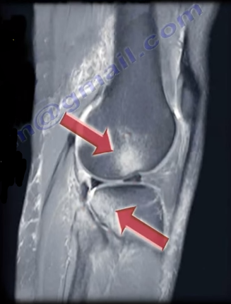
easier. MRI of the knee joint will show the hematoma, and it may show bone
lesions or bruising in the typical location which is characteristic with tears
of the ACL. These injuries are typically located at the middle of the femoral
condyle and posterior part of the tibia laterally. You may find a triple injury
within the MRI (O’Donoghue’s Unhappy Triad). The O’Donoghue’s Unhappy Triad
include an anterior cruciate ligament (ACL) injury, a medial cruciate ligament
(MCL) injury, and a lateral meniscus injury. In chronic ACL tears, the posterior
horn of the medial meniscus is the most commonly injured structure. In acute
ACL tear, send the patient for therapy for range of motion, brace the patient
and allow the MCL to heal and reconstruct the ACL later if needed. Patients
should do stress hamstring therapy in ACL tears. The patient will probably
complain of instability immediately or later on.The tibia can be pulled forward more than
normal (anterior translation). The examiner will have a sense of increased
movement and lack of a solid end point. For the pivot shift test, the patient
should be lying supine and totally relaxed. With pivot shift, the knee is in
the subluxed position when the knee is in full extension. The pivot shift
starts with extension of the knee and you can feel a “clunk” at 20-30 degrees
of flexion. To perform, hold the knee in full extension then add valgus force
plus internal rotation of the tibia to increase the rotational instability of
the knee. Then take the knee into flexion. A palpable clunk is very specific of
an ACL tear. The iliotibial band will reduce the tibia and create the clunk on
the outside of the knee. Always compare with the other side. The ACL prevents
anterior translation of the tibia. It is a secondary restraint to tibial
rotation and varus and valgus stress. The ACL consists of two bundles: the posterolateral
bundle and the anteromedial bundle. The posterolateral bundle prevents pivot
shift, contributes to rotational stability, prevents internal rotation of the
tibia with the knee in near extension, and increases the anterior translation
and tibial rotation at 30
The ACL
keeps the tibia from sliding out in front of the femur and provides rotational
stability to the knee. Rupture of the ACL causes anterolateral rotatory
instability. The tibia moves anterolaterally in extension; however, when you
flex the knee, the IT band becomes a flexor of the knee. The IT band pulls back
and reduces the tibia. The pivot shift test goes from extension (tibia
subluxed) to flexion, with the tibia reduced by the iliotibial band. Both the
Lachman’s tests and the Pivot shift test are associated with 20-30 degrees of
knee flexion. The Lachman’s test starts at 20-30 degrees of flexion. With the
Pivot shift test, you feel the clunk at 20-30 degrees of flexion. 20-30 degrees
of flexion is important for examination of the ACL. For the Lachman’s test, the
femur is stabilized with one hand and the other hand pulls the tibia anteriorly
and posteriorly against the femur.
Tibial Spine fractures are similar to ACL injuries in
adults. It occurs due to hyperextension of the knee and is commonly seen during
falls from a bicycle. An injured child with a swollen knee and who has fallen
from a bicycle should alert the clinician to the possibility of a tibial spine
fracture. A meniscal injury may also occur, especially with the medial
meniscus. The interposition of the meniscus or rotation of the fracture may
prevent a closed reduction.
Tibial spine fractures are classified using the Meyers and
McKeever Classification and are separated into three types. Type I
classifications are nondisplaced, Type II fractures are identified as being
minimally displaced with an intact posterior hinge, and Type III fractures are
classified as being completely displaced.
The presentation and examination is similar to an ACL tear
with immediate swelling as well as a positive Lachman’s Test or Anterior Drawer
Test. An x-ray will show the fracture and a CT scan will help in planning for
surgery. An MRI may be required to show a trapped or a meniscal injury.
Treatment
Treatment will consist of an aspiration of the large
hematoma. Nonoperative treatment is used for Type I fractures and reducible
Type II fractures; a closed reduction and immobilization in 0-20 of flexion. Surgery
is performed in Type II fractures and unreducible Type II fractures. An ORIF or
arthroscopic reduction and fixation will be performed. During surgery, the
trapped meniscus will be moved out of the way and the surgeon will use sutures
or screws for fixation. It is important for the surgeon to remember to avoid
the physis.
Complication
ACL laxity is common but not clinically significant. Stiffness
or arthrofibrosis occurs with surgical fixation. Growth arrest is rare.
The anterior cruciate ligament is located at the front of
the knee. Rupture of the anterior cruciate ligament (ACL) is a condition
commonly seen in sports, usually due to a non-contact pivoting injury. The
Pivot Shift test is a specific test for an ACL deficient knee (ACL injury).
A pivot shift is pathognomonic for an ACL tear and is best demonstrated in a
chronic setting. The Lachman’s test is the most sensitive examination test for
an ACL injury.
The ACL keeps the tibia from sliding out in front of the femur
and provides rotational stability to the knee. Rupture of the ACL causes
anterolateral rotatory instability. The tibia moves anterolaterally in
extension; however, when you flex the knee the IT band becomes a flexor of the
knee and pulls back, reducing the tibia. The Pivot Shift Test goes
from extension (tibia subluxed) to flexion, with the tibia reduced by the
iliotibial band.
Both the Lachman’s test and the Pivot Shift test are
associated with 20-30 degrees of knee flexion. The Lachman’s test starts at
20-30 degrees of flexion, but with the Pivot Shift test, you will feel the clunk at
20-30 degrees of flexion. Remember: 20-30 degrees of flexion is important for
examination of the ACL. The femur is stabilized with one hand and the other
hand pulls the tibia anteriorly and posteriorly against the femur. The tibia
can be pulled forward more than normal (anterior translation). The examiner
will have a sense of increased movement and lack of a solid end point.
When performing the Pivot Shift test, the patient should be
totally relaxed and lying supine. The knee is in the
subluxed position when in full extension. The pivot shift starts
with extension of the knee and you can feel a “clunk” at 20-30 degrees of
flexion. The physician will hold the knee in full extension, then add valgus force, and internal
rotation of the tibia to increase the rotational instability of the knee. Then
the physician will take the knee into flexion. A palpable clunk is very specific of an
ACL tear. The iliotibial band will reduce the tibia and create the clunk on the
outside of the knee. The physician should always compare the results with the
other side.
The ACL prevents anterior translation of the tibia. It is a
secondary restraint to tibial rotation and varus and valgus. The ACL consists
of two bundles:
The Posterolateral Bundle
Anteromedial Bundle
The Posterolateral bundle prevents the pivot shift and
contributes to rotational stability. This bundle also prevents internal
rotation of the tibia with the knee in near extension (tight in extension,
loose in flexion). If it is sectioned, it increases the anterior translation
and tibial rotation at 30° of flexion. The Anteromedial bundle is tight in
flexion and if sectioned, it increases the anterior translation at 90° of
flexion.
The Lachman’s test is the most sensitive test, especially in
acute settings. The examiner will find no end point with anterior translation
of the knee and the physical examination can be difficult or limited due to
pain. With the Pivot Shift test, the patient must be completely relaxed. The
test is helpful in chronic situations, especially if the patient complains of
the knee giving way.
During the Pivot Shift, the knee subluxes in extension and reduces
at 20-30 degrees of flexion. The Pivot Shift correlates closely with patient
satisfaction of their reconstructed knee. It is also a measure of functional
instability following ACL reconstruction. Verticle femoral tunnel placement
will cause rotational instability seen as a positive pivot shift, and the
malposition of the bone tunnel will be seen in an AP view x-ray of the knee. The
9 or 10 o’clock position is better than the 12 o’clock. A vertical position is
bad.
The patient with an ACL injury usually has a non-contact
pivoting injury even with:
Awkward landing
Feeling a “Pop” sensation
Immediate swelling
Aspiration usually shows blood in the knee (75%
chance of ACL tear with hemorrhage in the knee)
Positive Lachman’s Test (may be hard to examine
due to pain)
An MRI is going to be the best imaging technique. An MRI of
the knee joint will show bone lesions or bruising in the typical location
associated with tears of the ACL. These injuries are typically located at the
middle of the femoral condyle and posterior part of the tibia laterally. It is
also possible to find a triple injury within the MRI (O’Donoghue’s Unhappy
Triad).
O’Donoghue’s Unhappy Triad consists of:
Tear of the Lateral Meniscus
Anterior Cruciate Ligament Injury (ACL tear)
Medial Collateral Ligament Injury
In chronic ACL tears, the posterior horn of the medial
meniscus is the most commonly injured structure. In acute ACL tears, send the
patient for therapy for range of motion, brace the patient, and allow the MCL
to heal and reconstruct the ACL later if needed. It is important to stress
hamstring therapy in ACL tears. The patient will probably complain of
instability immediately or later on.