Showing posts with label children. Show all posts

Tuesday, March 6, 2018

Tibial Spine Fractures in Children


Tibial Spine fractures are similar to ACL injuries in adults. It occurs due to hyperextension of the knee and is commonly seen during falls from a bicycle. An injured child with a swollen knee and who has fallen from a bicycle should alert the clinician to the possibility of a tibial spine fracture. A meniscal injury may also occur, especially with the medial meniscus. The interposition of the meniscus or rotation of the fracture may prevent a closed reduction.
Tibial spine fractures are classified using the Meyers and McKeever Classification and are separated into three types. Type I classifications are nondisplaced, Type II fractures are identified as being minimally displaced with an intact posterior hinge, and Type III fractures are classified as being completely displaced.
The presentation and examination is similar to an ACL tear with immediate swelling as well as a positive Lachman’s Test or Anterior Drawer Test. An x-ray will show the fracture and a CT scan will help in planning for surgery. An MRI may be required to show a trapped or a meniscal injury.

Treatment


Treatment will consist of an aspiration of the large hematoma. Nonoperative treatment is used for Type I fractures and reducible Type II fractures; a closed reduction and immobilization in 0-20 of flexion. Surgery is performed in Type II fractures and unreducible Type II fractures. An ORIF or arthroscopic reduction and fixation will be performed. During surgery, the trapped meniscus will be moved out of the way and the surgeon will use sutures or screws for fixation. It is important for the surgeon to remember to avoid the physis.

Complication


ACL laxity is common but not clinically significant. Stiffness or arthrofibrosis occurs with surgical fixation. Growth arrest is rare.

Thursday, February 8, 2018

Bone Growth in Children


A Special Thanks to Miranda Ebraheim for assisting with this article


There are growth plates within the long bones which contributes to the development of the bones in children.

The growth distribution in the humerus is about 80% in the proximal and 20% in the distal area. Displaced fractures of the proximal humerus in children are usually treated without surgery.

In regards to the ulna, the growth distribution is about 80% proximal and 20% distal. Growth arrest is common in fractures involving the distal ulna and occur approximately 50% of the time. Within the radius, it is about 25% proximal and 75% distal. Fractures at the distal radius usually heals and corrects its angulation after a closed reduction. Surgery is rarely necessary. Fractures involving the growth plate of the distal radius rarely involves growth arrest.

Within the femur, the growth rate distribution is about 30% proximal and 70% distal. Fractures involving the growth plate of the distal femur may cause major growth disturbances. It is expected that a child grows 1 cm per year from the distal femur growth plate. Boys will continue to grow up until 16 years old, while girls stop growing at the age of 14.


Finally, there are the bones of the tibia and fibula. The growth distribution in the tibia is about 55% proximal and 45% distal. Within the fibula, it is about 60% proximal and 40% distal.

A growth spur occurs at the time of puberty. Puberty typically occurs in females around 8-13 years of age, and at 10-15 years of age in males. Fractures near the growth plate remodel well.

Thursday, November 30, 2017

Barlow & Ortolani Signs—DDH, Congenital Hip Dislocation


In order to remember these tests, remember that “B” comes before “O” in the alphabet and will go in this order for these procedures as well. The “B” stands for Barlow and “O” for Ortolani. When reading the word “barlow” think: “We are going out tonight!”, because the hip can be popped out of the acetabulum with this maneuver. To remember Ortolani, think: “Once we have been out, now it is time to go home!”—during the Ortolani maneuver, the femoral head is reduced back into the acetabulum.
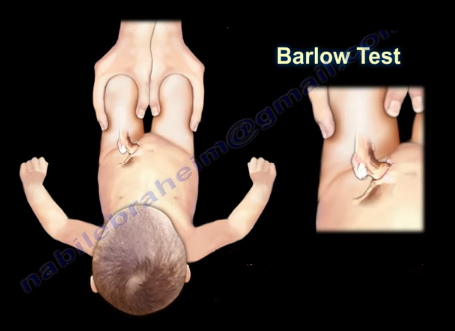
The Barlow maneuver identifies the unstable hip that is in a reduced position that the clinician can passively dislocate. When performing the Barlow Test, the examiner will flex the hip and knees to 90 degrees. The maneuver is performed by bringing the thigh towards the midline (adducting the hip). Mild pressure is then placed on the knee while directing the force posteriorly. The femoral head will be pushed out of the socket. The Barlow Test is considered positive if the hip can be popped out of socket with this maneuver. The dislocation will be palpable.


The Ortolani Test is used to confirm the findings of the Barlow test. The Ortolani maneuver is performed following the Barlow test to determine if the hip is actually dislocated. The Ortolani test is performed by the examiner flexing the hips and knees to 90 degrees. Reduction is done by abduction of the hip and pushing the thigh anteriorly. The test is positive is a palpable and audible clunk is heard from the hip being reduced. A hip click is a nonspecific finding.
In summary, The Barlow Test is performed when the hip is reduced and is used to dislocate the hip. The Ortolani Test is performed only after the Barlow Test has been performed and the hip has been dislocated. The Ortolani Test will reduce the hip.
Both of these tests are used for screening newborns during the neonatal period. The hips are examined one at a time and usually the hips are flexed during these maneuvers. Early diagnosis by these tests and preferably with ultrasound is essential to detect hip instability and dislocation in the neonatal period.

Treatment is directed at stabilizing the hip that has positive Barlow and Ortolani Signs. The first born female with a breach presentation and a positive family history are at risk of developing developmental dysplasia of the hip (DDH). If the hip remains dislocated for weeks, these two tests are usually not reliable. Barlow and Ortolani are not positive after 3 months due to the soft tissue contracture around the hip region. Limitation of abduction becomes the most consistent clinical findings. Children older than 12 months will have other findings which include asymmetry of hip abduction, a positive Trendelenburg gait and a positive Galeazzi sign.