Showing posts with label reduction. Show all posts

Tuesday, June 19, 2018

Isolated Fibular Fractures


Fibular fractures are usually associated with a complex injury, however they can be an isolated fracture. Complex injuries where a fibula fracture can occur include: fracture of the fibula and tibia, ankle fracture, pilon fracture, and Maisonneuve fractures.

Maisonneuve fractures involve a fracture of the proximal fibula associated with an occult injury of the ankle. Isolated fibular fractures are rare and usually the result of direct trauma. The fibula carries about 15% of the axial load and is the site of muscle attachment for the peroneus muscles and the flexor hallucis longus muscle. Check the patient who has a fibular fracture and no other fracture involving the tibia to rule out a possible Maisonneuve fracture, especially if there is no history of direct trauma to the leg. A high index of suspicion is necessary to diagnose and treat this injury. For high fibular fractures, the physician should look for signs of syndesmotic injury. Syndesmotic injury may include an unexplained increase in the medial clear space or the tibiofibular clear space is widened (should be less than 5mm). The x-ray will show the fracture to be rotational or oblique. Maisonneuve fractures require surgery to fix the syndesmosis.


Treatment will consist of reduction and fixation. It is important to determine if the injury is a Maisonneuve fracture or an isolated fibular fracture. An isolated fibular fracture will not need surgery.

Thursday, November 30, 2017

Barlow & Ortolani Signs—DDH, Congenital Hip Dislocation


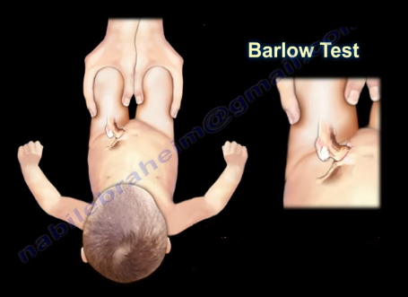
In order to remember these tests, remember that “B” comes before “O” in the alphabet and will go in this order for these procedures as well. The “B” stands for Barlow and “O” for Ortolani. When reading the word “barlow” think: “We are going out tonight!”, because the hip can be popped out of the acetabulum with this maneuver. To remember Ortolani, think: “Once we have been out, now it is time to go home!”—during the Ortolani maneuver, the femoral head is reduced back into the acetabulum.
The Barlow maneuver identifies the unstable hip that is in a reduced position that the clinician can passively dislocate. When performing the Barlow Test, the examiner will flex the hip and knees to 90 degrees. The maneuver is performed by bringing the thigh towards the midline (adducting the hip). Mild pressure is then placed on the knee while directing the force posteriorly. The femoral head will be pushed out of the socket. The Barlow Test is considered positive if the hip can be popped out of socket with this maneuver. The dislocation will be palpable.


The Ortolani Test is used to confirm the findings of the Barlow test. The Ortolani maneuver is performed following the Barlow test to determine if the hip is actually dislocated. The Ortolani test is performed by the examiner flexing the hips and knees to 90 degrees. Reduction is done by abduction of the hip and pushing the thigh anteriorly. The test is positive is a palpable and audible clunk is heard from the hip being reduced. A hip click is a nonspecific finding.
In summary, The Barlow Test is performed when the hip is reduced and is used to dislocate the hip. The Ortolani Test is performed only after the Barlow Test has been performed and the hip has been dislocated. The Ortolani Test will reduce the hip.
Both of these tests are used for screening newborns during the neonatal period. The hips are examined one at a time and usually the hips are flexed during these maneuvers. Early diagnosis by these tests and preferably with ultrasound is essential to detect hip instability and dislocation in the neonatal period.

Treatment is directed at stabilizing the hip that has positive Barlow and Ortolani Signs. The first born female with a breach presentation and a positive family history are at risk of developing developmental dysplasia of the hip (DDH). If the hip remains dislocated for weeks, these two tests are usually not reliable. Barlow and Ortolani are not positive after 3 months due to the soft tissue contracture around the hip region. Limitation of abduction becomes the most consistent clinical findings. Children older than 12 months will have other findings which include asymmetry of hip abduction, a positive Trendelenburg gait and a positive Galeazzi sign.