Tuesday, July 17, 2018

Intra-articular Extensile Approach for Tibial Plateau Fractures


Several types of tibial plateau fractures are a complex management problem. The knee joint may have a significant comminution and depression, and the physician may need to take an extensile approach for reduction and fixation of this fracture. Personally, I use the intra-articular extensile approach for tibial plateau fracture reduction and fixation. In general, fracture of the tibial plateau is a complicated problem.


A vascular evaluation is necessary. The ankle-brachial index (ABI) is needed in some types, such as in medial plateau fractures or in severe types, such as Schatzker Type V or Type VI. The ABI should be more than 0.9. Usually, medial tibial plateau fractures are considered to be a knee dislocation. A fasciotomy may be needed if compartment syndrome occurs. The soft tissue condition may be bad, and an external fixator may be initially used until the soft tissue condition improves.

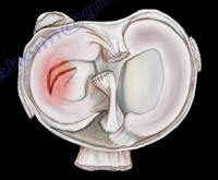
The association between tibial plateau fractures and meniscal tear is not uncommon. A lateral plateau fracture will create a lateral meniscal tear, while the medial plateau fracture will cause a medial meniscal tear. A tear of the meniscus is usually peripheral. It should be recognized and dealt with. The physician may want to look at the x-ray and see if there is a depression or separation of more than 6mm, as this indicates a high chance of meniscal tear.

The posteromedial fragment is another problem with tibial plateau fractures which needs to be fixed separately. When an extensive comminuted displaced tibial plateau fracture occurs, the physician may need excellent exposure of the articular surface to allow for anatomic reduction of the joint and visualization and repair or debridement of the meniscus if it is torn. This extensile exposure is important, especially if the posterior part of the plateau is involved. The traditional way to see the articular cartilage of the tibial plateau is to use the submeniscal approach by cutting the coronary ligament, but the exposure is limited. Other extensile approaches are also developed; however, we use the extensile intra-articular approach for complex, comminuted tibial plateau fractures. This involves anterior detachment and retraction of the meniscus to improve visualization of the tibial articular surface. This approach can be utilized for lateral or medial tibial plateau fractures and it is especially helpful in diagnosing and repairing the torn meniscus. This allows for inspection of the meniscus pathology in fractures of the articular surface. This improves reduction of the fracture and the torn meniscus is repaired and reattached to the coronary ligament. Incision and reflection of the meniscus allows great exposure and inspection of the joint which is followed by reattachment and suturing of the anterior horn of the meniscus to its normal position which is followed by reattachment of the meniscotibial (coronary) ligament. The sutures are tied to the sides of the patellar tendon on the opposite side of the meniscus.

Tuesday, July 10, 2018

McMurray's Test- Meniscal Tear


Meniscal injuries are very common. The McMurray’s Test is a rotational maneuver of the knee that is frequently used to aid in the diagnosis of meniscal tears. With a meniscal tear, the patient usually complains of knee pain localized to the lateral or medial side of the knee joint. The patient will have locking, clicking, pain, or effusion.


During the physical examination, joint line tenderness is the most sensitive finding. Swelling of the knee and a possible extension lag (locked knee) is also a common finding. Pain at a higher level is usually associated with the medial collateral ligament. Pain at a lower level is usually associated with the pes anserine bursa.


What is the McMurrays test?             


The McMurray’s test is a knee examination test that provokes pain or a painful click as the knee is brought from flexion to extension with either internal or external rotation. The McMurray’s test uses the tibia to trap the meniscus between the femoral condyles of the femur and the tibia. When performing the test, the patient should be lying supine with the knee hyperflexed. The examiner then grasps the patient’s heel with one hand and places the other hand over the knee joint. To test the medial meniscus, the knee is fully flexed, and the examiner then passively externally rotates the tibia and places a valgus force. The knee is then extended in order to test the medial meniscus. To test the lateral meniscus, the examiner passively internally rotates the tibia and places a varus force. The knee is then extended in order to test the lateral meniscus. A positive test is indicated by pain, clicking or popping within the joint and may signal a tear of either the medial or lateral meniscus when the knee is brought from flexion to extension.


How reliable is the McMurray’s test?


There are mixed reviews for the validity of this test. An MRI is a very sensitive exam and makes the diagnosis easier, while excluding other associated injuries.


Tuesday, July 3, 2018

Hip Dislocation Following Total Hip Surgery


There are two types of hip joint dislocations: posterior and anterior. The position of the leg is important in determining the type of hip dislocation. When the hip is dislocated, the leg is usually shortened and it assumes a different position than the normal leg (the other leg). If the dislocation is posterior, the leg will be in adduction and internal rotation. If the dislocation is anterior, the leg will be in abduction and external rotation. Notice that the affected extremity is shortened and externally rotated. Leg shortening can also be seen in hip fractures and the leg will be shortened and externally rotated.


Dislocation of the hip following total hip surgery may require revision surgery, but it is rare. The majority of hip dislocations after total hip dislocations are posterior, and they are usually treated without surgery. Most occur within the first month of THA; 1-4% in primary, 16% in revision. There is more incidence of dislocation in revision hip replacement.


Causes & Risk Factors:

  • Posterior Approach (try to repair the capsule adequately)
  • Malposition of the component
    • Ideally, the normal cup component will be in 20° of anteversion and 40° of abduction
    • When the hip dislocates posterior, always check for retroversion of the cup.

  • Prior hip fracture surgery, especially in the elderly
  • Weakness of the abductor muscle—must achieve soft tissue tension and function
  • Alcohol abuse
  • Improper neck length—looseness of the hip

The patient should be careful to avoid all activities that cause dislocation after total hip surgery. The patient should use a pillow between the legs while sleeping on their back and they should be careful to not cross their legs in their sleep. Patients cannot sleep on their sides as well. The patient should not bend the body at the waist farther than 90°. When sitting, the patient must avoid chairs that make it difficult to stand up, and sit at more than a 90° angle. The patient must not sit with their legs crossed in the chair. The patient must be made aware that if the leg is changed from its usual position, or becomes shortened, then the hip is probably dislocated and their doctor should be consulted.


X-rays of the dislocated total hip should include AP and lateral views. Look for eccentric wear and look for the position of the prosthesis. CT scans may be needed before or after reduction of the dislocation to check the version of the components. Treatment is variable and depends on the situation. The treatment should be tailored for each case. The majority of these cases with early dislocations can be treated successfully with closed reduction and immobilization.

The treatment should start with closed reduction of the total hip and immobilization. Hip stability is checked after reduction of the dislocation. Immobilization can be done by a brace or a hip spica. Trochanteric osteotomy and advancement of the trochanter and tensioning the abductor muscle. Screws or wires can be used. The prosthesis must be in good alignment for this procedure to work. Constrained acetabular components are used when the abductor muscle is deficient and the component position is good. Revision total hip is done in recurrent dislocation with malposition of the component or polyethylene wear.

Tuesday, June 26, 2018

Distal Femur Fractures & Nonunion- Use of Fibular Graft


The physician may be faced with some complex distal femur fractures or nonunion where the bony stock is not adequate or the fixation may have failed. It may also be a situation where a bone graft cannot be obtained from the patient. In some of these cases, I use an intramedually fibular graft in addition to plate fixation. The intramedullary fibular graft technique can be used in: complicated cases, comminuted fractures with osteoporosis, failure of traditional method of fixation, complex nonunion, and complex supracondylar periprothetic fractures.


How do you perform the technique?


The physician must find the starting point, which is the center of the intercondylar notch just
superior to the Blumensaat’s Line. The physician will insert a guide wire after reduction of the fracture. Then, the physician will ream over the guide wire to the appropriate size of the fibular graft, which you may need to fashion slightly. The physician needs to be sure to change the beaded guide wire to a smooth one and put the fibular graft through the guide wire into the medullary canal across the fracture of the nonunion. If the medullary canal of the fibula is small and it will not go through the guide wire, then place the fibular graft free hand. The physician should be sure that the fibular graft is not prominent through the joint. Next, fix the fracture or nonunion with a plate preferably a locking plate. You can augment the fixation with bone graft, allograft, or bone graft substitute. This procedure can also be helpful in periprosthetic fractures of the distal femur. If the prosthesisi is stable, you will do fixation of the fracture of the nonunion. It will be ideal to use a plate fixation after insertion of an intramedullary fibular graft, especially if the bony stock is very poor and if you can pass the fibular graft through the femoral component.

Tuesday, June 19, 2018

Isolated Fibular Fractures


Fibular fractures are usually associated with a complex injury, however they can be an isolated fracture. Complex injuries where a fibula fracture can occur include: fracture of the fibula and tibia, ankle fracture, pilon fracture, and Maisonneuve fractures.

Maisonneuve fractures involve a fracture of the proximal fibula associated with an occult injury of the ankle. Isolated fibular fractures are rare and usually the result of direct trauma. The fibula carries about 15% of the axial load and is the site of muscle attachment for the peroneus muscles and the flexor hallucis longus muscle. Check the patient who has a fibular fracture and no other fracture involving the tibia to rule out a possible Maisonneuve fracture, especially if there is no history of direct trauma to the leg. A high index of suspicion is necessary to diagnose and treat this injury. For high fibular fractures, the physician should look for signs of syndesmotic injury. Syndesmotic injury may include an unexplained increase in the medial clear space or the tibiofibular clear space is widened (should be less than 5mm). The x-ray will show the fracture to be rotational or oblique. Maisonneuve fractures require surgery to fix the syndesmosis.


Treatment will consist of reduction and fixation. It is important to determine if the injury is a Maisonneuve fracture or an isolated fibular fracture. An isolated fibular fracture will not need surgery.

Tuesday, June 12, 2018

Congenital Dislocation of the Knee


Congenital Dislocation of the knee is rare and may occur due to a contracture of the quadriceps. This condition usually occurs in patients with myelo, arthrogryposis, or Larsen’s syndrome. The patient with a congenital dislocation of the knee may have developmental dysplasia of the hip (DDH) and club foot. On examination, the patient will have a hyperextended knee at birth. They may have their foot placed against their face and there will be limited flexion at the knee. The patient may have a dimple or skin crease at the anterior aspect of the knee. You must examine the hip to rule out ipsilateral hip dislocation. 50% or more patients will have hip dysplasia. The etiology is not known; however, it could be due to fetal positioning or congenital absence of the cruciate.
There are grades, or a spectrum, for this deformity. Grade I deformities are referred to as Severe Genu Recurvatum, and the knee is hyperextended. If the range of passive flexion is more than 90°, it is considered to be a simple recurvatum. Grade II deformities are identified by subluxation with a range of 30-90° in passive flexion. Grade III deformities are complete dislocations with a range of passive flexion being less than 30°.

Congenital dislocation of the knee will take priority over treatment of hip dysplasia or club foot. The Pavlik harness and club foot cast will require knee flexion, so the physician will need to treat the knee dislocation first. With Grade I deformities, the initial treatment will be stretching of the knee and serial casting with the knee in flexion. In serial stretching and casting, the goal is to obtain at least 90° of flexion and reduction of the deformity over the course of several weeks. The physician should avoid pseudo-correction through an iatrogenic fracture of the proximal tibial physis. The prognosis is usually good if reduction is achieved without surgery. With Grade II deformities, if the infant is less than 1 month old, you will do serial casting first followed by percutaneous quadriceps recession, especially if the flexion is less than 90°. In Grade III deformities, a V-Y quadricepsplasty with above the knee cast is done in Grade III (frank dislocation), especially if nonsurgical treatment fails to reduce the tibia on the femur.   The result of open surgery is better when it is done in children younger than 6 months. In general, open reduction is reserved for children who did not respond to stretching and cast immobilization. It is important that the hip dysplasia is recognized and the knee dislocation is corrected early. This will help in early reduction of the hip.

Tuesday, June 5, 2018

Martin-Gruber Anastomosis


Martin-Gruber Anastomosis is median to ulner anastomosis in the forearm. It occurs through a communicating nerve branch between the median nerve and the ulnar nerve in the forearm. This connection carries motor nerve fibers. It can be confusing clinically and also on an EMG. It has a clinical significance for understanding the median nerve lesions and carpal tunnel syndrome. The axons will leave the median nerve or the anterior interosseous nerve crossing through the forearm to join the main trunk of the ulnar nerve, innervating the intrinsic muscles of the hand. The lesion above the communicating branch will affect the median nerve muscles. A lesion below the anastomosis (connecting branch) will not affect the median nerve muscles, it will spare the thenar motor intrinsic muscles of the hand. An isolated ulnar nerve lesion at the elbow will produce an unusual pattern for intrinsic muscle paralysis. Martin-Gruber Anastomosis is the most common anastomosis anomaly between the two nerves. In cases of nerve lesions of the median or ulnar nerve, this anastomosis serves as a conduit or an alternative innervation of parts of the hand and the forearm (it is really a detour). This can be a good explanation of difficult challenges, especially in the differential diagnosis. Incidence is high (about 15%). The physician should factor Martin-Gruber anastomosis into the differential diagnosis and the diagnosis.


If the communicating nerve arises from the anterior interosseous nerve, then a patient with anterior interosseous nerve palsy may present with hand intrinsic weakness, normally supplied by the ulnar nerve. Damage of the ulnar nerve at the wrist will lead to severe deficit of the intrinsic hand function greater than expected. There are other anastomoses available and reported as well as many variations that are possible.
There are three common anastomoses:

  1. Ulnar to median anastomosis in the forearm-reverse of Martin-Gruber (Marinacci anastomosis)
  2. Ulnar to median anastomosis in the hand (Riche-Cannieu anastomosis)
    1. Connection between the deep branch of the ulnar nerve and the recurrent branch of the median nerve
    2. It carries motor fibers and this anastomosis usually occurs in the region of the thenar and adductor pollicis muscles.
  3. Berrettini Anastomosis
    1. Communication between the digital nerves (sensory nerves) arising from the ulnar and median nerves in the hand
    2. Most common nerve anastomosis pattern

When the examination does not make sense and it is confusing, you can consider Martin-Gruber anastomosis.