Tuesday, September 4, 2018

SLAP Tear- Symptoms, Diagnosis, and Treatment


A SLAP tear is a tear that occurs where the biceps tendon inserts into the superior labrum. A SLAP tear is different from a Bankart lesion. SLAP tears are not common and can be hard to diagnose. Symptoms of a SLAP tear include: pain deep within the shoulder or in the back of the shoulder, as well as catching, popping, or clicking sensations. The patient may also experience pain when throwing a ball with a decrease in velocity and the feeling of having a dead arm after pitching. Patients will also experience pain with overhead activity which mimics impingement syndrome. This typically affects throwing athletes. When the biceps tendon is involved, pain may also be located at the front of the shoulder. A SLAP tear can be an isolated lesion or it can be associated with internal impingement, articular sided cuff tear, or instability.
A SLAP tear is diagnosed with a clinical examination and testing. The O’Brien’s test is the most commonly used test. Multiple tests are usually used including the anterior slide test and the clunk test. An MRI with contrast is the best imaging technique. When performing the O’Brien’s test, the patient is standing or sitting with the arm at 90° of flexion, 10° of adduction, and full internal rotation with the forearm pronated.  The examiner applies pressure to the forearm and instructs the patient to resist the applied downward force. Pain at the shoulder joint suggests a SLAP lesion. Decrease in pain of the shoulder joint on supination of the arm is suggestive of a SLAP tear.
Treatment consists of physical therapy, anti-inflammatory medications, injections, and surgery (when conservative treatment fails). If surgery is necessary, a labral debridement will be performed for minor tearing and fraying. Biceps Tenodesis is becoming popular, as it is a procedure that cuts the biceps tendon where it attaches to the labrum and reinserts it in another area, usually in front of the shoulder. A biceps tenotomy is a procedure that cuts the biceps tendon from the glenoid, releasing the long head of the biceps tendon from its attachment allowing it to fall into the upper arm out of the shoulder joint. A biceps tenotomy is probably best suitable for some elderly patients. A SLAP repair is a procedure which uses sutures to anchor the torn labrum to the glenoid. This repair is usually done for athletes and patients under the age of 40 years.

Tuesday, August 28, 2018

Massive Rotator Cuff Tear


To view my Youtube video, 'Massive Rotator Cuff Tear- Classic', click here.


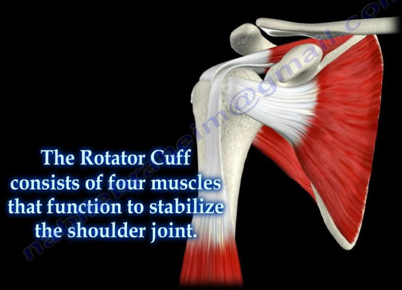
Massive rotator cuff tears are a disabling problem. These tears can cause pain, weakness, and sometimes swelling of the shoulder. The rotator cuff consists of four muscles that function to stabilize the shoulder joint: supraspinatus rotator cuff tendon, subscapularis tendon, infraspinatus rotator cuff tendon, and the teres minor rotator cuff tendon. The supraspinatus tendon is the most common of the rotator cuff tendons to become ruptured. Massive tears of the rotator cuff that are greater than 5cm usually involving both the supraspinatus and infraspinatus tendons.


Massive tears of the rotator cuff are defined as tears greater than 5cm, usually involving both the supraspinatus and infraspinatus tendons. Retraction of the rotator cuff tendons along with muscle atrophy and fatty infiltration can occur. This makes surgical reconstruction difficult with the surgical outcome being unpredictable and less than satisfactory.

Treatment varies from physiotherapy to replacement of the humeral head. Arthroscopic or open repair is usually the selected treatment. Reconstruction can be done in selected cases. A rotator cuff arthropathy is performed on massive cuff tears that are associated with superior migration of the humeral head as well as instability and arthritis of the shoulder. The patient will have pseudoparalysis and an x-ray will show shift of the humerus proximally. An MRI will show massive cuff tear with retraction at the level of the glenoid with atrophy of the muscle and fatty infiltration. A reverse shoulder is the treatment of choice for the elderly with rotator cuff arthropathy as it improves the pain and function. Hemiarthroplasty is the treatment for younger patients. A standard head or a big humeral head can be selected.


A patient with a massive tear of the cuff usually develops weakness of the shoulder and becomes unable to actively lift the arm without assistance. Fluid collection within the shoulder may occur with a massive tear of the rotator cuff.

Tuesday, August 21, 2018

Toe Deformities


Deformities of the toes are not uncommon and can occur from muscle imbalance, or other causes such as rheumatoid arthritis, diabetes, compartment syndrome, synovitis, or neurological disorders. Hammer toe occurs as flexion of the proximal interphalangeal (PIP) joint. Hammer toe is similar to the Boutonniere deformity of the finger. Claw Toe is a hyperextension deformity of the MTP joint and flexion of the PIP and DIP, resembling a pirate hook. Claw toe is similar to an intrinsic minus deformity of the hand, or “claw hand”. Mallet toe is similar in appearance to mallet finger, and is a flexion deformity of the DIP joint.

Tuesday, August 14, 2018

Triplane Fracture of the Distal Tibia


A triplane fracture of the distal tibia usually occurs during adolescence and occurs before complete closure of the distal tibial physis. The distal tibial physis (growth plate) is a weak area which closes from central to medial, with the lateral side being the last part to close. A Triplane fracture is a Salter-Harris Type IV Fracture, involving all three planes, the coronal (metaphysis), transverse (growth plate), and sagittal (epiphysis). The fracture has several variations and occurs due to external rotation forces. This fracture typically occurs in patients between 12-15 years of age.


Triplane fractures are complicated three-dimensional fractures. A two-part fracture is a Salter-Harris Type IV. A Three-part fracture is a combination of Salter-Harris Type III in an AP view and a Type II in a lateral view. CT scans are helpful.


An ORIF is necessary if there is displacement of the fragments of more than 2 mm.

Wednesday, August 8, 2018

Discoid Meniscus


The meniscus is a cushion structure made of cartilage which fits within the knee joint between the tibia and the femur. The medial meniscus is C-shaped and the lateral meniscus in the more circular. The meniscus is made up of type I collagen that provides shock absorption and stability to the knee joint. The meniscus helps to protect the knee joint, allowing the bones to slide freely on each other. Discoid meniscus is a rare variation of the meniscus that usually affects the lateral meniscus of the knee in less than 5% of the population and could be bilateral in about 25% of the cases.
Discoid meniscus is a large meniscus with abnormal attachment causing increased mobility of the meniscus. It causes a pop, click, or snapping with locking and pain. There will be loss of full knee extension with tenderness on the lateral joint space. Symptoms occur more during extension of the knee. The discoid meniscus occurs due to the abnormal development and increase in size of the meniscus. An x-ray could show increased widening of the joint space. An MRI will show the “bow tie” sign in three or more sagittal continuous cuts. The coronal MRI will show a thick and flat meniscus extending beyond the halfway point of the condyle.


Watanabe Classification of Discoid Lateral Meniscus


  • Type I: Block-shaped stable
  • Type II: Block-shaped, stable, partial meniscus (has good peripheral attachment)
  • Type III: Unstable meniscus with stability arising only form the ligament of Wrisberg. (no posterior meniscal tibial attachment).

Treatment


An asymptomatic patient will be treated with observation. A symptomatic patient may receive a partial meniscectomy and saucerization with repair of type III (no posterior tibial meniscal attachment)

Tuesday, July 31, 2018

Examination of the Acromioclavicular Joint


The acromioclavicular joint is located at the top of the shoulder, where the acromion of the scapula and the clavicle join together. The AC joint is a small synovial gliding joint that can be affected by arthritis and osteoarthritis. The oblique orientation of the joint’s articular surfaces may allow the acromion to be driven underneath the clavicle when the AC joint is injured. The condition could be subtle. Injuries of the acromioclavicular joint most commonly occur due to separation of the AC joint. Falling directly onto the shoulder can injure the ligaments that stabilize the AC joint. The AC ligament provides anterior-posterior stability of the AC joint. The posterior and superior AC ligaments are most important for stability. The coracoclavicular ligaments provide superior-inferior stability. Activity related pain with overhead activity and arm adduction.
During the physical examination, in order to test for injury to the AC joint, the physician will begin by palpating the AC joint. They should check to see if pain is present with direct palpation of the AC joint. If pressing down onto the AC joint causes pain, this is a sign of an AC joint problem such as distal clavicle osteolysis, arthritis, sprain of the AC ligament, or separation. Osteolysis of the distal clavicle is a localized area of inflammation, hyperemia, microfracture, bone resorption, and eventually arthritis of the AC joint. When pulling down on the shoulder, if there is a separation of the AC joint, the clavicle will rise and a bump will be seen in the area of the joint. Sometimes, this is demonstrated by adding weights and comparing both sides. The cross body adduction test can also be done by bringing the shoulder across the body. This squeezes the acromion and clavicle together, causing pain directly in the area of the joint if an AC joint separation or arthritis is present.

The acromioclavicular joint is best evaluated using the Zanca view radiograph. Using the Zanca view, the x-ray beam is directed with a cephalad angle of 15 degrees. Clavicular osteolysis can be assessed using the Zanca view. The acromion will be normal with the abnormality isolated to the distal clavicle. The Zanca view is also used for diagnosis of arthritis of the AC joint. It can show osteophytes and joint space narrowing. The patient’s symptoms may not correlate with the x-ray findings. An MRI will show an increased signal and edema in the AC joint.

Tuesday, July 24, 2018

Lumbosacral Plexus


The sciatic nerve is the key nerve of the lumbosacral plexus. It arises from the spinal nerves of L4 through S3. The sciatic nerve is the cornerstone of the lumbosacral plexus. Knowledge of the lumbosacral plexus starts with the sciatic nerve because it is the most important nerve. The lumbosacral plexus is comprised of the sciatic nerve and S4. The branches of the lumbosacral plexus are the superior gluteal nerve (L4-S1), the inferior gluteal nerve (L5-S2), the posterior cutaneous nerve of the thigh (S1-S3), and the pudendal nerve (S2-S4) (SIPP).