Tuesday, February 27, 2018

Sciatica, Disc Herniation, and Piriformis Syndrome


The sciatic nerve is a large nerve that comes from the lumbosacral plexus. The sciatic nerve has five nerve roots, L4, L5, S1, S2, and S3. The sciatic nerve runs from the lower spine, through the buttock to the lower leg and foot. The sciatic nerve initially emerges from the pelvis and exits the greater sciatic notch anteriorly and deep to the piriformis muscle, exiting below the piriformis muscle.
The sciatic nerve then enters the thigh between the ischial tuberosity and the greater trochanter of the femur. In about 10% of patients, the sciatic nerve is separated by all or part of the piriformis muscle. The sciatic nerve enters the thigh beneath the lower border of the gluteus maximus muscle. The nerve then runs down and branches out within the posterior aspect of the thigh, down to the leg and foot. The sciatic nerve gives multiple sensory and motor branches to specific areas and muscles in the leg and foot. The two main branches of the sciatic nerve are the posterior tibial nerve and the common peroneal nerve.
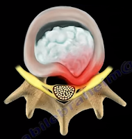
Irritation of the sciatic nerve may occur at multiple sites. The first site that we need to look at is the spine—which is where irritation may occur, usually from lumbar disc herniation. This is considered true sciatica or lumbar radiculopathy. Another site for irritation of the sciatic nerve is at the piriformis muscle. The sciatic nerve may become compressed by the piriformis muscle (piriformis syndrome). Piriformis Syndrome is a diagnosis of exclusion! If the patient has symptoms of sciatica, then there must be an MRI of the spine that is negative, proving that the symptoms are not associated with a possible disc problem. Once the MRI is negative, then you can say that the condition of sciatica may come from piriformis syndrome.
Disc Herniation

Tuesday, February 20, 2018

Jefferson Fractures


Fifty percent of patients with Jefferson fractures will have associated spine injuries. The canal is wide with a low risk of spinal cord injuries unless the transverse ligament is disrupted. It is difficult to view Jefferson Fractures on an x-ray (usually seen on the lateral side”. This fracture is considered a “Junctional Fracture” and could be missed. The classic Jefferson fracture is a burst fracture that results from an axial load. It could be a four part fracture with bilateral fractures of the anterior and posterior arch. There are variations which include two and three part fractures and incomplete formations of the posterior arch can be mistaken as a fracture.
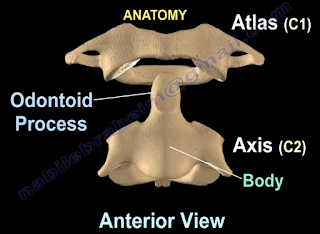
When speaking of Jefferson fractures, it is important to be familiar with the structures that may be involved. These bony structures include: The Atlas (C1), Axis (C2), and the odontoid process. C1 and C2 are stabilized together by the transverse ligament and C1 and C2 provide a 50% of rotation of the neck. The C1 is a ring. At the upper cervical region, the spinal canal is 2.5 times larger than the cord size. The stability and treatment of Jefferson fractures depends on the integrity of the transverse ligament and the displacement of the fracture. You need to know about the important ligaments related to the Jefferson fracture. These ligaments include: the transverse ligament, the apical ligament, and the Alar ligament.

Diagnosing ligamentous injury


In order to determine a ligamentous injury, the physician will want to check the Atlanto-dens interval (A.D.I). Normally, this interval should be less than 3mm in adults and less than 5mm in children. If the ADI is between 3-5mm, this indicates an injury to the transverse ligament; the transverse ligament holds the odontoid and C1 together, alar and apical ligaments will be intact. If the A.D.I measures greater than 5mm, then there is an injury to the transverse, alar, and apical ligaments.


Fracture Types


A bony injury with the intact transverse ligament and a lateral mass displacement less than 7mm and the A.D.I is less than 3mm is considered a stable fracture. Nondisplaced fractures of this nature should be treated with a rigid orthosis. If the fracture is displaced, a halo will need to be used.
Another type of fracture can occur at C1 with a transverse ligament tear. The Atlanto-dens interval will be more than 3 mm in adults. The treatment will depend on the type of injury to the transverse ligament. With bony avulsions of the transverse ligament, the halo will need to be used cautiously. However, some surgeons prefer to do a fusion of C1 and C2. If there is an intrasubstance tear of the transverse ligament, the surgeon will perform a fusion at C1-C2. The surgeon will need to do early surgery as this is a significant injury with a risk of spinal cord compression.


In regards to “Open Mouth Views”, the normal overhang is visible during an “Open Mouth View”. If it is just a bony injury Jefferson fracture, the combined overhang will be less than 7mm and the transverse ligament is intact and it is a stable fracture. If a Jefferson fracture has a combined overhang of more than 7mm, then the transverse ligament is probably torn and there is an unstable fracture present.

Radiological Studies


A CT scan is probably the best study in diagnosing the characteristics of the bony injury. An MRI is the best study in diagnosing any associated transverse ligament injuries.

Tuesday, February 13, 2018

Pronator Teres Syndrome


The nerve that is involved in pronator teres syndrome is the median nerve. Pronator Teres Syndrome is caused by a compression of the median nerve at the level of the elbow which occurs more in women. In the forearm, the median nerve runs between the two heads of the pronator teres muscle and then it lies between the flexor digitorum superficialis and flexor digitorum profundus muscles. This syndrome may be associated with medial epicondylitis. The principle symptoms of numbness in the radial 3 ½ fingers as well as thenar weakness which may be mistakenly attributed to carpal tunnel syndrome.
The most common cause of entrapment is due to compression of the median nerve between the two heads of the pronator teres muscle. This commonly occurs in people who perform repetitive forceful pronation of the forearm. Compression may be due to the thickening of the bicipital aponeurosis. The aponeurosis crosses from lateral to medial over the antecubital fossa and may irritate the median nerve. Compression of the nerve may also occur due to the fibrous arch of the origin of the flexor digitorum superficialis (FDS).


The median nerve runs down the medial side of the arm and passes 2 ½ to 4 cm below the level of the medial epicondyle before it enters between the two heads of the pronator teres. About 1% of
patients have a medial supracondylar humeral spur about 5cm proximally to the medial epicondyle. The ligament of Struthers is attached to this bony projection which connects the process to the medial epicondyle. The bony process points towards the elbow joint and the median nerve can become compressed by the supracondylar spur. The median nerve can also become trapped by the ligament of Struthers that extends from the supracondylar process to the medial epicondyle. The ligament of Struthers is different from the arcade of Struthers, which deals with the compression of the ulnar nerve around the elbow.


Paresthesia in these lateral 3 ½ fingers may occur with the compression of the median nerve at the elbow region or at the carpal tunnel region. These symptoms are similar to carpal tunnel syndrome but the symptoms are worse with rotation of the forearm. The patient will complain of dull aching pain over the proximal forearm with no nighttime symptoms. The pain is usually worsened by repetitive or forceful pronation. Tenderness of palpation to the pronator teres muscle will be detected. The median nerve gives off a palmar cutaneous branch before entering the carpal tunnel. Sensory disturbances over the palm of the hand occur due to involvement of the palmar cutaneous branch of the medial nerve and this occurs proximal to the carpal tunnel. Sensory disturbances in this area indicates median nerve problems proximal to the carpal tunnel. This differentiates between carpal tunnel syndrome and pronator teres syndrome.

There are specific provocative tests that produce the pain and distal paresthesia that are used to localize the site of compression. The Tinel’s sign at the wrist and the Phalen’s test will be negative. The Median nerve compression tests are negative at the carpal tunnel; however, there will be a positive Tinel’s sign at the proximal forearm. There will be abnormal sensation in the “palmar triangle”. When compression of the nerve involves the supracondylar process, the test is considered positive if symptoms of tingling worsen while tapping on the spur.
Occassionally, the spur can be felt. The pronator teres muscle can be assessed as the cause of the median nerve compression in different ways. Resisted forearm pronation with elbow flexion will test for compression at the two heads of the pronator teres muscle. During this test, the patient’s forearm is held in resisted pronation and flexion. While remaining in a pronated position, the forearm is gradually extended. Compression of the median nerve may also be tested by: resisted elbow flexion with forearm supination (compression at the bicipital aponeurosis) and resisted contraction of the FDS to the middle finger (compression at the FDS arch).


Differential Diagnosis

C6/C7 Radiculopathy occurs due to involvement of the nerves at these levels which will cause numbness of the thumb, index, and long fingers, as well as weakness of the muscles of the forearm that are innervated by the median nerve. The radial nerve part of C6-C7 will show normal function of the wrist extensors and the triceps.

X-rays, imaging and nerve conduction studies may be helpful in the diagnosis.

Treatment typically consists of rest, splints, and NSAIDs. Surgical decompression of the median nerve through all 4 or 5 possible sites of compression when non-operative management fails for 3-6 months. The results of surgery are variable. Full recovery is not always seen in all patients as only about 80% of patients improve from surgery. The skin incision may leave an unsatisfactory scar.

Thursday, February 8, 2018

Bone Growth in Children


A Special Thanks to Miranda Ebraheim for assisting with this article


There are growth plates within the long bones which contributes to the development of the bones in children.

The growth distribution in the humerus is about 80% in the proximal and 20% in the distal area. Displaced fractures of the proximal humerus in children are usually treated without surgery.

In regards to the ulna, the growth distribution is about 80% proximal and 20% distal. Growth arrest is common in fractures involving the distal ulna and occur approximately 50% of the time. Within the radius, it is about 25% proximal and 75% distal. Fractures at the distal radius usually heals and corrects its angulation after a closed reduction. Surgery is rarely necessary. Fractures involving the growth plate of the distal radius rarely involves growth arrest.

Within the femur, the growth rate distribution is about 30% proximal and 70% distal. Fractures involving the growth plate of the distal femur may cause major growth disturbances. It is expected that a child grows 1 cm per year from the distal femur growth plate. Boys will continue to grow up until 16 years old, while girls stop growing at the age of 14.


Finally, there are the bones of the tibia and fibula. The growth distribution in the tibia is about 55% proximal and 45% distal. Within the fibula, it is about 60% proximal and 40% distal.

A growth spur occurs at the time of puberty. Puberty typically occurs in females around 8-13 years of age, and at 10-15 years of age in males. Fractures near the growth plate remodel well.

Tuesday, February 6, 2018

Stem Cells and Orthopaedics


Stem cells may help tissues that are injured or damaged to renew and regenerate themselves. Depending on the treatment and medium, stem cells have the ability to become different types of cells such as bone, cartilage, and blood vessels. There are several conditions in which stem cells are used as treatment, including: avascular necrosis, arthritis, and nonunion.
When Avascular Necrosis of the femoral head occurs due to the diminished blood supply, there is a death of a segment of bone, which is considered necrotic. The surgeon can inject stem cells into this area to revive this area by drilling into the bone. When using stem cells to treat AVN, the surgeon will need to create a channel for new blood vessels to form into the area that lacks blood supply. After the channel is created, the stem cells are injected into the necrotic femoral head.
Stem cell treatments for joint pain and arthritis is not proven to be effective. However, there is some use in knee arthritis for cartilage regeneration.
 

The best use of stem cells in Orthopaedics is its treatment for nonunion fractures. A nonunion fracture is classified as a fracture that does not heal after a reasonable period of time or a fixation failure. Nonunion may also be due to motion of the bony ends and incomplete healing of the fracture; fractures of this nature will need a lot of assistance. Two elements are needed for treatment of nonunions: vascularity—which improve the local conditions to facilitate healing; and stability—in the form of fixation such as a rod or plate.

The most common causes of nonunion are smoking (5 times more common), diabetes, obesity, osteoporosis, unstable fixation, infection (most common), open fractures, and the severe displacement of the fracture.

 Options available for treatment:

  1. Bone Morphogenetic Protein—very expensive
  2. Bone Graft—donor site morbidity
  3. Stem Cells

Stem cells must be extracted from the bone marrow and are aspirated and harvested from the anterior iliac crest. This procedure is performed with an outlet view under fluoroscopy. Once extracted, the bone marrow is prepared to be centrifuged. After centrifuging the bone marrow, a good sample is extracted for injection.

The surgeon will mark and localize the area for injection and the trocar is placed. The sample will then be injected into the fracture area—occasionally, two areas of nonunion are treated. Adult mesenchymal stem cells are special cells that can copy themselves, divide, and multiply. They can differentiate into bone cells that heal the nonunion and lay down new bone. This process can be monitored by alkaline phosphatase activity or by the genes of the stem cells. The whole cellular mechanism can help increase the vascularity of the nonunion.


It is important to note that adult mesenchymal stem cells are not embryonic stem cells. There is a large amount of information in regards to stem cells that is lacking or misleading. Cells should probably be combined with some type of matrix. Additionally, surgeons need a better delivery system and localization during the injection of the stem cells due to the fact that the dye kills the cells. It is beneficial to allow the cells to expand and grow in the culture prior to injection. Moreover, the effect of certain medications such as aspirin, Plavix, and Coumadin, should be studied further.