Tuesday, June 12, 2018

Congenital Dislocation of the Knee


Congenital Dislocation of the knee is rare and may occur due to a contracture of the quadriceps. This condition usually occurs in patients with myelo, arthrogryposis, or Larsen’s syndrome. The patient with a congenital dislocation of the knee may have developmental dysplasia of the hip (DDH) and club foot. On examination, the patient will have a hyperextended knee at birth. They may have their foot placed against their face and there will be limited flexion at the knee. The patient may have a dimple or skin crease at the anterior aspect of the knee. You must examine the hip to rule out ipsilateral hip dislocation. 50% or more patients will have hip dysplasia. The etiology is not known; however, it could be due to fetal positioning or congenital absence of the cruciate.
There are grades, or a spectrum, for this deformity. Grade I deformities are referred to as Severe Genu Recurvatum, and the knee is hyperextended. If the range of passive flexion is more than 90°, it is considered to be a simple recurvatum. Grade II deformities are identified by subluxation with a range of 30-90° in passive flexion. Grade III deformities are complete dislocations with a range of passive flexion being less than 30°.

Congenital dislocation of the knee will take priority over treatment of hip dysplasia or club foot. The Pavlik harness and club foot cast will require knee flexion, so the physician will need to treat the knee dislocation first. With Grade I deformities, the initial treatment will be stretching of the knee and serial casting with the knee in flexion. In serial stretching and casting, the goal is to obtain at least 90° of flexion and reduction of the deformity over the course of several weeks. The physician should avoid pseudo-correction through an iatrogenic fracture of the proximal tibial physis. The prognosis is usually good if reduction is achieved without surgery. With Grade II deformities, if the infant is less than 1 month old, you will do serial casting first followed by percutaneous quadriceps recession, especially if the flexion is less than 90°. In Grade III deformities, a V-Y quadricepsplasty with above the knee cast is done in Grade III (frank dislocation), especially if nonsurgical treatment fails to reduce the tibia on the femur.   The result of open surgery is better when it is done in children younger than 6 months. In general, open reduction is reserved for children who did not respond to stretching and cast immobilization. It is important that the hip dysplasia is recognized and the knee dislocation is corrected early. This will help in early reduction of the hip.

Tuesday, June 5, 2018

Martin-Gruber Anastomosis


Martin-Gruber Anastomosis is median to ulner anastomosis in the forearm. It occurs through a communicating nerve branch between the median nerve and the ulnar nerve in the forearm. This connection carries motor nerve fibers. It can be confusing clinically and also on an EMG. It has a clinical significance for understanding the median nerve lesions and carpal tunnel syndrome. The axons will leave the median nerve or the anterior interosseous nerve crossing through the forearm to join the main trunk of the ulnar nerve, innervating the intrinsic muscles of the hand. The lesion above the communicating branch will affect the median nerve muscles. A lesion below the anastomosis (connecting branch) will not affect the median nerve muscles, it will spare the thenar motor intrinsic muscles of the hand. An isolated ulnar nerve lesion at the elbow will produce an unusual pattern for intrinsic muscle paralysis. Martin-Gruber Anastomosis is the most common anastomosis anomaly between the two nerves. In cases of nerve lesions of the median or ulnar nerve, this anastomosis serves as a conduit or an alternative innervation of parts of the hand and the forearm (it is really a detour). This can be a good explanation of difficult challenges, especially in the differential diagnosis. Incidence is high (about 15%). The physician should factor Martin-Gruber anastomosis into the differential diagnosis and the diagnosis.


If the communicating nerve arises from the anterior interosseous nerve, then a patient with anterior interosseous nerve palsy may present with hand intrinsic weakness, normally supplied by the ulnar nerve. Damage of the ulnar nerve at the wrist will lead to severe deficit of the intrinsic hand function greater than expected. There are other anastomoses available and reported as well as many variations that are possible.
There are three common anastomoses:

  1. Ulnar to median anastomosis in the forearm-reverse of Martin-Gruber (Marinacci anastomosis)
  2. Ulnar to median anastomosis in the hand (Riche-Cannieu anastomosis)
    1. Connection between the deep branch of the ulnar nerve and the recurrent branch of the median nerve
    2. It carries motor fibers and this anastomosis usually occurs in the region of the thenar and adductor pollicis muscles.
  3. Berrettini Anastomosis
    1. Communication between the digital nerves (sensory nerves) arising from the ulnar and median nerves in the hand
    2. Most common nerve anastomosis pattern

When the examination does not make sense and it is confusing, you can consider Martin-Gruber anastomosis.

Tuesday, May 1, 2018

Patellar Tendon Rupture


A patellar tendon rupture is a rupture of the tendon that connects the patella to the tibia. Rupture often occurs at the lower pole insertion site of the patella and it could be associated with degenerative changes. Rupture most often occurs in patients younger than 40 years of age. When the tendon is ruptured, the quadriceps muscle pulls the patella upward. One way to measure the height of the patella is by measuring the Blumensaat’s line. The knee needs to be flexed at least 30 degrees, then a line can be drawn through the roof of the intercondylar notch and usually touches the tip of the patella. The patella moves upward with the patellar tendon rupture (patella alta).

Associated Risk Factors


  • Rheumatoid Arthritis
  • Diabetes
  • Chronic Renal Failure
  • Systemic Corticosteroid Therapy
  • Chronic Patellar Tendonitis
  • Degenerative Changes


During the radiographic evaluation, an AP and Lateral x-ray is necessary. The patella alta is seen on the lateral view (*patella superior to Blumensaat’s line). An MRI is effective in assessing the patellar tendon, especially if other intraarticular or soft tissue injuries are suspected.
Treatment consists of a surgical reattachment of the tendon. The patient will need to keep their knee in extension and in a knee immobilizer for about 4-6 weeks.

Tuesday, April 24, 2018

Tibial Bowing


Tibial bowing is not uncommon. The direction and the apex of the tibial bow can alert the clinician to the type of the deformity, its treatment, and its prognosis. Tibial bowing can occur anteriorly, laterally, anterolaterally, and posteromedially.
Anterior bowing is often associated with fibular hemimelia. This bowing may be associated with the loss of the lateral rays of the foot, equinovalgus foot deformity, tarsal coalition, and significant leg length discrepancy.


Lateral bowing is a common variation, which occurs bilaterally. This condition is mild and not associated with other problems.


Anterolateral bowing is a serious tibial bowing that may increase and lead to a fracture as well as pseudoarthrosis of the tibia. This type of bowing occurs early in infancy. Pseudoarthrosis is usually associated with neurofibromatosis. 10% of patients with neurofibromatosis will have anterolateral tibial bowing. Neurofibromatosis is found in 50% of the patients with ALB. The patient should be carefully examined for café-au-lait spots. In this type of bowing, bone ends are usually thin and the fibula may also be involved. Treatment of anterolateral bowing is bracing with total contact orthosis. In order to treat pseudoarthosis in the tibia, surgery is usually needed. Multiple options are available for surgery, and none of these options are perfect. 50% of patients may undergo amputation due to the inability to achieve healing of pseudoarthrosis.

Posteromedial bowing is a rare calcaneovalgus deformity of the foot plus leg length discrepancy. This condition usually resolves, but may have residual leg length discrepancy.

Tuesday, April 17, 2018

Freiberg's Disease


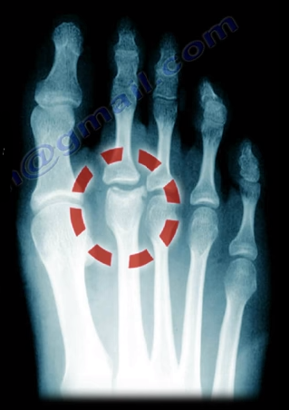
Freiberg’s Disease is caused by avascular necrosis of the head of the 2nd metatarsal. This condition is more common in patients who have a longer 2nd metatarsal bone relative to the 1st metatarsal. This leads to the transfer of excessive loads onto the 2nd metatarsal, which may interfere with the blood supply. This disease tends to occur more commonly in young females during growth spurts.

Freiberg’s Disease usually presents itself as pain and swelling at the 2nd metatarsophalangeal joint that is related to activities and walking in high heels. There may be point tenderness and swelling over the head of the 2nd metatarsal. There may also be limited range of motion in the 2nd metatarsophalangeal joint. Early in the disease, x-rays may only show minimal changes. Radiological evidence of the condition may only be clearly visible on an MRI and bone scan. In more severe cases, sclerosis, fragmentation, collapse of the metatarsal head, and severe arthritis, may make the condition easily visible on an x-ray.

Treatment


Conservative treatment consists of nonsteroidal anti-inflammatory medications, activity modification, orthotics, and immobilization with a short leg cast. Surgical intervention involving a joint debridement is indicated only after the failure of all conservative measures. Other procedures may be indicated depending on the complexity and severity of the case.

Tuesday, April 10, 2018

Pes Anserine Bursitis


Several bursa are seen around the knee area. These bursa include the suprapatellar, prepatellar, infrapatellar, and pes anserine. The pes answerine bursa is a small fluid filled sac located between the tibia and the three tendons of the Sartorius, Gracilis, and Semi-tendinosus.
These muscles are innervated by three separate nerves, the femoral, obturator, and the tibial branch of the sciatic nerve, respectively. Pes Anserine bursitis, or “breast stroke knee”, is an inflammatory condition of the medial knee at the pes anserine bursa that is common in swimmers.

What is the pes anserine?

The pes anserine is the common area of insertion for the three tendons along the proximal medial aspect of the tibia. This condition is also sometimes referred to as a “goosefoot” because the pes anserinus tendons resemble the shape of a goose foot. Pes Anserine bursitis is usually seen as causing pain, tenderness, and localized swelling after trauma or total knee replacement. The pain is seen below the joint line on the medial part of the proximal tibial with the bursa being deep to the tendons.


Treatment

Treatment consists of physical therapy, nonsteroidal anti-inflammatory medications, and injections. The physician will need to rule out meniscal tears, stress fractures, or osteonecrosis of the tibia, as these are all differential diagnosis.


Tuesday, April 3, 2018

Cauda Equina—Central Disc Herniation


Disc herniations usually occurs posterolaterally, but it may also be central. The cauda equina is composed of several nerves within the lower end of the spinal canal. The top of the spinal cord is a tubular bundle of nervous tissue extending from the brain.

The following section of the spinal cord is called the Conus Medullaris and is the lower end of the spinal cord. The Cauda Equina is made up of multiple nerve roots beginning at the level of L1.


The most common disc herniation is the Posterolateral Disc Herniation. This type of herniation is a nerve root injury, which will cause changes to both the sensory and motor skills as well as the reflexes. A posterolateral disc herniation usually affects the foot and ankle, and may cause unilateral leg pain and weakness. Observe for a positive straight leg raising test. These herniations are usually initially treated with conservative methods.  


A central disc herniation will cause cauda equina syndrome, which is a compression over the lumbosacral nerve roots. This compression will cause more back pain than leg pain, and bladder and bowel symptoms will be evident. This herniation is considered a surgical emergency.