Tibial bowing is not uncommon. The direction and the apex of
the tibial bow can alert the clinician to the type of the deformity, its
treatment, and its prognosis. Tibial bowing can occur anteriorly, laterally,
anterolaterally, and posteromedially.
Anterior bowing is often associated with fibular hemimelia.
Thisbowing may be associated with the
loss of the lateral rays of the foot, equinovalgus foot deformity, tarsal
coalition, and significant leg length discrepancy.
Lateral bowing is a common variation, which occurs
bilaterally. This condition is mild and not associated with other problems.
Anterolateral bowing is a serious tibial bowing that may
increase and lead to a fracture as well as pseudoarthrosis of the tibia. This
type of bowing occurs early in infancy. Pseudoarthrosis is usually associated
with neurofibromatosis. 10% of patients with neurofibromatosis will have
anterolateral tibial bowing. Neurofibromatosis is found in 50% of the patients
with ALB. The patient should be carefully examined for café-au-lait spots. In
this type of bowing, bone ends are usually thin and the fibula may also be
involved. Treatment of anterolateral bowing is bracing with total contact
orthosis. In order to treat pseudoarthosis in the tibia, surgery is usually
needed. Multiple options are available for surgery, and none of these options
are perfect. 50% of patients may undergo amputation due to the inability to
achieve healing of pseudoarthrosis.
Posteromedial bowing is a rare calcaneovalgus deformity of
the foot plus leg length discrepancy. This condition usually resolves, but may
have residual leg length discrepancy.
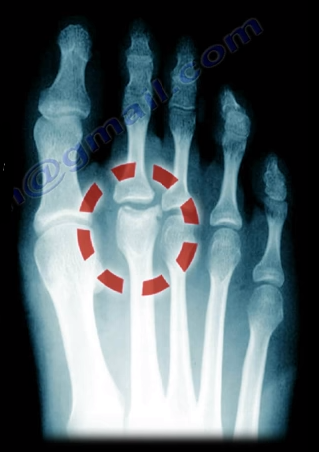
Freiberg’s Disease is caused by avascular necrosis of the
head of the 2nd metatarsal. This condition is more common in
patients who have a longer 2nd metatarsal bone relative to the 1st
metatarsal. This leads to the transfer of excessive loads onto the 2nd
metatarsal, which may interfere with the blood supply. This disease tends to
occur more commonly in young females during growth spurts.
Freiberg’s Disease usually presents itself as pain and
swelling at the 2nd metatarsophalangeal joint that is related to
activities and walking in high heels. There may be point tenderness and
swelling over the head of the 2nd metatarsal. There may also be
limited range of motion in the 2nd metatarsophalangeal joint. Early
in the disease, x-rays may only show minimal changes. Radiological evidence of
the condition may only be clearly visible on an MRI and bone scan. In more
severe cases, sclerosis, fragmentation, collapse of the metatarsal head, and
severe arthritis, may make the condition easily visible on an x-ray.
Treatment
Conservative treatment consists of nonsteroidal
anti-inflammatory medications, activity modification, orthotics, and immobilization
with a short leg cast. Surgical intervention involving a joint debridement is
indicated only after the failure of all conservative measures. Other procedures
may be indicated depending on the complexity and severity of the case.
Several bursa are seen around the knee area. These bursa
include the suprapatellar, prepatellar, infrapatellar, and pes anserine. The
pes answerine bursa is a small fluid filled sac located between the tibia and
the three tendons of the Sartorius, Gracilis, and Semi-tendinosus.
These
muscles are innervated by three separate nerves, the femoral, obturator, and
the tibial branch of the sciatic nerve, respectively. Pes Anserine bursitis, or
“breast stroke knee”, is an inflammatory condition of the medial knee at the
pes anserine bursa that is common in swimmers.
What is the pes anserine?
The pes anserine is the common area of insertion for the three
tendons along the proximal medial aspect of the tibia. This condition is also
sometimes referred to as a “goosefoot” because the pes anserinus tendons
resemble the shape of a goose foot. Pes Anserine bursitis is usually seen as
causing pain, tenderness, and localized swelling after trauma or total knee
replacement. The pain is seen below the joint line on the medial part of the
proximal tibial with the bursa being deep to the tendons.
Treatment
Treatment consists of physical therapy, nonsteroidal anti-inflammatory
medications, and injections. The physician will need to rule out meniscal
tears, stress fractures, or osteonecrosis of the tibia, as these are all
differential diagnosis.
Disc herniations usually occurs posterolaterally, but it may
also be central. The cauda equina is composed of several nerves within the
lower end of the spinal canal. The top of the spinal cord is a tubular bundle
of nervous tissue extending from the brain.
The following section of the spinal
cord is called the Conus Medullaris and is the lower end of the spinal cord.
The Cauda Equina is made up of multiple nerve roots beginning at the level of
L1.
The most common disc herniation is the Posterolateral Disc
Herniation. This type of herniation is a nerve root injury, which will cause
changes to both the sensory and motor skills as well as the reflexes. A
posterolateral disc herniation usually affects the foot and ankle, and may
cause unilateral leg pain and weakness. Observe for a positive straight leg
raising test. These herniations are usually initially treated with conservative
methods.
A central disc herniation will cause cauda equina syndrome,
which is a compression over the lumbosacral nerve roots. This compression will
cause more back pain than leg pain, and bladder and bowel symptoms will be
evident. This herniation is considered a surgical emergency.
Pain located at the heel can have several different causes.
It is important to make the correct diagnosis for the cause of the heel pain,
so that appropriate treatment can be given to the patient. Common causes of
heel pain include:
Baxter’s Nerve Compression
Plantar Fasciitis
Fat Pad Atrophy
Achilles Tendonitis
Stress Fractures of the Calcaneus
Lumbar Spine Radiculopathy
The sites and locations from different causes of the pain are
all very close to each other, making it difficult to determine the source of
the pain and to diagnose injuries.
In plantar fasciitis, irritation and swelling will occur at
the thick tissue on the bottom of the foot. This fascia can become inflamed and
painful, making walking more difficult. Plantar fasciitis is most severe in the
morning when patients first stand on their feet. Stretching exercises or
walking, often helps in relieving the painful tightening associated with
plantar fasciitis. Pain symptoms will intensify with prolonged exercise or
standing. Plantar fasciitis is usually
associated with a tight heel cord.
During fat pad atrophy, the fat that cushions the calcaneus
is thinned. This condition is common in elderly people and can cause
significant pain while walking. It also commonly occurs in patients with a
history of steroid injections.
Achilles Tendonitis is a chronic injury to the tendon that
joins the heel to the muscles of the lower leg, primarily occurring from
overuse. The Achilles tendon gives us the ability to rise up on our toes,
allowing for the act of walking. If a patient has Achilles tendonitis, walking
can be painful and difficult.
Stress fractures of the calcaneus can occur due to overuse
injuries. The patient will experience weight bearing pain. Stress fracture
injuries of the heel are typical in patients who engage in running sports, due
to the repetitive shock being placed on the heel. Side to side compression of
the calcaneus is painful.
Pain on the lateral side of the foot may be a result of
L5-S1 radiculopathy.
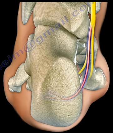
The Baxter’s nerve is the first branch of the lateral
plantar nerve. It also contributes to 20% of all heel pain causes. The Baxter’s
nerve provides motor innervation to the abductor digiti minimi muscle. When the
nerve is affected by compression, the symptoms are similar to planter
fasciitis. Entrapment of the Baxter’s nerve may cause up to 20% of heel pain,
but is still overlooked. The first branch of the lateral planter nerve is the
inferior calcaneal nerve (Baxter’s Nerve).
The nerve courses vertically between
the abductor hallucis and the quadratus plantae muscles, then makes a 90°
horizontal turn, coursing laterally beneath the calcaneus to innervate the
abductor digiti minimi muscle. The Baxter’s nerve can become entrapped within
the medial heel. The entrapment will result in heel pain, paresthesia, abnormal
sensations on the plantar aspect (bottom) of the heel, and medial heel
tenderness. There are two sites of entrapment that are typical with Baxter’s
nerve impingements. The first site is located between the fascia of the
abductor hallucis and quadratus plantae muscles. The second site is where the
nerve passes along the anterior aspect of the medial calcaneal tuberosity.
Treatment
Management is usually conservative and consists of heel
stretching exercises, rest, NSAIDs, and Orthotics. Surgery is done as a last
resort with neurolysis of the nerve after release of the deep fascia of the abductor
hallucis muscle. There is a good-excellent result in about 90% of patients
after decompression of the nerve. Involvement of the Baxter’s nerve may affect
running athletes causing pain on the medial plantar aspect of the foot.
It is difficult to reduce and maintain the reduction of
clavicle fractures as there will be deforming forces in the midshaft area of
the clavicle. The clavicle will heal regardless as to if a sling or figure 8
strap is used. Despite the presence of a bump, fracture displacement, and
deformity, healing of the fracture still occurs rapidly. Healing occurs in
about 85% of cases, however, the clavicle will not look aligned due to the
difficulty in reducing the fracture. It is hard to achieve a reduction of these
fracture without surgery. Without surgical reduction, the fracture may end with
some degree of malunion and possible shortening of the clavicle. The fragments
will not line up with the distal fragment appearing to be downward and
anteriorly rotated. Shortening is clinically significant because it alters the
dynamics of the muscles around the shoulder. It also narrows the
costoclavicular space. The patient may complain of decreased shoulder strength
and endurance if the patient had a displaced midshaft clavicle fracture that
healed with more than 2 cm of shortening.
What are the symptoms of clavicle malunion?
Pain, easy fatigability, cosmetic problems (especially in
females), neurological dysfunction (possible involvement of the brachial plexus—especially
the ulnar nerve), and the patient may have thoracic outlet syndrome.
X-rays should be done of both shoulders. Bilateral panoramic
views are beneficial in order to measure the shortening. The physician will
also want to check the amount of overlap at the ends of the clavicle.
Treatment
Surgical treatment of the malunion may be successful in
restoring the function and relieving the pain. A clavicle osteotomy is done in
the plane of the healed fracture. The fracture is recreated with correction of
the deformity and the length of the clavicle is restored. A local or iliac
crest bone graft can be used if needed.
If an osteotomy is done with reduction of the clavicle to
its preinjury position and leaves a significant bone defect, the physician will
need a tricortical piece of bone at the osteotomy area. It is probably better
to use a precontoured clavicle plate in the superior position with six
cortices, three placed on each side of the osteotomy.
An Elbow dislocation occurs when the radius and ulna bones
of the forearm move out of place from the humerus bone of the upper arm. There
are two basic types of elbow dislocations:
Simple
No fracture of the bones around the elbow
joint
Usually ligamentous injury
Complex
Fracture has occurred along with ligamentous
injury
Simple elbow dislocations typically occur when the patient
falls onto an outstretched hand. Injury progression from lateral to medial in
most patients. Posterolateral simple dislocations are the most common, occurring
approximately 90% of the time.
The proximal ulna and radius are displaced
posterolaterally relative to the distal humerus. Postemedial dislocations occur
at the proximal ulna and radius and are displaced posteromedially relative to
the distal humerus. In medial dislocations, the proximal ulna and radius are
displaced medially relative to the distal humerus. With lateral dislocations,
the proximal ulna and radius are displaced laterally relative to the distal
humerus. Anterior dislocations are rare, as they result from a direct force
applied to the posterior aspect of the forearm with the elbow in a flexed
position. Anterior dislocations occur when the proximal ulna and radius are
displaced anteriorly relative to the distal humerus. If stable, simple acute
fractures can be treated with a closed reduction and a splint for two to three
days (no more than two weeks) in addition to range-of-motion exercises and
physical therapy. Unstable simple fractures are rare, but can be stabilized by
ligament repair and/or by the use of an external fixator or cross pinning of
the joint in the elderly.
Chronic Elbow dislocations will need to be treated with an
open reduction and external fixator and is usually hinged. Recurrent elbow
dislocations (diagnosed by pivot shift) occurs due to a deficiency of the
lateral collateral ligament and is treated by a reconstruction of the
ulnohumeral ligament with a tendon graft.